Research Article | Volume 13, Issue 1, January, 2025

Characterization of the bioactive components of Chloris virgata Sw. and assessment of its antimicrobial and anti-arthritic potential

Dolly Kumari Priya Chaudhary Priya Yadav Pracheta Janmeda   

Open Access   

Published:  Nov 15, 2024

DOI: 10.7324/JABB.2024.195146
Abstract

The goals of this study are to characterize the bioactive ingredients present in the formulation of three parts of Chloris virgata Sw. and to assess their antimicrobial and anti-arthritic properties. C. virgata Sw. extracts (methanol, hydro-ethanol, and aqueous) were characterized using ultraviolet-visible (UV-Vis) spectroscopy and Fourier transform infrared spectroscopy (FTIR) techniques. Antimicrobial activity was measured with the disc diffusion method, and anti-arthritic activity was determined by an in vitro protein denaturation assay. The in-vivo study was performed on methanol extract, and the biochemical and hematological parameters were investigated by Freund’s complete adjuvant model. UV-visible spectroscopy revealed the presence of flavonoids, terpenoids, phenols, alkaloids, and saponins, whereas FTIR results show functional groups like carboxylic acids, alkenes, amides, carbohydrates and proteins, ether, and esters. Methanol and hydroethanol extracts showed higher antimicrobial activity at 400 μg/mL against Escherichia coli, Bacillus subtilis, and Aspergillus niger in comparison to the standards streptomycin and fluconazole at 10 μg/ mL. In-vitro results of the methanol extract clearly show 72.58% inhibition as compared to other extracts. In-vivo anti-arthritic evaluation was performed with methanol extract, and it showed remarkable paw reduction in the treated group on 21st day at a high dose (2.2 ± 0.1) as compared to indomethacin (1.74 ± 0.114). Hematological parameters such as hemoglobin, red blood cells (RBC), white blood cells (WBC), neutrophils, lymphocytes, and platelets, and biochemical parameters like alkaline phosphatase (ALP) (51.36 ± 9.113 IU/L), aspartate aminotransferase (AST) (64.65 ± 10.011 IU/L), and alanine aminotransferase (ALT) (129.35 ± 13.53 IU/L) were restored to normal in the treated. The findings indicated that C. virgata extracts under UV spectroscopy and FTIR revealed significant bioactive components, which are determined to be significant contributors to their antioxidant and antibacterial properties.


Keyword:     Anti-arthritic Antimicrobial Chloris virgata Sw. FTIR Rheumatoid arthritis UV-visible spectrometry


Citation:

Kumari D, Chaudhary P, Yadav P, Janmeda P. Characterization of the bioactive components of Chloris virgata Sw. and assessment of its antimicrobial and anti-arthritic potential. J App Biol Biotech. 2025; 13(1):135-142. http://doi.org/10.7324/JABB.2024.195146

Copyright: Author(s). This is an Open Access article distributed under the terms of the Creative Commons Attribution-NonCommercial-ShareAlike license.

HTML Full Text

1. INTRODUCTION

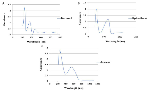
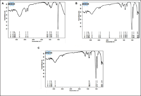
Rheumatoid arthritis (RA) is an inflammatory condition that mostly affects the joints. It causes mild swelling, and excessive and atypical synovial development, which finally alters the shape of the joint [1]. RA affects women more frequently than men, and although it can strike at any age, it usually appears between the ages of 50 and 60. Traditional remedies have been used as a form of healthcare since the prehistoric era and are frequently used to treat a wide range of disorders. Traditional remedies are safe for use at any age and have few negative effects [2]. To lead a healthy lifestyle, Ayurveda is extremely important. Animals, plants, and microorganisms all contribute to the synthesis of physiologically active phytochemical substances that are used in the creation of pharmaceuticals. These are classified as primary and secondary constituents depending on their role in plant metabolism. Many sophisticated techniques can be used to validate the presence of phytoconstituents in medicinal plants. Ultraviolet-Visible (UV-Vis) spectrometry and Fourier transform infrared spectroscopy (FTIR) are two effective techniques for identifying and determining the structure of phytochemicals [3].

Biologic agents, disease-modifying anti-rheumatic medicines (DMARDs), and non-steroidal anti-inflammatory drugs (NSAIDs) are commonly used to reduce inflammation and regulate symptoms [4], although they have been reported to have some side effects. According to estimates made by Arya et al. [5], about 43% of arthritis patients in India use herbal remedies to manage their condition. Chloris virgata Sw. of the Poaceae family is one of the weeds of warm weather in the eastern part of Australia, and its occurrence can be easily seen throughout the mainland of Australia. Resistance or tolerance to glyphosate, dispersal through flood and wind, and greater production of seeds are some factors that resulted in the prevalence of C. virgata Sw. in both non-cropping and cropping seasons [6,7]. Different transcriptomic analyses were done on C. virgata Sw. which reported the major genes responsible for their fastest growth and germination, especially in the major grasslands of Mongolia [8].

An intradermal or subdermal injection of Freund’s complete adjuvant (FCA) has been broadly used for the induction of arthritis in rats. However, its application to direct arthritis in mice has been reported occasionally, with less success [9]. Adjuvant directed arthritis has several limitations for an animal model; especially the disorder that resulted after the injection due to the resulting stress and severe discomfort. The polyarthritis caused by FCA is an attempt to result in less drastic arthritis by eliminating some of the complicating factors of this model. Major modifications include its injection locally, either around or into the tibiotarsal joint. These modifications allow the occurrence of localized arthritis without targeting the mobility of poor animals [10]. Panda et al. [11] reported that the decoction forms of C. virgata Sw. are employed by tribal people in Odisha, India, to treat a variety of rheumatic conditions. In rural Tamil Nadu, India, residents reportedly utilize C. virgata Sw. to treat rheumatism, inflammation, discomfort, and stomach aches. According to numerous studies, protein denaturation is a crucial factor in the development of rheumatoid arthritis. Therefore, the purpose of this study was to investigate all possible ideas and methods for future sources of herbal medicines to treat arthritis, as well as to characterize the bioactive components in C. virgata Sw. plant extracts using UV and FTIR.


2. MATERIALS AND METHODS

2.1. Plant Identification and Authentication

The three parts (leaves, stem, and roots) of C. virgata Sw. were collected from the Banasthali Vidyapith, Rajasthan, India, in 2019. The plant herbarium was submitted (voucher number: BURI 1392/2021; plantarum number: Fl. Ind. Occid. 1: 203 1797) and it was further identified and authenticated by the botanist of the Department of Bioscience and Biotechnology.

2.2. Preparation of Plant Extracts with Different Solvents

The plant sample was washed thoroughly and shade dried for one week, then ground into fine powder form and stored in an airtight container. About 30 g of powder (10 g from each leaf, stem, and root) was successively taken to carry out the successive extraction with different solvents [methanol, hydroethanol (50:50), and aqueous] using a soxhlet apparatus for 1-2 working days. The obtained extracts were filtered and dried using a rotary evaporator. The dried extracts were kept at 4°C for further experimental use [12]. For characterization and antimicrobial analysis, all three extracts (methanol, hydroethanol, and aqueous) of the whole plant C. virgata Sw. were taken. For in-vitro protein denaturation and in-vivo anti-arthritic studies, only the selected methanol extract of the whole plant of C. virgata Sw. was used.

2.3. Characterization of C. virgata Sw. Extracts

2.3.1. UV-Vis spectroscopic analysis

The plant extracts were dissolved in the respective solvent and centrifuged for 10 min at 3000 rpm. The supernatant was then filtered and diluted at a 1:10 ratio with the same solvent. The extracts were scanned with a Labman-UV1900 at a wavelength range of 200-1100 nm [13].

2.3.2. FTIR analysis

Subashini et al. [14] described a method for performing FTIR analysis. It was carried out in order to determine the functional groups present in C. virgata Sw. extracts and identify them based on peak values in the infrared radiation region.

2.4. Antimicrobial Activity of C. virgata Sw. Extracts

2.4.1. Test microorganisms and their inoculum preparation

The pure cultures of the test organisms were provided by the Department of Bioscience and Biotechnology, Banasthali Vidyapith, Rajasthan, India. Bacillus subtilis (B. subtilis; MTCC441), Escherichia coli, (E. coli; MTCC739), and Aspergillus niger (A. niger; MTCC282) were the bacterial and fungal strains employed in the investigation.

2.4.2. Antimicrobial activity (Disc diffusion method)

On the nutrition and potato dextrose agar plates, 250 µL of the standardized inoculum of microbial species was applied using a sterile cotton swab. On the infected agar plates, the extract-impregnated discs were deposited in varied quantities (ranging from 50 to 400 µg/mL) for antibacterial and antifungal purposes. Using the standards of fluconazole and streptomycin (10 µg/disc), the sensitivity of various bacterial and fungal species was assessed. For 24 h at 37°C (bacteria) and 72 h at 27°C (fungi), all petri dishes were incubated. The inhibitory zone diameter was measured in millimeters [15].

2.5. In-vitro Anti-arthritic Study

2.5.1. Protein denaturation inhibition assay

The method described by Djuichou et al. [16] was used to measure the proportion of denaturized protein that was inhibited by employing bovine serum albumin (BSA). At 660 nm, the absorbance was measured, and the inhibition percentage was computed using the following formula:

% Inhibition = (Control Abs -Sample Abs) ×100/ Control Abs.

2.6. In-vivo Anti-arthritic Study

2.6.1. Experimental animals

Male albino Wistar rats weighing between 150 and 200 g were purchased and in-vivo study was also done at Bilwal Medchem and Research Laboratory Pvt. Ltd. Rats were handled in accordance with the recommendations made by CPCSEA (BMRL/AD/CPCSEA/IAEC/2022/3/2).

2.6.2. Acute toxicity study

Organization for Economic Co-operation and Development (OECD) standards 423 were followed while evaluating the acute toxicity of the methanol extract of C. virgata Sw.

2.6.3. FCA-induced arthritis

For the purpose of studying FCA-induced arthritis, rats were divided into six groups, each containing five rats (n = 5). FCA was injected subcutaneously into the left hind paw in an amount of around 0.1 mL. The standard drugs indomethacin and methanol extract of C. virgata Sw. were subsequently administered daily until the 21st day following the second day of FCA injection. The body weight change was also noticed on the first, seventh, fourteenth, and twenty-first days [17]. Group 1- Normal control rats; Group 2- Arthritic control rats; Group 3- Arthritic rats + standard drug indomethacin at 10 mg/kg body weight (b.w.), orally (p.o.); Group 4- Arthritic rats + methanol extract of C. virgata Sw. at 200 mg/kg b.w., p.o.; Group 5- Arthritic rats + methanol extract of C. virgata Sw. at 300 mg/kg b.w., p.o.; and Group 6- Arthritic rats + methanol extract of C. virgata Sw. at 400 mg/kg b.w., p.o.

2.6.4. Evaluation of hematological and serological parameters

Blood samples were taken on the study’s last day (day 22) from each animal group using a retro-orbital puncture into two tubes. One tube is coated with EDTA. Shaking the samples must combine the blood and EDTA right away. Following that, an EDTA tube was sent through an automated Rayto, China hematoanalyzer to measure hemoglobin, red blood cells (RBCs), neutrophils, lymphocytes, platelets, and white blood cells (WBCs) [18]. To separate the serum, the anticoagulant-free blood tube was centrifuged at 2500 g for 10 min. Using the Abcam diagnostic kits, the levels of alkaline phosphatase (ALP), aspartate aminotransferase (AST), and alanine aminotransferase (ALT) in the serum sample were determined. The ALP, AST, and ALT concentrations were expressed as IU/L of serum.

2.7. Statistical Analysis

The mean and standard deviation (SD) of all data were calculated in triplicate. Using the IBM 8 SPSS (20) statistical program, one-way analysis of variance (ANOVA), post-hock analysis, and Dunnet’s test were performed for each of the factors under investigation. At p < 0.05 and p < 0.01, the values were deemed statistically significant.


3. RESULTS AND DISCUSSION

3.1. Characterization of C. virgata Sw. Extract by UV-Vis Spectroscopic

The primary bands were detected at absorption peaks, and the presence of flavonoids, phenols, saponins, terpenoids, and alkaloids in C. virgata Sw. extracts was indicated by the absorption bands [208 (0.628), 256 (2.268), 362 (1.083), 463 (0.436), and 622 (1.189)] obtained in the UV-Vis region [Figure 1]. The present study results were consistent with earlier research for alkaloids, flavonoids, terpenoids, and related glycosides by Rawat and Garg [19]. The current findings were also found to be compatible with the report of Yadav et al. [20]. The presence of various phytocompounds in C. virgata Sw. was also supported by the studies of Abbasi et al. [21] and Uddin et al. [22] using the leaf extracts of Rhamnus virgata and Berberis balochistanica.

Figure 1: UV-Vis spectrum of C. virgata extracts. (A). methanol; (B). hydroethanol; and (C). aqueous extract.


[Click here to view]

3.2. Characterization of C. virgata Sw. Extracts by FTIR Spectroscopy

Functional groups found in C. virgata Sw. at 3,676 and 3,421 (alcohol); 3,747 (hydroxyl); 1,017, 2,831, 3,327, and 1,114 (carboxylic acid); 1,454 and 2,997 (alkene); 1,649 (amide); 3,420 (carbohydrates and proteins); and 1,019 (ether and esters) are confirmed by FTIR analysis as depicted in Figure 2. The identified phytochemicals are advantageous to medical fields, such as alkenes, which are used as a starting point for the production of alcohols, while aldehydes are crucial for the fruit ripening process. Carboxylic acids have potent antibacterial properties and are also effective in treating headaches, fever, pain, rheumatic joint aches, ulcers, and jaundice, while amides are helpful in protein synthesis. The present study and the one conducted by Pharmawati and Wrasiati [23] are extremely comparable.

Figure 2: FTIR analysis of C. virgata extracts. (A). methanol extract; (B). hydroethanol; and (C). aqueous extract.


[Click here to view]

3.3. Antimicrobial Assay

3.3.1. Antibacterial activity of C. virgata Sw. extracts

Antibacterial results revealed that all three extracts of the experimental plant had the potential to inhibit the growth of bacteria. The methanol extract had the greatest zone of inhibition at 400 µg/mL for E. coli and B. subtilis, and no zone of inhibition was observed at 50 µg/mL for either of the two hydroethanol and aqueous extracts. However, the extracts zones of inhibition against E. coli and B. subtilis at 10 µg/mL were somewhat lower than those of standard streptomycin [Table 1 and Figures 3 and 4]. In support of the present study, the effectiveness of different extracts of Argemone mexicana against various pathogenic bacteria has also been investigated [24]. The conventional and green extracts of Salvia sclarea also represented the results in favor of the current study [25].

Table 1: Measure of zone of inhibition among different extracts of C. virgata against bacterial strains.

Plant extractsConcentration (1 mg/ml)Tested bacterial strains
E. coliB. subtilis
Methanol507.05 ± 3.046.07 ± 3.5
1008.1 ± 3.399.07 ± 5.23
2009.25 ± 4.0112.8 ± 7.40
4009.78 ± 0.0113.1 ± 6.40
Hydroethanol504.6 ± 2.655.32 ± 3.07
1005.32 ± 3.076.82 ± 3.94
2006.02 ± 3.478.35 ± 4.82
4008.37 ± 4.8312.12 ± 7.00
Aqueous50NZNZ
1004.6 ± 2.655.35 ± 3.09
2007.4 ± 0.086.82 ± 3.94
4006.77 ± 3.919.07 ± 5.23
Streptomycin (standard)10 µg/ml22 ± 0.87125 ± 0.654

Notes: All the results are expressed as mean ± SD (n = 5). NZ: no zone of inhibition.

Figure 3: Antibacterial study of C. virgata against E. coli- (A). standard (Streptomycin) and methanol, (B). aqueous, and (C). hydroethanol extract.


[Click here to view]
Figure 4: Antibacterial study of C. virgata against B. subtilis. (A). methanol, (B). aqueous, and (C). hydroethanol extracts.


[Click here to view]

3.3.2. Antifungal activity of C. virgata Sw. extracts

In this screening investigation, it was found that C. virgata Sw. methanol and hydroethanol extracts inhibited A. niger at concentrations between 50 and 400 µg/mL, but the aqueous extract did not exhibit any zone of inhibition at 50 µg/mL concentration. All extracts had negligible inhibitory effects when compared to standard fluconazole at 10 µg/mL, although methanol and hydroethanol extracts exhibited a greater zone of inhibition against A. niger at 400 µg/mL [Table 2 and Figure 5]. Comparable studies were carried out by Farjana et al. [26] on the effectiveness of several leaf extracts against different pathogens that cause disease and found somewhere similar results to the present findings. Similarly, the results of Okla et al. [27] in determining the antifungal activity of various parts of Avicennia marina were in favor of the outcomes of the current findings.

Table 2: Measure of zone of inhibition among different extracts of C. virgata against fungal strains.

Plant extracts/StandardConcentration (1 mg/ml)A. niger
Methanol505.32 ± 0.08
1006.82 ± 0.08
2009.07 ± 0.81
40012.1 ± 0.12
Hydroethanol504.57 ± 2.64
1006.1 ± 3.52
2008.35 ± 4.82
4009.82 ± 5.67
Aqueous50NZ
1003.97 ± 2.99
2004.57 ± 2.64
4005.35 ± 3.09
Fluconazole (standard)10 µg/ml21 ± 2.23

Notes: All the results are expressed as mean ± SD (n = 5). NZ: no zone of inhibition.

Figure 5: Antifungal activity of C. virgata against A. niger. (A). standard (Fluconazole) and methanol, (B). aqueous, and (C). hydroethanol extracts.


[Click here to view]

3.4. In-vitro Protein Denaturation Assay

As with usual medication, all of the extracts of C. virgata Sw. shown measurable anti-arthritic efficacy. The methanol extract denaturized the protein most efficiently (72.58 ± 0.022%) at the maximum dose (2000 µg/mL), while hydro-ethanol and aqueous extracts showed 68.91 ± 0.016% and 60.11 ± 0.012% inhibition, respectively [Table 3]. The denaturation of methanol extract was somehow less effective in comparison to standard diclofenac sodium at the maximum dosage, but it is quite close to it. In light of this, Khandelwal [28] showed a substantial anti-arthritic impact of Crossandra infundibuliformis leaves, which was consistent with the findings of this study. Similarly, Dharmadeva et al. [29] evaluated the in-vitro anti-inflammatory study of Ficus racemosa by using albumin denaturation methods, and the results obtained were quite comparable with the results of the current analysis.

Table 3: In-vitro protein denaturation of C. virgata extracts and standard diclofenac sodium.

Conc. (µg/ml)% inhibition
Diclofen ac sodiumMethanol extractHydroethanol extractAqueous extract
039.88 ± 0.01126.09 ± 0.02117.30 ± 0.00714.36 ± 0.021
10051.02 ± 0.02334.01 ± 0.01225.51 ± 0.01623.02 ± 0.012
25063.04 ± 0.03239.88 ± 0.01932.40 ± 0.01327.12 ± 0.010
50069.94 ± 0.02450.73 ± 0.02047.65 ± 0.01839.88 ± 0.020
100078.59 ± 0.03161.58 ± 0.01158.65 ± 0.01348.38 ± 0.015
200086.95 ± 0.02272.58 ± 0.02268.91 ± 0.01660.11 ± 0.012

Notes: All the results are expressed as mean ± SD (n = 5).

3.5. In-vivo Anti-arthritic Analysis of C. virgata Sw. Methanol Extract

3.5.1. Acute toxicity study

Methanol extract dosages up to 2000 mg/kg b.w. do not exhibit any hazardous or fatal effects. Rat eyes, skin, salivation, diarrhea, and behavioral patterns were not altered in any way. The relative organ weight of the rat did not significantly change. As a result, treatment dosages of 200, 300, and 400 mg/kg b.w. of C. virgata Sw. methanol extract were used in the experiment.

3.5.2. Change in body weight

A substantial decrease in body weight was seen in the arthritic control rats after normal control and arthritic control rats were compared. Rats given all three dosages of C. virgata Sw. methanol extract and the standard treatment indomethacin experienced a significant (p < 0.05) increase in body weight compared to the rats in the arthritic control group [Table 4]. High, medium, and low dosages of the methanol extract of C. virgata Sw. as well as standard medication significantly (p < 0.05) increased body weight compared to the healthy control group [Table 4]. The methanol extract and conventional medication induced rise in body weight may be connected to the intestine’s capacity to absorb nutrients. In a related study on Tridax procumbens, Petchi et al. [18] found that the arthritic control group displayed a substantial reduction in body weight compared to the control group. Similarly, the body weight was significantly lower in the arthritic rat, but during the last week of treatment with Nigella sativa, the weight of immunized rats tended to return to normal as observed in the present finding [30].

Table 4: Effect of C. virgata methanol extract on change in body weight in FCA-induced arthritic rats.

Groups0 day7th day14th day21st dayMean change in body weight
Normal control158.6 ± 2.516160.06 ± 2.081160.4 ± 1.504165.1 ± 0.8543.93%
Arthritic control133.8 ± 3.164a136.11 ± 3.200a140.07 ± 8.620a137.77 ± 8.161a2.9%
Standard indomethacin172.03 ± 5.078c180.28 ± 6.475d185.2 ± 10.362a192.88 ± 6.233c12.11%
Methanol extract of C. virgata (200 mg/kg)157.24 ± 14.89b149.42 ± 7.895b154.07 ± 10.625b165.9 ± 10.043b5.5%
Methanol extract of C. virgata (300 mg/kg)162.14 ± 12.485c164.95 ± 4.741c16.752 ± 10.043c171.77 ± 5.366b5.9%
Methanol extract of C. virgata (400 mg/kg)173.27 ± 13.047c174.08 ± 7.523d179.91 ± 8.963d187.08 ± 9.116c7.97%

Notes: All the results are expressed as mean ± SD (n = 5). ap < 0.05 vs. normal control, bp < 0.01 vs. normal control, cp < 0.05 vs. arthritic control, and dp < 0.01 vs. arthritic control.

3.5.3. Effect on paw diameter

Seven days later, there was a significant increase (p < 0.05) in the paw diameter of the arthritic control group of rats in contrast to the healthy control group. While the regular indomethacin group of rats showed a decline up until the last day. In comparison to the control group of arthritic rats [Table 5], the methanol extract of C. virgata Sw. action demonstrated (p < 0.05) a reduction of paw diameter at low, medium, and high doses, respectively. The inhibition by C. virgata Sw. methanol extract obtained on day 21 of treatment was effective and equivalent to the standard. This work is supported by Sarkar and Rai [17] investigation on the anti-arthritic properties of Trichosanthes dioica hydroalcoholic extract and Yadav et al. [31] of Heteropogon contortus methanol extract. Similarly, Singh et al. [32] also supported the significant effect of the methanol extract of Calotropis procera in reducing paw edema on the 17th, 21st, 24th, and 28th days.

Table 5: Effect of C. virgata methanol extract on paw edema thickness of rats.

Groups0 day7th day14th day21st day
Normal control5.33 ± 0.9295.54 ± 0.4135.64 ± 0.3655.75 ± 0.406
Arthritic control5.34 ± 0.181b7.84 ± 0.089a8.44 ± 0.181b8.92 ± 0.130c
Standard Indomethacin5.72 ± 0.192d (31%)5.95 ± 0.238c (54%)5.89 ± 0.083b (66%)5.78 ± 0.114a (80%)
Methanol extract of C. virgata (200 mg/kg)5.72 ± 0.192d (19%)6.9 ± 0.122c (24%)5.93 ± 0.238b (39%)5.86 ± 0.223a (51%)
Methanol extract of C. virgata (300 mg/kg)5.26 ± 0.270d (24%)6.16 ± 0.089c (34%)5.91 ± 0.151b (43%)5.84 ± 0.192a (58%)
Methanol extract of C. virgata (400 mg/kg)5.1 ± 0.141d (26%)5.98 ± 0.238c (39%)5.89 ± 0.187b (54%)5.81 ± 0.1a (75%)

Notes: All the results are expressed as mean ± SD (n = 5). ap < 0.05 vs. normal control, bp < 0.01 vs. normal control, cp < 0.05 vs. arthritic control, and dp < 0.01 vs. arthritic control.

3.5.4. Effect on hematological parameters

The total number of WBC, neutrophils, lymphocytes, and platelets in the arthritic control group II was significantly (p < 0.05) higher than in the healthy normal control group I. Nevertheless, the groups treated with indomethacin and methanol extract (GpIII to GpVI; Table 6) had significantly (p < 0.05) lower counts of neutrophils, lymphocytes, platelets, and total white blood cells. In the current investigation, rats with arthritis Gp II had lower RBC and Hb counts than control Gp I rats. The methanol extract of C. virgata Sw. for 21 days significantly (p < 0.05) improved the hemoglobin (Hb) content and RBC count towards normal in comparison to the arthritic control. This suggests that the extract has potential applications in arthritic disorders and indicates a significant improvement in the anemic state. Conditions that call for a WBC count include infectious and inflammatory disorders [33]. In their investigation, Rahman et al. [34] found that an ethanolic extract of Aquilaria agallocha leaves restored WBC and hemoglobin levels to normal, hence preventing rheumatoid arthritis. Similarly, treatment with crocetin raised hemoglobin, and red blood cells, along with a decrease in white blood cell count [35].

Table 6: Effect of C. virgata methanolic extract at three dosing levels and indomethacin on several parameters

GroupsHb (g/deci L)RBCs (106/mm3)WBCs (103/mm3)Neutrophils (103/mm3)Lymphocytes (103/mm3)Platelets (lakhs/mm3)
Normal control14.44 ± 0.389.14 ± 0.667.97 ± 0.3352.93 ± 0.245.24 ± 0.348563.3 ± 9.07
Arthritic control12.23 ± 0.016a6.82 ± 0.013d11.73 ± 0.0151d6.83 ± 0.023c10.41 ± 0.008e821.6 ± 18.52a
Standard indomethacin14.32 ± 0.008e10.46 ± 0.0151b6.54 ± 0.023b3.74 ± 0.0240a6.46 ± 0.0216c625.4 ± 10.01a
Methanol extract (200 mg/kg)12.82 ± 0.083b7.58 ± 0.0678c10.53 ± 0.0173c6.17 ± 0.0070b9.32 ± 0.032d7.69 ± 17.10a
Methanol extract (300 mg/kg)13.23 ± 0.015c8.46 ± 0.1673b6.26 ± 0.02b9.75 ± 0.025e5.55 ± 0.02b714 ± 17.38a
Methanol extract (400 mg/kg)14.03 ± 0.029d9.72 ± 0.0192a5.74 ± 0.015a8.84 ± 0.016d4.75 ± 0.013a654 ± 20.69a

Notes: All the results are expressed as mean ± SD (n = 5). ap < 0.05 vs. normal control, bp < 0.01 vs. normal control, cp < 0.05 vs. arthritic control, and dp < 0.01 vs. arthritic control.

3.5.5. Effect on serological parameters

ALP, AST, and ALT levels in all arthritic rats were significantly (p < 0.05) higher than those in the healthy control group. The enzyme levels were considerably (p < 0.05) lower in rats in Groups 3 (standard) and 6 (high dosage) than in rats in the arthritic control group, and just slightly lower in Groups 4 and 5. The standard group had the lowest levels of ALP, AST, and ALT, measuring 43.5 ± 7.844, 53.9 ± 10.34, and 121.5 ± 38.79 IU/L, respectively, as compared to the extract-treated groups (Gp IV to GP VI) [Table 7]. This demonstrates the anti-arthritic effectiveness of the therapy. Kamal et al. [36] found a notable and significant rise in the liver enzymes (ALP, AST, and ALT) with Aloe thraskii extract, which has protective benefits against rheumatoid arthritis. The findings of the current study are consistent with the other investigations determining the action of FCA-induced arthritis in rats along with its treatment using phytocompounds such as silibinin [37]. Similarly, treatment with Nyctanthes arbor-tristis restored the elevated levels of AST and ALT in arthritis-induced rats [38].

Table 7: Effect of standard and methanol extract of C. virgata on various serological parameters.

GroupsALP (IU/L)AST (IU/L)ALT (IU/L)
Normal control44.13 ± 1.724b57.76 ± 1.569a53.2 ± 1.708b
Arthritic control144.71 ± 9.763c113.0 ± 25.78c226.3 ± 25.71c
Standard indomethacin (10 mg/kg)43.5 ± 7.844a53.9 ± 10.34a121.5 ± 38.79a
Methanol extract of C. virgata (200 mg/kg)87.63 ± 16.08c104.2 ± 12.49c205.9 ± 26.53b,c
Methanol extract of C. virgata (300 mg/kg)73.72 ± 10.92b78.45 ± 9.783b161.96 ± 11.86b
Methanol extract of C. virgata (400 mg/kg)51.36 ± 9.113a,b64.65 ± 10.011a,b129.35 ± 13.53a

Notes: All the results are expressed as mean ± SD (n = 5). ap < 0.05 vs. normal control, bp < 0.01 vs. normal control, cp < 0.05 vs. arthritic control, and dp < 0.01 vs. arthritic control.


4. CONCLUSION

Findings concluded that the extracts of C. virgata Sw. under FTIR and UV spectroscopy revealed the presence of significant bioactive components that were found to be important contributors to its antibacterial and anti-arthritic activities. Treatment with methanolic extract at a dose of 400 mg/kg of C. virgata possesses potentially useful anti-arthritic activity since it gives a positive result in controlling inflammation in rats induced with FCA. It has reduced body weight, paw edema swelling, and decreased inflammatory cytokine levels. The study validated the promising anti-arthritic activity of C. virgata Sw.; however, more research is needed to standardize the plant extract before it can be considered a viable treatment option alongside conventional medicines.


5. ACKNOWLEDGMENTS

The authors sincerely thank the Vice-Chancellor of the Banasthali Vidyapith for making provisions of research facilities for us and also acknowledge the Bioinformatics Centre, which is supported by the DBT and DST for providing computation as well as networking provision at the Department of Bioscience and Biotechnology, Banasthali Vidyapith, Rajasthan.


6. AUTHOR CONTRIBUTIONS

All authors made substantial contributions to conception and design, acquisition of data, or analysis and interpretation of data; took part in drafting the article or revising it critically for important intellectual content; agreed to submit to the current journal; gave final approval of the version to be published; and agree to be accountable for all aspects of the work. All the authors are eligible to be an author as per the international committee of Medical Journal Editors (ICMJE) requirements/guidelines.


7. ETHICAL APPROVALS

The study protocol was approved by the Institutional Animal Ethics Committee of Bilwal Medchem and Research Laboratory Pvt. Ltd., Rajasthan (Approval No.: BMRL/AD/CPCSEA/IAEC/2022/3/2).


8. CONFLICTS OF INTEREST

The authors declare no conflicts of interest associated with this study.


9. FINANCIAL SUPPORT AND SPONSORSHIP

This study is not supported by any funding agency.


10. DATA AVAILABILITY

Data will be available from the corresponding author on special request.


11. USE OF ARTIFICIAL INTELLIGENCE (AI)-ASSISTED TECHNOLOGY

The authors confirm that there was no use of artificial intelligence (AI)-assisted technology for assisting in the writing or editing of the manuscript and no images were manipulated using AI.


12. PUBLISHER’S NOTE

This journal remains neutral with regard to jurisdictional claims in published institutional affiliation.


REFERENCES

1.  Wang Q, Kuang H, Su Y, Sun Y, Feng J, Guo R, Chan K. Naturally derived anti-inflammatory compounds from Chinese medicinal plants. J Ethnopharmacol. 2013;146(1):9-39. [CrossRef]

2.  Shinwari ZK. Medicinal plants research in Pakistan. J Med Plant Res. 2010;4(3):161-76.

3.  Altemimi A, Lakhssassi N, Baharlouei A, Watson DG, Lightfoot DA. Pytochemicals: extraction, isolation, and identification of bioactive compounds from plant extracts. Plants. 2017;6(4):42. [CrossRef]

4.  Rajendran R, Krishnakumar E. Anti-arthritic activity of Premna serratifolia (Linn.), wood against adjuvant induced arthritis. Avicenna J Med Biotechnol. 2010;2(2):101-6.

5.  Arya V, Gupta VK, Kaur R. A review on plants having antiarthritis potential. Int J Pharm Sci Rev Res. 2011;7(2):131-6.

6.  Mahajan G, Chauhan BS. Evaluation of preemergent herbicides for Chloris virgata control in Mungbean. Plants. 2021;10(8):1632. [CrossRef]

7.  Rojas-Sandoval J. Chloris virgata (feather finger grass). CABI Compendium. 2022. [CrossRef].

8.  Bolortuya B, Kawabata S, Yamagami A, Davaapurev B-O, Takahashi F, Inoue K, et al. Transcriptome analysis of Chloris virgata, which shows the fastest germination and growth in the major Mongolian grassland plant. Front Plant Sci. 2021;12:684987. [CrossRef].

9.  Liu C, Li Y, Wen C, Olatunji OJ, Yin Z. Dehydrozingerone alleviates hyperalgesia, oxidative stress and inflammatory factors in complete Freund’s adjuvant-induced arthritic rats. Drug Design Develop Therapy. 2022;16:3015-22. [CrossRef]

10.  Naz R, Ahmed Z, Shahzad M, Shabbir A, Kamal F. Amelioration of rheumatoid arthritis by anacardium occidentale via inhibition of collagenase and lysosomal enzymes. Evid Based Complementary Altern Med. 2020;2020:8869484. [CrossRef]

11.  Panda SS, Sahu SC, Dhal N K. Documentation of tribal claims for rheumatism in Odisha, India. Int J Pharm Sci. 2015;7:379-84.

12.  Hossen SM, Hossain MS, Yusuf ATM, Chaudhary P, Emon NU, Janmeda P. Profiling of phytochemical and antioxidant activity of wild mushrooms: evidence from the in vitro study and phytoconstituent’s binding affinity to the human erythrocyte catalase and human glutathione reductase. Food Sci Nutr. 2022;10(1):88-102. [CrossRef].

13.  Karpagasundari C, Kulothungan S. Analysis of bioactive compounds in Physalis minima leaves using GC MS, HPLC, UV-VIS and FTIR techniques. J Pharmacogn Phytochem. 2014;3(4):196-201.

14.  Subashini MS, Rajendran P, Ashok G, Kanthesh BM. TLC, FTIR and GCMS analysis of leaves of Gymnema sylvestre R. Br from Kolli Hills, Tamil Nadu, India. Int J Curr Microbiol App Sci. 2015;4(7):757-64.

15.  Chew AL, Jessica JJA, Sasidharan S. Antioxidant and antibacterial activity of different parts of Leucas aspera. Asian Pac J Trop Biomed. 2012;2(3):176-80. [CrossRef]

16.  Djuichou Nguemnang SF, Tsafack EG, Mbiantcha M, Gilbert A, Atsamo AD, Yousseu Nana W, et al. In vitro anti-inflammatory and in vivo antiarthritic activities of aqueous and ethanolic extracts of Dissotis thollonii Cogn. (Melastomataceae) in rats. Evid Based Complementary Altern Med. 2019;3612481. [CrossRef]

17.  Sarkar AK, Rai AP. Phytochemical investigation and anti-arthritic activity of hydroalcoholic extracts of Trichosanthes dioica. MAP. 2020;9(5):356. [CrossRef]

18.  Petchi RR, Vijaya C, Parasuraman S. Anti-arthritic activity of ethanolic extract of Tridax procumbens (Linn.) in Sprague Dawley Rats. Pharmacogn Res. 2013;5(2):113-7. [CrossRef]

19.  Rawat B, Garg AP. Characterization of phytochemicals isolated from Cucurbita pepo seeds using UV–Vis and FTIR spectroscopy. Plant Arch. 2021;21(1):892-9. [CrossRef]

20.  Yadav P, Chaudhary P, Kumari D, Janmeda P. Assessment of phytochemical screening and antioxidant potential of Heteropogon contortus (L.) whole plant. T Appl Biol Chem. 2023;3(3):62-70. [CrossRef]

21.  Abbasi BA, Iqbal J, Khan Z, Ahmad R, Uddin S, Shahbaz A, et al. Phytofabrication of cobalt oxide nanoparticles from Rhamnus virgata leaves extract and investigation of different bioactivities. Microscop Res Tech. 2020;84(2):192-201. [CrossRef]

22.  Uddin S, Safdar LB, Iqbal J, Yaseen T, Laila S, Anwar S, et al. Green synthesis of nickel oxide nanoparticles using leaf extract of Berberis balochistanica: Characterization, and diverse biological applications. Microscop Res Tech. 2021;84(9):2004-16. [CrossRef]

23.  Pharmawati M, Wrasiati LP. Phytochemical screening and FTIR spectroscopy on crude extract from Enhalus acoroides leaves. Malaysian J Anal Sci. 2020;24(1):70-7.

24.  Bhattacharjee I, Chatterjee SK, Chatterjee S, Chandra G. Antibacterial potentiality of Argemone mexicana solvent extracts against some pathogenic bacteria. Mem Inst Oswaldo Cruz. 2006;101:645-8.

25.  Quradha MM, Duru ME, Kucukaydin S, Tamfu AN, Lqbal M, Bibi H, et al. Comparative assessment of phenolic composition profile and biological activities of green extract and conventional extracts of Salvia sclarea. Sci Rep. 2024;14:1885. [CrossRef]

26.  Farjana A, Zerin N, Kabir MS. Antimicrobial activity of medicinal plant leaf extracts against pathogenic bacteria. Asian Pac J Trop Dis. 2014;4: S920-3. [CrossRef]

27.  Okla MK, Alatar AA, Al-amri SS, Soufan WH, Ahmad A, Abdel-Maksoud MA. Antibacterial and antifungal activity of the extracts of different parts of Avicennia marina (Forssk.) Vierh Plants. 2021;10(2):252. [CrossRef]

28.  Khandelwal KR. Practical pharmacognosy. Technique and experiments. 9th ed. Pune, India: Nirali Publications; 2000.

29.  Dharmadeva S, Galgamuwa LS, Prasadinie C, Kumarasinghe N. In vitro anti-inflammatory activity of Ficus racemosa L. bark using albumin denaturation method. Ayu. 2018;39(4):239-42. [CrossRef]

30.  Nasuti C, Fedeli D, Bordoni L, Piangerelli M, Servili M, Selvaggini R, Gabbianelli R. Anti-inflammatory, anti-arthritic and anti-nociceptive activities of nigella sativa oil in a rat model of arthritis. Antioxidants (Basel). 2019;8(9):342. [CrossRef]

31.  Yadav P, Chaudhary P, Singh D, Chaudhary S, Prakash A, Janmeda P. Pharmacognostic, antimicrobial and antiarthritic properties of heteropogon contortus (L.). Biochem. Cell. Arch. 2023;23(1):651-61. [CrossRef]

32.  Singh VS, Dhawale SC, Shakeel F, Faiyazuddin M, Alshehri S. Antiarthritic potential of Calotropis procera leaf fractions in FCA-induced arthritic rats: involvement of cellular inflammatory mediators and other biomarkers. Agriculture. 2021;11(1):68. [CrossRef]

33.  Hu F, Hepburn HR, Li Y, Chen M, Radloff SE, Daya S. Effect of ethanol and water extracts of Propolis (bee glue) on acute inflammatory animal models. J. Ethnopharmacol. 2005;100(3):276-83. [CrossRef]

34.  Rahman H, Eswaraiah MC, Dutta AM. Anti-arthritic activity of leaves and oil of Aquilaria agallocha. Saudi J Life Sci. 2016;1(1):34-43.

35.  Li Y, Kakkar R, Wang J. In vivo and in vitro approach to anti-arthritic and anti-inflammatory effect of crocetin by alteration of nuclear factor-e2-related factor 2/hem oxygenase (HO)-1 and NF-κB expression. Front Pharmacol. 2018;9:1341. [CrossRef]

36.  Kamal RM, Sabry MM, Aly ZY, Hifnawy MS. Phytochemical and in- vivo anti-arthritic significance of Aloe thraskii baker in combined therapy with methotrexate in adjuvant-induced arthritis in rats. Molecules. 2021;26(12):3660. [CrossRef]

37.  Khawaja G, El-Orfali Y. Silibinin’s effects against methotrexate-induced hepatotoxicity in adjuvant-induced arthritis rat model. Pharmaceuticals. 2024;17(4):431. [CrossRef]

38.  Sharma A, Goel A, Lin Z. Analysis of anti-rheumatic activity of Nyctanthes arbor-tristis via in vivo and pharmacovigilance approaches. Front Pharmacol. 2023;14:1307799. [CrossRef]

Reference

1. Wang Q, Kuang H, Su Y, Sun Y, Feng J, Guo R, Chan K. Naturally derived anti-inflammatory compounds from Chinese medicinal plants. J Ethnopharmacol. 2013;146(1):9-39. https://doi.org/10.1016/j.jep.2012.12.013

2. Shinwari ZK. Medicinal plants research in Pakistan. J Med Plant Res. 2010;4(3):161-76.

3. Altemimi A, Lakhssassi N, Baharlouei A, Watson DG, Lightfoot DA. Pytochemicals: extraction, isolation, and identification of bioactive compounds from plant extracts. Plants. 2017;6(4):42. https://doi.org/10.3390/plants6040042

4. Rajendran R, Krishnakumar E. Anti-arthritic activity of Premna serratifolia (Linn.), wood against adjuvant induced arthritis. Avicenna J Med Biotechnol. 2010;2(2):101-6.

5. Arya V, Gupta VK, Kaur R. A review on plants having antiarthritis potential. Int J Pharm Sci Rev Res. 2011;7(2):131-6.

6. Mahajan G, Chauhan BS. Evaluation of preemergent herbicides for Chloris virgata control in Mungbean. Plants. 2021;10(8):1632. https://doi.org/10.3390/plants10081632

7. Rojas-Sandoval J. Chloris virgata (feather finger grass). CABI Compendium. 2022. https://doi.org/10.1079/cabicompendium.113265

8. Bolortuya B, Kawabata S, Yamagami A, Davaapurev B-O, Takahashi F, Inoue K, et al. Transcriptome analysis of Chloris virgata, which shows the fastest germination and growth in the major Mongolian grassland plant. Front Plant Sci. 2021;12:684987. https://doi.org/10.3389/fpls.2021.684987

9. Liu C, Li Y, Wen C, Olatunji OJ, Yin Z. Dehydrozingerone alleviates hyperalgesia, oxidative stress and inflammatory factors in complete Freund's adjuvant-induced arthritic rats. Drug Design Develop Therapy. 2022;16:3015-22. https://doi.org/10.2147/DDDT.S374827

10. Naz R, Ahmed Z, Shahzad M, Shabbir A, Kamal F. Amelioration of rheumatoid arthritis by anacardium occidentale via inhibition of collagenase and lysosomal enzymes. Evid Based Complementary Altern Med. 2020;2020:8869484. https://doi.org/10.1155/2020/8869484

11. Panda SS, Sahu SC, Dhal N K. Documentation of tribal claims for rheumatism in Odisha, India. Int J Pharm Sci. 2015;7:379-84.

12. Hossen SM, Hossain MS, Yusuf ATM, Chaudhary P, Emon NU, Janmeda P. Profiling of phytochemical and antioxidant activity of wild mushrooms: evidence from the in vitro study and phytoconstituent's binding affinity to the human erythrocyte catalase and human glutathione reductase. Food Sci Nutr. 2022;10(1):88-102. https://doi.org/10.1002/fsn3.2650

13. Karpagasundari C, Kulothungan S. Analysis of bioactive compounds in Physalis minima leaves using GC MS, HPLC, UV-VIS and FTIR techniques. J Pharmacogn Phytochem. 2014;3(4):196-201.

14. Subashini MS, Rajendran P, Ashok G, Kanthesh BM. TLC, FTIR and GCMS analysis of leaves of Gymnema sylvestre R. Br from Kolli Hills, Tamil Nadu, India. Int J Curr Microbiol App Sci. 2015;4(7):757-64.

15. Chew AL, Jessica JJA, Sasidharan S. Antioxidant and antibacterial activity of different parts of Leucas aspera. Asian Pac J Trop Biomed. 2012;2(3):176-80. https://doi.org/10.1016/S2221-1691(12)60037-9

16. Djuichou Nguemnang SF, Tsafack EG, Mbiantcha M, Gilbert A, Atsamo AD, Yousseu Nana W, et al. In vitro anti-inflammatory and in vivo antiarthritic activities of aqueous and ethanolic extracts of Dissotis thollonii Cogn. (Melastomataceae) in rats. Evid Based Complementary Altern Med. 2019;3612481. https://doi.org/10.1155/2019/3612481

17. Sarkar AK, Rai AP. Phytochemical investigation and anti-arthritic activity of hydroalcoholic extracts of Trichosanthes dioica. MAP. 2020;9(5):356.

18. Petchi RR, Vijaya C, Parasuraman S. Anti-arthritic activity of ethanolic extract of Tridax procumbens (Linn.) in Sprague Dawley Rats. Pharmacogn Res. 2013;5(2):113-7. https://doi.org/10.4103/0974-8490.110541

19. Rawat B, Garg AP. Characterization of phytochemicals isolated from Cucurbita pepo seeds using UV-Vis and FTIR spectroscopy. Plant Arch. 2021;21(1):892-9. https://doi.org/10.51470/PLANTARCHIVES.2021.v21.no1.122

20. Yadav P, Chaudhary P, Kumari D, Janmeda P. Assessment of phytochemical screening and antioxidant potential of Heteropogon contortus (L.) whole plant. T Appl Biol Chem. 2023;3(3):62-70. https://doi.org/10.52679/tabcj.2022.0007

21. Abbasi BA, Iqbal J, Khan Z, Ahmad R, Uddin S, Shahbaz A, et al. Phytofabrication of cobalt oxide nanoparticles from Rhamnus virgata leaves extract and investigation of different bioactivities. Microscop Res Tech. 2020;84(2):192-201. https://doi.org/10.1002/jemt.23577

22. Uddin S, Safdar LB, Iqbal J, Yaseen T, Laila S, Anwar S, et al. Green synthesis of nickel oxide nanoparticles using leaf extract of Berberis balochistanica: Characterization, and diverse biological applications. Microscop Res Tech. 2021;84(9):2004-16. https://doi.org/10.1002/jemt.23756

23. Pharmawati M, Wrasiati LP. Phytochemical screening and FTIR spectroscopy on crude extract from Enhalus acoroides leaves. Malaysian J Anal Sci. 2020;24(1):70-7.

24. Bhattacharjee I, Chatterjee SK, Chatterjee S, Chandra G. Antibacterial potentiality of Argemone mexicana solvent extracts against some pathogenic bacteria. Mem Inst Oswaldo Cruz. 2006;101:645-8. https://doi.org/10.1590/S0074-02762006000600011

25. Quradha MM, Duru ME, Kucukaydin S, Tamfu AN, Lqbal M, Bibi H, et al. Comparative assessment of phenolic composition profile and biological activities of green extract and conventional extracts of Salvia sclarea. Sci Rep. 2024;14:1885. https://doi.org/10.1038/s41598-024-51661-z

26. Farjana A, Zerin N, Kabir MS. Antimicrobial activity of medicinal plant leaf extracts against pathogenic bacteria. Asian Pac J Trop Dis. 2014;4: S920-3. https://doi.org/10.1016/S2222-1808(14)60758-1

27. Okla MK, Alatar AA, Al-amri SS, Soufan WH, Ahmad A, Abdel- Maksoud MA. Antibacterial and antifungal activity of the extracts of different parts of Avicennia marina (Forssk.) Vierh Plants. 2021;10(2):252. https://doi.org/10.3390/plants10020252

28. Khandelwal KR. Practical pharmacognosy. Technique and experiments. 9th ed. Pune, India: Nirali Publications; 2000.

29. Dharmadeva S, Galgamuwa LS, Prasadinie C, Kumarasinghe N. In vitro anti-inflammatory activity of Ficus racemosa L. bark using albumin denaturation method. Ayu. 2018;39(4):239-42. https://doi.org/10.4103/ayu.AYU_27_18

30. Nasuti C, Fedeli D, Bordoni L, Piangerelli M, Servili M, Selvaggini R, Gabbianelli R. Anti-inflammatory, anti-arthritic and anti-nociceptive activities of nigella sativa oil in a rat model of arthritis. Antioxidants (Basel). 2019;8(9):342. https://doi.org/10.3390/antiox8090342

31. Yadav P, Chaudhary P, Singh D, Chaudhary S, Prakash A, Janmeda P. Pharmacognostic, antimicrobial and antiarthritic properties of heteropogon contortus (L.). Biochem. Cell. Arch. 2023;23(1):651- 61. https://doi.org/10.51470/bca.2023.23.1.651

32. Singh VS, Dhawale SC, Shakeel F, Faiyazuddin M, Alshehri S. Antiarthritic potential of Calotropis procera leaf fractions in FCA-induced arthritic rats: involvement of cellular inflammatory mediators and other biomarkers. Agriculture. 2021;11(1):68. https://doi.org/10.3390/agriculture11010068

33. Hu F, Hepburn HR, Li Y, Chen M, Radloff SE, Daya S. Effect of ethanol and water extracts of Propolis (bee glue) on acute inflammatory animal models. J. Ethnopharmacol. 2005;100(3):276- 83. https://doi.org/10.1016/j.jep.2005.02.044

34. Rahman H, Eswaraiah MC, Dutta AM. Anti-arthritic activity of leaves and oil of Aquilaria agallocha. Saudi J Life Sci. 2016;1(1):34-43.

35. Li Y, Kakkar R, Wang J. In vivo and in vitro approach to anti-arthritic and anti-inflammatory effect of crocetin by alteration of nuclear factor-e2-related factor 2/hem oxygenase (HO)-1 and NF-κB expression. Front Pharmacol. 2018;9:1341. https://doi.org/10.3389/fphar.2018.01341

36. Kamal RM, Sabry MM, Aly ZY, Hifnawy MS. Phytochemical and in- vivo anti-arthritic significance of Aloe thraskii baker in combined therapy with methotrexate in adjuvant-induced arthritis in rats. Molecules. 2021;26(12):3660. https://doi.org/10.3390/molecules26123660

37. Khawaja G, El-Orfali Y. Silibinin's effects against methotrexate-induced hepatotoxicity in adjuvant-induced arthritis rat model. Pharmaceuticals. 2024;17(4):431. https://doi.org/10.3390/ph17040431

38. Sharma A, Goel A, Lin Z. Analysis of anti-rheumatic activity of Nyctanthes arbor-tristis via in vivo and pharmacovigilance approaches. Front Pharmacol. 2023;14:1307799. https://doi.org/10.3389/fphar.2023.1307799

Article Metrics
77 Views 58 Downloads 135 Total

Year

Month

Related Search

By author names

Similar Articles

Bioactivity assessment of endophytic fungi associated with Centella asiatica and Murraya koengii

Archana Nath, Jyoti Pathak and SR Joshi

Antibacterial activity of Ferula asafoetida: a comparison of red and white type

Richa Bhatnager, Reena Rani, Amita Suneja Dang

Medicinal and Functional Values of Thyme (Thymus vulgaris L.) Herb

Eqbal M. A. Dauqan, Aminah Abdullah

Probiotic potential of thermotolerant lactic acid bacteria isolated from “Gari “a cassava-based African fermented food

Bertrand Tatsinkou Fossi, Robert Ndjouenkeu

Chemical composition, antioxidant and antimicrobial activities of the essential oil of Vetiveria nigritana (Benth.) Stapf roots from Burkina Faso

Zenabou Semde, Jean Koudou, Cheikna Zongo, Gilles Figueredo, Marius K. Somda, Leguet Ganou, Alfred S. Traore

Production, characterization and optimization of fermented tomato and carrot juices by using Lysinibacillus sphaericus isolate

Naga Sivudu Seelam, Harika Akula, Umamahesh Katike, Vijaya Sarathi Reddy Obulam

Antimicrobial applications of sophorolipid from Candida bombicola: A promising alternative to conventional drugs

Victoria Akemi Itakura Silveira, Christiane Aparecida Urzedo Queiroz Freitas, Maria Antonia Pedrine Colabone Celligoi

Antibacterial and antifungal activities of leaf and stem of Marsilea minuta L. against selected microbial pathogens

Govindaraj Sabithira, Rajangam Udayakumar

Antibacterial potential of Caesalpinia bonducella extracts and their isolated phytoconstituents: in vitro and in silico analysis

S R Santosh Kumar, Sudhesh L Shastri, Venkatesh R, K Pradeepa, V Krishna

Phytochemical analysis, antimicrobial and antioxidant activities of Aidia borneensis leaf extracts

Zulhamizan Awang-Jamil, Aida Maryam Basri, Norhayati Ahmad, Hussein Taha

Biological activities and phytochemicals of Hyptis capitata grown in East Kalimantan, Indonesia

Irawan Wijaya Kusuma, Rahmini, Enos Tangke Arung, Arif Yudo Pramono, Erwin, Supomo

Partial purification and characterization of antimicrobial peptide from the hemolymph of cockroach Periplaneta americana

Reymend Encily Martin, Yogita Raosaheb Channe

Prevalence and antimicrobial susceptibility profile of Mycoplasma hominis and Ureaplasma urealyticum in female population, Gabon

Mohamed Ag Baraïka, Richard Onanga, Berthold Bivigou-Mboumba, Arsène Mabika-Mabika, Ulrick Jolhy Bisvigou, Fousseyni S. Touré Ndouo, N. Coumba Touré Kane

Enhanced production of a bioactive molecule from a symbiotic marine bacterium, Paenibacillus macerans SAM 9 isolated from the sea anemone, Heteractis aurora

Thayanithi Bharathi, Kathirvelu Sambandan, Kandasamy Sivasubramani

Antimicrobial, anti-inflammatory, and anticancer activities of leaves extracts of Filicium decipiens

Atiyaparveen I Basarikatti, Sanjay Mishra, Vijaykumar Uppar, Basavaraj Padmashali

The comparative antimicrobial and anticancer of chemical extract from in vitro and in vivo Peperomia pellucida plantlet

Lydia Teoh, Nareshwaran Gnanasegaran, Ahmad Faris Mohd Adnan, Rosna Mat Taha

Screening of synergistic and antimicrobial effect of Cr (VI) and Ni (II) tolerant bacteria Bacillus cereus

Subhayan Dutta, Khaleda Firdous, Swati Chakraborty

Antimicrobial activity of the lichens Parmotrema andium and Dirinaria applanata

Bijayananda Sahoo, Satyabrata Dash, Sabyasachy Parida, Jayanta Kumar Sahu, Biswajit Rath

Production of bioactive compounds by Streptomyces sp. and their antimicrobial potential against selected MDR uropathogens

Archana Singh, Padma Singh

Diversity and antimicrobial potential of endophytic fungi from aromatic plants of Bhadra Wildlife Sanctuary, Western Ghats, Karnataka

Rajeshwari Jagadish, Srinivas Chowdappa

A review on fish peptides isolated from fish waste with their potent bioactivities

Ayusman Behera, Rajashree Das, Pranati Patnaik, Jyotirmaya Mohanty, Gargee Mohanty

Antimicrobial peptide coding gene of thermophilic bacteria isolated from crater hot spring in mountains around West Java

Emma Rachmawati, Sinta Asarina, Gabriel Bagus Kennardi, Ratu Safitri, Toto Subroto, Ani Melani Maskoen

Leveraging monoclonal antibodies as therapeutics to address antimicrobial resistance in bacteria

Vaishali Verma

Antimicrobial potential of Indian cooperative spider, Stegodyphus sarasinorum, and Golden orb spider, Nephila pilipes hemolymph against pathogenic bacteria

Dhanapal Sangavi, Padur Sankaranarayanan Anisha, Douglas J. H. Shyu, Parthasarathy Thiruchenthil Nathan

A novel trypsin inhibitor peptide MoCh I with antimicrobial activity derived from Momordica charantia

Samriti Dogra, Rajesh Biswas, Rupinderjeet Kaur, Sangeeta Sharma, Kakoli Biswas

Isolation of yeast endophytes from healthy seeds of Capsicum annuum L. and assessment of their antimicrobial activity

Barbi Bhuyan, Purthimi Kungri Hansepi, Suraiya Akhtar, Raja Ahmed, Rafiul Amin Laskar, Kumanand Tayung

Metabolic profile, bioactivities, and variations in chemical constituents of essential oils of twenty mango ginger (Curcuma amada) accessions

Jyotirmayee Lenka, Snehalata Khuntia, Basudeba Kar, Suprava Sahoo

Investigating plasmid-mediated antimicrobial resistance in gut microbes: A focus on Prevotella and Bifidobacterium

Jayavigneshwar Natarajan, Sorna Lakshmi S, Ishaan Ramachandran, Devika Sharma, Bhuvaneshwaran Rajaguru, Priya Swaminathan

Bioactive properties of the extracts of peels, pomace, seeds, and essential oils of Citrus limon and Citrus aurantifolia

Folasade Oluwatobi, Olakunle Afolabi, Pius Okiki, Funmilayo Adeniyi, Oghenerobor Akpor

Solid-state fermentation of pigment producing endophytic fungus Fusarium solani from Madiwala lake and its toxicity studies

Bhoomika Prakash Poornamath, Suma Sarojini, Saranya Jayaram, Soma Biswas, Anand Kaloor, Mridul Umesh

Antimicrobial peptide and their role in sperm cryopreservation

Manish Kumar,, Ravi Ranjan, Alok Bhardwaj

The medicinal plant Berberis aristata and its endophytes for pharmacological applications: Current research and future challenges

Sheetal Sharma, M. V. N. L. Chaitanya, Sarika Sharma, Sanjeev Kumar, Sarvesh Rustagi, Sangram Singh, Sheikh Shreaz, Ashutosh Kumar Rai, Rajeshwari Negi, Ajar Nath Yadav,

Phenotypic and genotypic detection of Beta-lactamase producing Pseudomonas aeruginosa isolated from Haryana, India

Khem Chand,, Amit Lath, Aparna Yadav, Madhu Sharma, Nater Pal Singh, Sanjay Kumar, Rajiv Yadav, Sawraj Singh, Anita Rani Santal

Secondary metabolite profiles, antimicrobial and antioxidant activities of callus, and leaves extract of Piper sarmentosum Roxb.

Junairiah Junairiah, Listijani Suhargo, Tri Nurhariyati, Nabilah Istighfari Zuraidassanaaz

Exploring Bougainvillea glabra flowers: a promising source of natural antimicrobial and anticancer agents

Wanchat Sirisarn, Auemphon Mordmuang, Kankamol Kerdkumthong, Sompop Saeheng,,

Production of recombinant OXA-23 carbapenemase: a target for developing antibody-based diagnostics against carbapenem-resistant Acinetobacter baumannii

Abhishek Kasana, Brinda Bhardwaj, Vaishali Verma

Antimicrobial peptides as new antibiotics: A comprehensive review

Amanpreet Thakur, Puja Gupta

Identification, antimicrobial activity, and mycochemicals of Lentinus spp. cultivated in papaya peel medium

Alivia Nur Maysaroh, Lucia Dhiantika Witasari, Sari Darmasiwi

Potential of FTIR spectroscopy in chemical characterization of Termitomyces Pellets

Rosy Agnes D#;Souza, Nandkumar Mukund Kamat

Evaluation of thermogravimetric analysis as a rapid tool for the detection of rhizobacteria biostimulants used in precision agriculture

Eugene Carmichael, Juluri R. Rao

Identification of Orthosiphon stamineus from different phytogeographical zones in Indonesia by FTIR-fingerprinting and chemometrics

Kartini Kartini, Khusnul Khotimah, Nikmatul Ikhrom Eka Jayani, Finna Setiawan, Nina Dewi Oktaviyanti, Mochammad Arbi Hadiyat

Characterization and metabolomic analysis of Plant-derived Extracellular Vesicles (PdEVs) isolated from indigenous medicinal plants

Harris Ravitchandirane, Varsha Singh, Archana Rajavel, Raja Natesan Sella

Optimization of process variables for the preparation of oat milk using the Box–Behnken response surface model and studying the effect of enzyme hydrolysis on structural and thermal properties of oat starch

Sanika Bhokarikar, P. Gurumoorthi, K. A. Athmaselvi, Heartwin A. Pushpadhas

HLA-B*27 subtypes and its clinical implications characterization by studying sequence-specific alleles

Narotam Sharma, Mohd Anas, Ahmad Rabbani, Priyam Bharti, Shitanshu Kaundal, Vijay Kumar, Bhageshwari Mahato

Comparative transcriptome analysis to identify common genes involved in the progression of Sjögren’s syndrome and rheumatoid arthritis

Khushboo Choudhury, Navjyoti Chakraborty, Monika Gandhi, Sayan Chatterjee, Ram Singh Purty