
1. INTRODUCTION
Ferns and their allies are in a major division of the plant kingdom called Pteridophyta, which have been appeared for millions of years. There are over 250 different genera of ferns and about 12,000 species. It has been observed that pteridophytes are not infected by microbial pathogens [1]. Medicinal plants play a key role in curing a variety of diseases. Bacteria and fungi cause severe infections in humans as well as animals. Many bacterial species are expressing resistance to commercial synthetic antimicrobial agents [2]. The development of new drugs from secondary metabolites produced by medicinal plants is more important [3]. Medicinal plants are readily available, accessible, affordable, potent, and relatively lower incidence of adverse reactions compared to modern synthetic drugs [4]. The researchers have intensified to screen the medicinal plants to provide a documented scientific backing and ultimately recommend them as novel sources of future antimicrobial agents [5].
Plants can be used as a valuable resource for the isolation of novel bioactive molecules to combat microbial diseases as they have been used since ancient times as a natural product for maintaining human health. The most important bioactive compounds from plants are alkaloids, flavonoids, tannins, and phenolics [6]. The extensive use of antibiotics is associated with several unwanted consequences such as toxicity, irritability, and hypersensitive reactions. Indiscriminate use of antibiotics may lead to the emergence of antibiotic-resistant strains.
The lower vascular plants such as mosses and ferns are used in different traditional Indian Systems of Medicine [7]. Many studies have been conducted on medicinal plants in different areas to evaluate their properties [8]. Food as Medicine is one of the basic concepts of traditional Siddha System of Indian Medicine [9]. The house hold recipes containing greens are part of Indian culture. Even today one can find preparations containing greens in the regular diet of South Indians. This tradition is passed through generations because of the immense medicinal properties including antibacterial, antifungal and antioxidant activities of greens [10].
Marsilea minuta (L.) belongs to the family of Marseliaceae and the genus Marsilea has 65 known species, among these nine species are most widely distributed in all over India [11]. M. minuta is a perennial fern with slender, rooted, creeping, branching rhizomes bearing erect leaves (sterile fronds) along their length [12]. The species of Marsilea are distributed in all parts of the world, but more common in the warmer parts of the world such as tropical regions of Africa and Australia. The medicinal plant M. minuta is usually found near the edge of ponds and channels and as a weed in wet rice fields. Marsiline is a macrocyclic ketone isolated from the leaves of M. minuta, and it possesses sedative and convulsant properties [13]. Marsiline has immense utility in psychopathy, diarrhea, cough, skin diseases, dyspepsia, fever, and insomnia. The leaf and stem of M. minuta are used as green vegetables in India [14,15].
M. minuta is considered as an important plant species in Ayurvedic System of Medicine with high medicinal value. M. minuta is reported to possess anti-infertility [16], antibacterial [17], anxiolytic [18], anticonvulsant and sedative [13], analgesic and anti-inflammatory [19], antidepressant [20], adaptogenic and antistress [14], hypocholesterolemic [21], and hepatoprotective [22] activities. Antimicrobial activity of the rhizome [23] and aerial part [24] of M. minuta has also been reported. However, there is not any study on antimicrobial activity of aqueous, methanol, ethanol, and diethyl ether extracts of the leaf and stem of M. minuta against selected microorganisms such as Bacillus subtilis, Escherichia coli, Klebsiella pneumoniae, Pseudomonas fluorescens, Streptococcus pyogenes, Aspergillus niger, Aspergillus flavus, Aspergillus terreus, Trichoderma viride, and Fusarium solani. Hence, the present study aimed to determine the antimicrobial activity of leaf and stem of M. minuta against above-mentioned selected human pathogenic microorganisms.
2. MATERIALS AND METHODS
2.1. Collection and Preparation of Plant Material
Healthy and disease-free plants of M. minuta were collected from natural habitats of Uppur Village, Thiruvarur District, Tamil Nadu, India, during October 2015. The collected plant was identified by Rev. Dr. S. John Britto, Director, Rapinat Herbarium and Centre for Molecular Systematics, St. Joseph’s College, Tiruchirappalli, Tamil Nadu, India, and deposited in the herbarium (Voucher specimen number KG 002). The plants were washed thoroughly with running tap water to remove the soil particles and adhered debris and then finally washed with sterile distilled water. The leaf and stem of M. minuta were separated, dried under shade, and then ground well into powder. The powdered materials were stored in air-tight containers until the time of use.
2.2. Preparation of Different Solvent Extracts of M. minuta
50 g of the leaf and stem powder of M. minuta was soaked in 500 ml of aqueous, methanol, ethanol, and diethyl ether separately and then kept in an orbital shaker for 48 h at room temperature. After 48 h, the mixture was filtered through a clean muslin cloth, and the filtrates were filtered again through Whatman no.1 filter paper. Then, the filtrates were concentrated and dried using a rotary evaporator at 37°C till a sticky mass was obtained [25]. After evaporation, the dried extracts were stored at 4°C until the time of further use.
2.3. Microorganisms
The bacterial strains B. subtilis (microbial type culture collection [MTCC] 441), E. coli (MTCC 25922), K. pneumonia (MTCC 15380), P. fluorescens (MTCC 27853), and S. pyogenes (MTCC 29212) and the fungal strains A. niger (MTCC 281), A. flavus (MTCC 277), A. terreus (MTCC 1782), T. viride (MTCC 167), and F. solani (MTCC 350) were used in this study. The microorganisms were obtained from MTCC and Gene Bank, Chandigarh, India.
2.4. Antimicrobial Activity
The antimicrobial screening of aqueous, methanol, ethanol, and diethyl ether of leaf and stem extracts of M. minuta was carried out by well diffusion method as described by Cheesbrough [26]. 5% (w/v) test solution of the leaf and stem of M. minuta extracts was prepared by dissolving 250 mg of each solvent extract separately in 5 ml of sterile dimethyl sulfoxide (DMSO). From this, 50, 75, and 100 μl of the extracts containing 2.5, 3.75, and 5 mg, respectively, were taken for antimicrobial test. The extracts of leaf and stem of M. minuta were loaded in the well on preinoculated Mueller-Hinton agar plates with respective bacterial cultures and then incubated at 37°C for 24 h. Streptomycin (30 μg) was used as a positive control of M. minuta activity. The same procedure was followed for fungal species using potato dextrose agar medium, and the plates were incubated at 27°C for 48 h. In this antifungal study of M. minuta, amphotericin-B (50 μg) was used as a positive control. The solvent DMSO was used as a negative control in both antibacterial and antifungal experiments. After incubation, the diameter of zone of inhibition (mm) around the well was measured using zone reader. All the experiments were developed by triplicate.
2.5. Statistical Analysis
The observed results of this study were subjected to statistical analysis and the results were expressed as mean ± standard deviation of triplicate. The statistical significance was evaluated by analysis of variance (ANOVA) and the values were considered statistically significant at 5% level (P < 0.05).
3. RESULTS
The antibacterial activity of different solvents such as aqueous, methanol, ethanol, and diethyl ether leaf extracts of M. minuta at different concentrations such as 2.5, 3.75, and 5 mg was analyzed against B. subtilis, E. coli, K. pneumoniae, P. fluorescens, and S. pyogenes by well diffusion method. The observed results were measured as diameter of zone of inhibition (mm), and the results are presented in Table 1. Aqueous, methanol, ethanol, and diethyl ether extracts of the leaf of M. minuta showed antibacterial activity against selected bacterial species. The positive control streptomycin showed that zone of inhibition ranges between 25 ± 0.6 mm and 26 ± 0.4 mm against all bacteria included in the study. The antibacterial activity of different solvent extracts of leaf at various concentrations was compared with the activity of positive control. The levels of antibacterial activity 25 ± 0.8 mm diameter of zone of inhibition in the aqueous leaf extract against B. subtilis followed by diethyl ether leaf extract against E. coli 25 ± 0.7 mm, aqueous leaf extract against P. fluorescens 25 ± 0.5 mm, and methanol leaf extract against B. subtilis 24 ± 1.3 mm at the concentration of 5 mg/100 μl were observed as like as the activity of positive control. The minimum levels of zone of inhibition were observed in the diethyl ether leaf extract against P. fluorescens at 3.75 mg and 5 mg of concentrations as 9 ± 0.6 mm and 10 ± 0.9 mm, respectively. However, there was no zone of inhibition observed at 2.5 mg of concentration.
 | Table 1: Antibacterial activity of aqueous, methanol, ethanol, and diethyl ether leaf extracts of Marsilea minuta [Click here to view] |
Similarly, the levels of antibacterial activity of different solvent extracts of the stem of M. minuta were also assessed, and the results are shown in Table 2. All bacteria included in the study showed sensitive against all tested solvent extracts of the stem of M. minuta except 2.5 mg concentration of ethanol stem extract against K. pneumoniae. E. coli was more sensitive to aqueous, methanol, ethanol, and diethyl ether stem extracts of M. minuta than other bacterial species. The antibacterial activity of different solvent extracts of the stem at various concentrations was also compared with the activity of the positive control streptomycin. The maximum levels of zone of inhibition in aqueous stem extract against K. pneumonia 25 ± 0.9 mm and E. coli 25 ± 0.8 mm, ethanol stem extract against P. fluorescens 25 ± 0.8 mm, methanol stem extract against S. pyogenes 25 ± 0.4 mm, and diethyl ether stem extract against E. coli 25 ± 0.3 mm were observed. The minimum levels of zone of inhibition in ethanolic stem extracts against K. pneumonia 10 ± 0.6 mm and methanolic stem extract against P. fluorescens 10 ± 1.2 mm and E. coli 13 ± 0.4 mm were observed. The results of antibacterial activity of the leaf and stem of M. minuta revealed that all bacterial species included in the study are sensitive to the extracts and it was varied based on the solvents and concentrations.
 | Table 2: Antibacterial activity of aqueous, methanol, ethanol, and diethyl ether stem extracts of Marsilea minuta [Click here to view] |
The statistical analysis showed that the levels of diameter of zone of inhibition were significantly (P < 0.05) varied within all tested categories such as bacterial species, parts of plant, solvents, concentrations, and their interactions. The results of the statistical analysis and significance are presented in Table 3. Among the bacterial species, B. subtilis, E. coli, K. pneumoniae, P. fluorescens, and S. pyogenes, a significant (P < 0.05) variation in the formation of zone of inhibition for both leaf and stem extracts of M. minuta was observed. The different concentrations were also exhibited significant (P < 0.05) variation in antibacterial activity by the formation of zone of inhibition against selected bacterial species for both leaf and stem extracts of M. minuta. Similarly, the aqueous, methanol, ethanol, and diethyl ether extracts showed significant (P < 0.05) variation in antibacterial activity by the formation of zone of inhibition against selected bacterial species for leaf and stem extracts of M. minuta. Among these, the aqueous and diethyl ether extracts showed similar antibacterial activity against selected bacterial species. Hence, based on the bacterial species, solvents, and concentrations, the antibacterial activity varied significantly (P < 0.05) for leaf and stem extracts of M. minuta, and the results of statistical analysis are shown in Table 4.
 | Table 3: ANOVA to test the validity of relationship between antimicrobial activity by zone of inhibition and indicated variables such as bacterial species, plant parts, solvents, and concentrations [Click here to view] |
 | Table 4: DMRT to rank the mean values of zone of inhibition (mm) based on bacterial species, solvents, and concentrations of extracts [Click here to view] |
The antifungal activity of different solvents such as aqueous, methanol, ethanol, and diethyl ether leaf and stem extracts of M. minuta against A. niger, A. flavus, A. terreus, T. viride, and F. solani was analyzed. All fungal species included in the study showed sensitive against all tested solvent extracts of the leaf of M. minuta, and the results are shown in Table 5. The positive control amphotericin-B showed that zone of inhibition ranges between 22 ± 0.6 mm and 26 ± 0.5 mm against all tested fungal species. The antifungal activity of different solvent extracts of leaf at various concentrations was compared with the activity of the positive control. Similar levels of zone of inhibition 25 ± 0.9 mm in the aqueous leaf extract against T. viride, followed by diethyl ether leaf extract against A. niger 25 ± 0.6 mm, diethyl ether leaf extract against A. terreus 25 ± 0.5 mm and F. solani 25 ± 0.5 mm, ethanolic leaf extract against F. solani 25 ± 0.4 mm, and aqueous leaf extract against F. solani 25 ± 0.2 mm were observed. The minimum levels of zone of inhibition in the methanolic leaf extract against A. terreus 10 ± 0.5 mm and F. solani 10 ± 0.5 mm, aqueous leaf extract against A. terreus 10 ± 0.6 mm, and ethanolic leaf extract against A. terreus 10 ± 0.8 mm were observed.
 | Table 5: Antifungal activity of aqueous, methanol, ethanol, and diethyl ether leaf extracts of Marsilea minuta [Click here to view] |
All fungal species selected in this study showed sensitive against all tested solvent extracts of stem of M. minuta, and the results are presented in Table 6. The antifungal activity of different solvent extracts of the stem at various concentrations was compared with the activity of positive control amphotericin-B. Similar levels of zone of inhibition in ethanol, aqueous, methanol, and diethyl ether stem extracts against F. solani as 25 ± 0.2 mm, 24 ± 1.2 mm, 24 ± 0.9 mm, and 24 ± 0.8 mm, respectively, were also observed. Similarly, the aqueous stem extract showed zone of inhibition against A. terreus 24 ± 0.8 mm. The minimum levels of the zone of inhibition in the methanolic stem extract against A. terreus 7 ± 0.8 mm, diethyl ether stem extract against A. niger 10 ± 0.9 mm, and methanolic stem extract against T. viride 11 ± 0.8 mm and A. niger 12 ± 0.4 mm were observed. The results of antifungal activity of leaf and stem of M. minuta revealed that all fungal species included in the study are sensitive to the extracts, and it was varied based on the solvents and concentrations.
 | Table 6: Antifungal activity of aqueous, methanol, ethanol, and diethyl ether stem extracts of Marsilea minuta [Click here to view] |
The statistical analysis showed that the levels of diameter of zone of inhibition were significantly (P < 0.05) varied within all tested categories such as fungal species, parts of plant, solvents, concentrations, and their interactions. The results of the statistical analysis and significance are presented in Table 7. Among the fungal species, A. niger, A. flavus, A. terreus, T. viride, and F. solani, a significant (P < 0.05) variation in the formation of zone of inhibition for both leaf and stem extracts of M. minuta was observed. However, A. flavus and F. solani are shown similar level of sensitivity for both leaf and stem extracts. Similarly, the different concentrations were exhibited significant (P < 0.05) variation in the antifungal activity by the formation of zone of inhibition against selected fungal species for both leaf and stem extracts. The aqueous, methanol, ethanol, and diethyl ether extracts were also shown significant (P < 0.05) variation in antifungal activity by the formation of zone of inhibition against selected fungal species for leaf and stem of M. minuta. Hence, based on the fungal species, solvents, and concentrations, the antifungal activity varied significantly (P < 0.05) for leaf and stem extracts of M. minuta, and the results of statistical analysis are shown in Table 8.
 | Table 7: ANOVA to test the validity of relationship between antimicrobial activity by zone of inhibition and indicated variables such as fungal species, plant parts, solvents, and concentrations [Click here to view] |
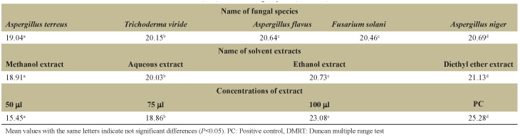 | Table 8: DMRT to rank the mean values of zone of inhibition (mm) based on fungal species, solvents, and concentrations of extracts [Click here to view] |
4. DISCUSSION
The synthetic antimicrobial agents have adverse and side effects, but the plant-derived compounds are generally safer and often more effective substitutes for the synthetic antimicrobial agents. The herbal medicines are believed to be much safer for use and proved as an elixir in the treatment on various illness and diseases [27]. Hence, there is a need to scientifically validate medicinally useful plants because of the appearance of drug resistance to antimicrobial agents and more effort is being made to find alternative antimicrobial components [28]. Plants are the storehouse and source of safer and cheaper chemicals which are pharmacologically active. The phytocompounds such as alkaloids, tannins, saponins, flavonoids, and phenolics protect the plants from their invaders such as fungi, bacteria, viruses, and nematodes [29]. The pteridophytic plants are being used ethnobotanically by various tribal communities. The antibacterial activity of 12 important pteridophytic plant extracts was reported [30].
In the present study, the extracts of leaf and stem of M. minuta showed antibacterial activity against both selected Gram-positive bacteria K. pneumoniae, E. coli, and P. fluorescens and Gram-negative bacteria B. subtilis and S. pyogenes. The present study results are in accordance with the other studies, in that they reported the antimicrobial activities of leaf of Marsilea quadrifolia [31-33] and whole plant of M. minuta [34]. In our previous study, the antibacterial activity of leaf and stem of M. quadrifolia against K. pneumoniae, E. coli, P. fluorescens, B. subtilis, and S. pyogenes was reported [35]. Similarly, the in vitro antibacterial activities of medicinal plants against human pathogenic microbes were reported by several researchers [36-38].
The antibacterial activity of the extracts of leaf and stem of M. minuta was observed at varying degrees based on solvents, bacterial strains, concentrations of extract, and parts of plant. The four different solvent extracts of the leaf and stem of M. minuta exhibited antibacterial activity against all five tested bacterial species. However, the antibacterial activity of extracts against all five tested bacterial species was varied with the solvents. In the present study, the antimicrobial activity of leaf and stem extracts of M. minuta was observed as dose dependent. Because, the present study results revealed that the higher dose of extract (5 mg) exhibited higher zone of inhibition than the lower doses (2.5 mg and 3.75 mg) of extract of leaf and stem of M. minuta. Hence, this study proved that the different solvent extracts of leaf and stem of M. minuta possess different levels of antibacterial activity against selected bacterial species at different concentrations such as 2.5, 3.75, and 5 mg.
The present study showed that the antifungal activity of extracts of leaf and stem of M. minuta against selected fungal pathogens such as A. niger, A. flavus, A. terreus, T. viride, and F. solani. These results are consistent with other studies, in that the antimicrobial activity of rhizome and frond extracts of M. minuta [39] and the antibacterial and antifungal activities of the leaf, fruits, and latex of Croton bonplandianum [40] were reported. Thus, the broad spectrum of antimicrobial activity of extracts of leaf and stem of M. minuta is consistent with the previous study in that the root, flower, and leaf of Aerva lanata (L.) showed zone of inhibition against selected microbial pathogens [41]. There was also observed different levels of antifungal activity of leaf and stem extracts of M. minuta based on solvents, fungal species, concentrations, and parts of plant. The leaf and stem extracts exhibited antifungal activity against all fungal species selected in the study, but it was varied with the solvents. The antifungal activity of leaf and stem extracts of M. minuta was also dose dependent. The higher dose of extract exhibited higher zone of inhibition than the lower doses of extract of leaf and stem of M. minuta. Hence, the present study revealed the variation in antifungal activity of leaf and stem of M. minuta against selected fungal species based on concentrations and solvents of extracts.
Fungal diseases are represented as a critical problem to health, and they are one of the main causes of morbidity and mortality worldwide [42]. The present study showed that the wide spectrum antibacterial and antifungal properties of different solvent extracts of leaf and stem of M. minuta against selected bacterial and fungal species. Medicinal plants either individually or collectively may be effective as antimicrobials for Gram-positive as well as Gram-negative bacteria, fungi, actinomycetes, protozoa, etc. [43]. In our previous study, there are many secondary metabolites screened in the leaf and stem of M. minuta by GC-MS analysis including the compounds possess antimicrobial properties such as hexanal, 2,3-dihydro-3,5-dihydroxy-6-methyl-4H-pyran-4-one, benzene carboxylic acid, 2,3-dihydro benzofuran, tetradecanoic acid, octanoic acid, and 10-octadecenoic acid [44]. Hence, the antibacterial and antifungal activities of leaf and stem of M. minuta may be due to the presence of bioactive compounds with antimicrobial properties.
The antimicrobial properties of leaf and stem extracts of M. minuta are of great interest in both academic research and herbal industry. So, the use of M. minuta as natural food supplement may be emerged from a growing tendency towards to replace synthetic antimicrobial agents by natural ones. Hence, this study results introduce a unique natural source, which possesses strong antimicrobial substances, and this seems to confirm the traditional therapeutic claims of M. minuta.
5. CONCLUSION
The results confirmed that the leaf and stem of M. minuta possess antibacterial and antifungal activities, and this study is also concluded that it may be useful in the treatment of infectious diseases caused by bacteria and fungi. It is also hoped that this study would lead to the establishment of some bioactive compounds, which may be useful to formulate new and more potent antimicrobial agents from M. minuta for the treatment of bacterial and fungal diseases.
6. ACKNOWLEDGMENTS
This research work was financially supported by the University Grants Commission [UGC-MRP-F. No. 42-638/2013 (SR)], New Delhi, India.
7. REFERENCES
1. Chang HC, Gupta SK, Tasay HS. Studies on folk medicinal fern: An example of “Gu-Sui-Bu”. In: Fernández H, Kumar A, Revilla MA, editors. Working with Ferns, Issues and Applications. New York, Dordrecht, Heidelberg, London: Springer; 2011. p. 285-304. Crossref
2. Evans CE, Banso A, Samuel OA. Efficacy of some new medicinal plants against Salmonella typhi: An in vitro study. J Ethnopharmacol 2002;80:21-4. Crossref
3. Tshibangu JN, Chifundera K, Kaminsky R, Wright AD, König GM. Screening of African medicinal plants for antimicrobial and enzyme inhibitory activity. J Ethnopharmacol 2002;80:25-35. Crossref
4. Adomi PO. Screening of leaves of three Nigerian plants for antibacterial activity. Afr J Biotechnol 2008;7:2540-64.
5. Akinsade KA, Olukoya DK. Vibroidal activities of some local herbs. J Diarrhoeal Dis Res 1995;13:127-9.
6. Chauhan NS. Medicinal and Aromatic Plants of Himachal Pradesh. 2nd ed. New Delhi: Indus Publishing Company; 2006.
7. Alves RR, Rosa IM. Biodiversity, traditional medicine and public health: Where do they meet? J Ethnobiol Ethnomed 2007;3:14. Crossref
8. Parinitha M, Srinivasa BH, Shivanna MB. Medicinal plant wealth of local communities in Shimoga District of Karnataka, India. J Ethnopharmacol 2005;98:307-12. Crossref
9. Thomas MW, Merish S, Tamizhamuthu M. Review of Alternanthera sessilis with reference to traditional Siddha medicine. Int J Pharm Phytochem Res 2014;6:249-54.
10. Kadharnivas R, Boominathan M. Antimicrobial evaluation of selected South Indian medicinal plants against Streptococcus pneumonia. Int J Adv Res Biol Sci 2015;2:262-6.
11. Parrotta JA. Healing Plants of Peninsular India. Vol. 06. New York: CAB International Publishing; 2001. p. 24-5. Crossref
12. Kostermans AJ, Wirjahardja S, Dekker RJ. The weeds: Description, ecology and control. In: Weeds of rice in Indonesia. Jakarta: Balai Pustaka; 1987. p. 24-565.
13. Chatterjee A, Dutta CP, Chaudhury B, Dey PK, Dey CD, Chatterjee C, et al. The chemistry and pharmacology of marceline: A sedative and anticonvulsant principle isolated from Marsilea minuta Linn. and Marsilea rajastanensis Gupta. J Exp Med Sci 1963;7:53-67.
14. Tiwari OP, Bhattamisra SK, Singh PN, Kumar V. Adaptogenic anti-stress activity of standardised extract of Marsilea minuta L. Pharmacol Online 2009;1:290-9.
15. Singh AP, Rawat VK, Behera SK, Khare PB. Perspectives of Pteridophytes Biodiversity: A Source of Economy Elevation. Lucknow: National Conference on Biodiversity, Development and Poverty Alleviation, National Botanical Research Institute; 2010.
16. Bhardwaja TN, Garg A. The antifertility effect of an Australian species of the aquatic fern Marsilea minuta (L.). Indian Fern J 1984;6:75-82.
17. Parihar P, Daswani L, Bohra A. Toxic effect of plant of Marsilea minuta (L.) on the growth of Staphylococcus aureus. Indian Fern J 2003;20:48-50.
18. Bhattamisra SK, Singh PN, Singh SK, Kumar V. Anxiolytic activity of Marsilea minuta (L.) in rodents. J Herbal Med Toxicol 2007;1:15-20.
19. Bhattamisra SK, Singh PN, Singh SK. Anti-inflammatory and Analgesic Activity of Ethanolic Extract of Marsilea minuta Linn. In rodents. Drug lines; 2009. In press.
20. Bhattamisra SK, Khanna VK, Agrawal AK, Singh PN, Singh SK. Anti-depressant activity of standardized extract of Marsilea minuta Linn. J Ethnopharmacol 2008;117:51-7. Crossref
21. Gupta RS, Kumar P, Sharma A, Bhardwaja TN, Dixit VP. Hypocholesterolemic activity of Marsilea minuta Linn. in gerbils. Fitoterapia 2000;70:113-7. Crossref
22. Praneetha P, Rani VS, Kumar BR. Hepatoprotective activity of methanolic extract of leaves of Marsilea minuta (Linn.) against CCl4 induced hepatic damage in rats. Glob J Pharmacol 2011;59:164-71.
23. Revathi M, Sara SC. Preliminary phytochemical analysis and antimicrobial activity of rhizome extract of Marsilea minuta. Int J Sci Res 2014;3:46-8.
24. Panda SS, Sahoo K, Rana M, Rout NC, Dhal NK. Antimicrobial activities and phytochemical investigation of some native pteridophytes. Asian J Pharm Clin Res 2014;7:43-5.
25. Ogu GI, Williams TO, Nwachukwu PU, Igere BE. Antimicrobial and phytochemical evaluation of the leaf, stem, bark and root extracts of Cyathula prostrata (L.) Blume against some human pathogens. J Intercul Ethnopharmacol 2012;1:35-43. Crossref
26. Cheesbrough M. District Laboratory Practical in Tropical Countries, Part 2. Cambridge, UK: Cambridge University Press; 2006. p. 137-50. Crossref
27. Ghosh A. Herbal folk remedies of Bankura and Medinipur District, West Bengal. J Tradit Knowl 2003;2:393-6.
28. Farnsworth NR. Ethnopharmacology and Drug Discovery in CIBA Foundation Symposium 185. Chichester: Wiley; 1994. p. 42-59.
29. Mithraja MJ, Antonisamy JM, Mahesh M, Paul ZM, Jeeva S. Chemical diversity analysis on some selected medicinally important pteridophytes of Western Ghats, India. Asian Pac J Trop Biomed 2012;2:S34-9. Crossref
30. Subhas VP, Satyajit S, Swami EV. Ethnomedicinally pteridophytic plants uses: Antibacterial activity of fronds (leaves). Int J Microbiol Res Rev 2015;12:39-145.
31. Sethi P. Antibacterial activity of Marsilea quadrifolia Linn. Int Res Plant Sci 2014;4:60-2.
32. Sivagurunathan A, Innocent BX. Phytochemical analysis and antimicrobial efficiency of Marsilea quadrifolia Linn. (Aquatic fern). Int J Pharm Res Sch 2014;3:2277-7873.
33. Gini TG, Jothi GJ. In vitro screening of antibacterial and antifungal activity of Marsilea quadrifolia (Marsileaceae) (Linn.) extract. Am J Phytomed Clin Ther 2015;3:313-29.
34. Sharma D, Bhatia VK, Patil S, Sharma PC. Antimicrobial activity of selected cryptogams from Solan region. Int J Biol Pharm Res 2013;4:448-54.
35. Gopalakrishnan K, Udayakumar R. Antimicrobial activity of Marsilea quadrifolia (L.) against some selected pathogenic microorganisms. Br Microbiol Res J 2014;4:1046-56. Crossref
36. Al-Harbi AH, Uddin MN. Quantitative and qualitative study of the bacterial flora of farmed fresh water prawn (Macrobrachium rosenbergii) larvae. J Appl Ichthyol 2004;20:461-5. Crossref
37. Devendra BN, Srinivas N, Solmon KS. A comparative pharmacological and phytochemical analysis of in vivo and in vitro propagated Crotalaria species. Asian Pac J Trop Med 2012;5:37-41. Crossref
38. Saranya SR, Krishna PJ, Singh RK, Gaanappriya M, Dhivya E, Rajasekar S. Screening and phytochemical analysis of pharmacologically active compounds from Abutilon indicum and Phyllanthus niruri and assessing their in vitro antimicrobial activity against pathogens. Int J Adv Biotechnol Res 2013;4:496-504.
39. Nagarajan G, De Britto AJ. Evaluation of antibacterial activity of different parts of medicinal fern-Marsilea minuta. Life Sci Leaf 2014;50:34-7.
40. Vennila V, Udayakumar R. Antibacterial activity of Croton bonplandianum (Bail.) against some bacterial isolates from infected wounds. Br Microbiol Res J 2015:5:83-93. Crossref
41. Vidhya R, Udayakumar R. Antibacterial potential of different parts of Aerva lanata (L.) against some selected clinical isolates from urinary tract infections. Br Microbiol Res J 2015;7:35-47. Crossref
42. CSIR. Wealth of India, Publications and Information Directory. New Delhi, India: CSIR; 1998. p. 164.
43. Dixit RD. Ferns-a much neglected group of medicinal plants. Int J Res Indian Med 1974;9:74-90.
44. Sabithira G, Udayakumar R. GC-MS Analysis of methanolic extracts of leaf and stem of Marsilea minuta (Linn.). J Complement Altern Med Res 2017;3:1-13. Crossref