REFERENCES
1. O’Brien K, Breyne K, Ughetto S, Laurent LC, Breakefield XO. RNA delivery by extracellular vesicles in mammalian cells and its applications. Nat Rev Mol Cell Biol. 2020;21(10):585-606.
2. Regente M, Corti-Monzón G, Maldonado AM, Pinedo M, Jorrín J, de la Canal L. Vesicular fractions of sunflower apoplastic fluids are associated with potential exosome marker proteins. FEBS Lett. 2009;583(20):3363-6.
3. Lian MQ, Chng WH, Liang J, Yeo HQ, Lee CK, Belaid M, Tollemeto M, Wacker MG, Czarny B, Pastorin G. Plant-derived extracellular vesicles: recent advancements and current challenges on their use for biomedical applications. J Extracell Vesicles. 2022;11(12):12283.
4. Mesmar J, Abdallah R, Badran A, Maresca M, Shaito A, Baydoun E. Ziziphus nummularia: a comprehensive review of its phytochemical constituents and pharmacological properties. Molecules. 2022;27(13):4240.
5. Woith E, Fuhrmann G, Melzig MF. Extracellular vesicles—connecting kingdoms. Int J Mol Sci. 2019;20(22):5695.
6. Dash M, Palaniyandi K, Ramalingam S, Sahabudeen S, Raja NS. Exosomes isolated from two different cell lines using three different isolation techniques show variation in physical and molecular characteristics. Biochim Biophys Acta Biomembranes. 2021;1863(2):183490.
7. Biswas K, Chattopadhyay I, Banerjee RK, Bandyopadhyay U. Biological activities and medicinal properties of neem (Azadirachta indica). Curr Sci. 2002;82(11):1336-45.
8. Morton JF. Country borage (Coleus amboinicus Lour.): a potent flavoring and medicinal plant. J Herbs Spices Med Plants. 1992;1(1-2):77-90.
9. Shackelford L, Mentreddy SR, Cedric S. Determination of total phenolics, flavonoids and antioxidant and chemopreventive potential of basil (Ocimum basilicum L. and Ocimum tenuiflorum L.). Int J Cancer Res. 2009;5(4):130-43.
10. Samanta SK, Kandimalla R, Gogoi B, Dutta KN, Choudhury P, Deb PK, et al. Phytochemical portfolio and anticancer activity of Murraya koenigii and its primary active component, mahanine. Pharmacol Res. 2018;129:227-36.
11. Rutter BD, Innes RW. Extracellular vesicles isolated from the leaf apoplast carry stress-response proteins. Plant Physiol. 2017;173(1):728-41.
12. Gentzel I, Giese L, Zhao W, Alonso AP, Mackey D. A simple method for measuring apoplast hydration and collecting apoplast contents. Plant Physiol. 2019;179(4):1265-72.
13. Gallart-Palau X, Serra A, Wong AS, Sandin S, Lai MK, Chen CP, et al. Extracellular vesicles are rapidly purified from human plasma by PRotein Organic Solvent PRecipitation (PROSPR). Sci Rep. 2015;5(1):14664.
14. Gallart-Palau X, Serra A, Sze SK. Enrichment of extracellular vesicles from tissues of the central nervous system by PROSPR. Mol Neurodegen. 2016;11(1):41.
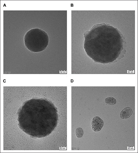
15. Malenica M, Vukomanovic M, Kurtjak M, Masciotti V, Dal Zilio S, Greco S, et al. Perspectives of microscopy methods for morphology characterisation of extracellular vesicles from human biofluids. Biomedicines. 2021;9(6):603.
16. Schneider CA, Rasband WS, Eliceiri KW. NIH Image to ImageJ: 25 years of image analysis. Nat Methods. 2012;9(7):671-5.
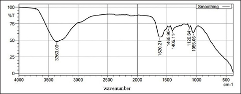
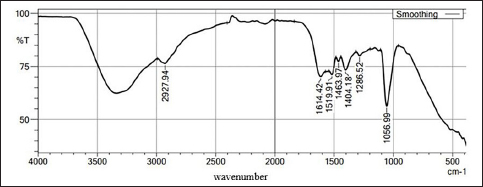
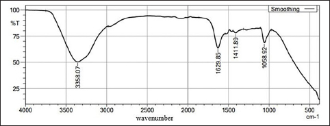
17. Di Santo R, Vaccaro M, Romanò S, Di Giacinto F, Papi M, Rapaccini GL, et al. Machine learning-assisted FTIR analysis of circulating extracellular vesicles for cancer liquid biopsy. J Pers Med. 2022;12(6):949.
18. Nandiyanto AB, Oktiani R, Ragadhita R. How to read and interpret FTIR spectroscope of organic material. Indonesian J Sci Technol. 2019;4(1):97-118.
19. Kluyver T, Ragan-Kelley B, Pérez F, Granger BE, Bussonnier M, Frederic J, et al. Jupyter notebooks-a publishing format for reproducible computational workflows. Int Conf Electronic Publishing. 2016;2016:87-90.
20. Vallejo F, Yuste JE, Teruel-Montoya R, Luengo-Gil G, Bohdan N, Espín S, et al. First exploratory study on the metabolome from plasma exosomes in patients with paroxysmal nocturnal hemoglobinuria. Thrombosis Res. 2019;183:80-5.
21. Wojakowska A, Zebrowska A, Skowronek A, Rutkowski T, Polanski K, Widlak P, et al. Metabolic profiles of whole serum and serum-derived exosomes are different in head and neck cancer patients treated by radiotherapy. J Pers Med. 2020;10(4):229.
22. Halket JM, Waterman D, Przyborowska AM, Patel RK, Fraser PD, Bramley PM. Chemical derivatization and mass spectral libraries in metabolic profiling by GC/MS and LC/MS/MS. J Exp Botany. 2005;56(410):219-43.
23. Irudhyaraj DF, Smila VN, Janani S. GC-MS analysis and antioxidant study on methanolic leaf extract of endemic medicinal plant Crotalaria paniculata willd. Adv Zool Bot. 2023;11(6):434-42.
24. Hussain S, Javed W, Tajammal A, Khalid M, Rasool N, Riaz M, et al. Synergistic antibacterial screening of Cymbopogon citratus and Azadirachta indica: phytochemical profiling and antioxidant and hemolytic activities. ACS Omega. 2023;8(19):16600-11.
25. Patil A, Jadhav V. GC-MS analysis of bioactive components from methanol leaf extract of Toddalia asiatica (L.). Int J Pharm Sci Rev Res. 2014;29(1):18-20.
26. El-Fayoumy EA, Shanab SM, Hassan OM, Shalaby EA. Enhancement of active ingredients and biological activities of Nostoc linckia biomass cultivated under modified BG-11o medium composition. Biomass Conv Biorefinery. 2021;13(7):6049-66.
27. Rizwana H, Al Otibi F, Al-Malki N. Chemical composition, FTIR studies and antibacterial activity of Passiflora edulis f. edulis (Fruit). J Pure Appl Microbiol. 2019;13(4):2489-98.
28. Ertürk Ö, Ayvaz MÇ, Çil E, Bagdatli E. Gas chromatography-mass spectrometry analysis and antimicrobial and antioxidant activities of some orchid (Orchidaceae) species growing in Turkey. Braz Arch Biol Technol. 2023;66:e23210265.
29. Waluyo J, Wahyuni D. Antibacterial effects of Pheretima javanica extract and bioactive chemical analysis using gas chromatography mass spectrum. In: Journal of Physics: Conference Series 2021, Vol. 1751. Bristol: IOP Publishing; 2021. p. 12055.
30. Tamil Muthu P, Selvaraj D. Analysis of bioactive constituents from the flesh of Turbo brunneus (Roding, 1798) by GCMS. Int J Fish Aquatic Stud. 2015;3(1):257-9.
31. Addai ZR, Abood MS, Hlail SH. GC-MS profiling, antioxidants and antimicrobial activity of prickly pear (Opuntiaficus-indica) pulp extract. Pharmacognosy J. 2022;14(2):262-7.
32. Salim SA. Identification of active pharmaceutical ingredients in Thevetia neriifolia juss leaf callus using analysis of GC-MS. Indian J Public Health Res Dev. 2018;9(12):1019-23.
33. Zhou J, Guo Z, Yu W, Li S, Qiao W. Clinical evaluation of preoperative radiotherapy combined with FOLFOX chemotherapy on patients with locally advanced colon cancer. Am Surg. 2019;85(4):313-20.
34. Elbaz HA, Stueckle TA, Tse W, Rojanasakul Y, Dinu CZ. Digitoxin and its analogs as novel cancer therapeutics. Exp Hematol Oncol. 2012;1(1):1.
35. Fan W, Qian MC. Identification of aroma compounds in Chinese ‘Yanghe Daqu’liquor by normal phase chromatography fractionation followed by gas chromatography [sol] olfactometry. Flavour Fragr J. 2006;21(2):333-42.
36. Gilbert KC, Sundareshan V, Bass RM, Lin SY. Antibacterial properties of additives used in injection immunotherapy. In: International Forum of Allergy & Rhinology, Vol. 2. Hoboken (NJ): Wiley Subscription Services, Inc., A Wiley Company; 2012 Mar. p. 135-8.
37. Mebude OO, Adeniyi B. GC-MS analysis of phyto components from the stem bark of Cola nitida Schott & Endl. J Plant Sci. 2017;5(4):99-103.
38. Lykholat YV, Khromykh NO, Didur OO, Drehval OA, Sklyar TV, Anishchenko AO. Chaenomeles speciosa fruit endophytic fungi isolation and characterization of their antimicrobial activity and the secondary metabolites composition. Beni-Suef Univ J Basic Appl Sci. 2021;10:83.
39. Ali A, Ali A, Warsi MH, Ahmad W. Chemical characterization, antidiabetic and anticancer activities of Santolina chamaecyparissus. Saudi J Biol Sci. 2021;28(8):4575-80.
40. Zhu Q, Tao C. Pharmacological class data representation in the Web Ontology Language (OWL). In: 2014 IEEE International Conference on Big Data (Big Data), Washington, DC, USA. IEEE; 2014 Oct 27 (pp. 77-84).
41. Cerda-García-Rojas CM, Burgueño-Tapia E, Román-Marín LU, Hernández-Hernández JD, Agulló-Ortuño T, González-Coloma A, et al. Antifeedant and cytotoxic activity of longipinane derivatives. Planta Med. 2010;76(03):297-302.
42. Sangeetha C, Krishnamoorthy AS, Amirtham D. Antifungal bioactive compounds from Chinese caterpillar fungus (Ophiocordyceps sinensis (Berk.) GH Sung et al.) against plant pathogens. Madras Agricult J. 2015;102:1.
43. Hugar AL, Londonkar RL. GC-MS profiling of bioactive components from aqueous extract of Pterocarpus marsupium. Int J ChemTech Res. 2017;10(9):557-64.
44. Das S, Barnwal P, Ramasamy A, Sen S, Mondal S. Lysergic acid diethylamide: a drug of ‘use’? Ther Adv Psychopharmacol. 2016;6(3):214-28.
45. Sandison RA, Spencer AM, Whitelaw JD. The therapeutic value of lysergic acid diethylamide in mental illness. J Ment Sci. 1954;100(419):491-507.
46. Al Bratty M, Makeen HA, Alhazmi HA, Syame SM, Abdalla AN, Homeida HE, et al. Phytochemical, cytotoxic, and antimicrobial evaluation of the fruits of miswak plant, Salvadora persica L. J Chem. 2020;2020:11.
47. Katariya D, Ashid M, Sharma BK, Joshi A. Synthesis, characterization and biological activity of some indole substituted propanoic acid. J Chem Chem Sci. 2019;9:206-13.
48. Kalsum N, Sulaeman A, Setiawan B, Wibawan IW. Phytochemical profiles of propolis Trigona spp. from three regions in Indonesia using GC-MS. J Biol Agricult Healthcare. 2016;6(14):31-7.
49. Abd Alzahra EM. Detecting of chemical compounds of aqueous and alcoholic extracts of damas Conocarpus lancifolus Engl. leaves using GC-MS technique. J Educ Pure Sci Univ Thi-Qar. 2022;12(1):29-39.
50. Huang WC, Tsai TH, Chuang LT, Li YY, Zouboulis CC, Tsai PJ. Anti-bacterial and anti-inflammatory properties of capric acid against Propionibacterium acnes: a comparative study with lauric acid. J Dermatol Sci. 2014;73(3):232-40.
51. Selvan PS, Velavan S. Analysis of bioactive compounds in methanol extract of cissus vitiginea leaf using GC-MS. Rasayan J Chem. 2015;8(4):443.
52. Hema R, Kumaravel S, Alagusundaram K. GC/MS determination of bioactive components of Murraya koenigii. J Am Sci. 2011;7(1):80-3.