Research Article | Volume 14, Issue 2, March, 2026

Steviol glycoside biosynthesis pathway gene expression profiling of transformed and non-transformed plant leaf tissues of Stevia rebaudiana (Bertoni)

Priya Singh Amol S. Phule Heena Tabassum Minal Wani   

Open Access   

Published:  Jan 25, 2026

DOI: 10.7324/JABB.2026.250346
Abstract

Stevia rebaudiana Bertoni, a member of the Asteraceae family, is recognized for its sweetened leaves, which are remarkably sweeter than sucrose by 200–300 times. This astounding property is because of steviol glycosides (SGs), a class of diterpenoid secondary metabolites that primarily consist of stevioside and rebaudioside A. These compounds are formed through a specific SG biosynthetic pathway that contains several key genes. In the present work, gene expression profiling of 15 core genes of SG biosynthetic pathway, along with metabolite analysis, was conducted in three groups of S. rebaudiana plants: In vitro regenerated non-transformed plantlets (NP), in vitro regenerated transformed plantlets (TP) via hairy root cultures using Rhizobium rhizogenes mediated transformation, and control plants (CP). Quantitative real-time polymerase chain reaction results showed that in NP and TP there was upregulation of 13 genes. Both NP and TP showed downregulated SrDXR and SrCDPS in comparison to CP, whereas SrUGT74G1 had higher expression in NP than TP. High-performance liquid chromatography chromatographic studies on SGs showed that stevioside content followed the order TP > NP > CP. These findings demonstrate that transformation enhances SG biosynthesis and support the use of genetically modified S. rebaudiana lines for increased natural sweetener production. Further studies are warranted to elucidate regulatory mechanisms and optimize metabolic engineering approaches.


Keyword:     Stevia rebaudiana Secondary metabolites Steviol glycosides pathway Gene expression Quantitative real-time polymerase chain reaction High-performance liquid chromatography


Citation:

Singh P, Phule AS, Tabassum H, Wani M. Steviol glycoside biosynthesis pathway gene expression profiling of transformed and non-transformed plant leaf tissues of Stevia rebaudiana (Bertoni). J Appl Biol Biotech 2026;14(2):204-211. http://doi.org/10.7324/JABB.2026.250346

Copyright: Author(s). This is an Open Access article distributed under the terms of the Creative Commons Attribution-NonCommercial-ShareAlike license.

HTML Full Text

1. INTRODUCTION

Stevia rebaudiana (Bertoni) is a perennial shrub that belongs to the Asteraceae family of flowering plants. It is native to Paraguay and is noted for having a sweet taste due to a particular type of tetracyclic diterpenoids, called steviol glycosides (SGs), found in the leaves [1]. These diterpenoids are naturally occurring, low-calorie sweeteners found in three species of plants, Angelica keiskei, Chinese Rubus suavissimus, and S. rebaudiana [2]. SGs are primarily located in the leaves of Stevia plants, and they are later transported to other parts of the plant [3]. To date, more than 60 SGs have been found in Stevia [4,5]. The first glycoside to be extracted from Stevia leaves was Stevioside, which was then followed by the isolation of rebA, Dulcoside A, rebB, C, D, E, F, I, M, and Steviol-bioside [6].

S. rebaudiana typically contains Stevioside (5–10%), Rebaudioside A (2–5%), Rebaudioside C (1%), Dulcoside A (0.5%), and trace amounts of Rebaudiosides D, E, F (0.2%) and Steviol-bioside (0.1%) [7]. Among these, Stevioside and Rebaudioside A are the crucial SGs because they are the final products of the pathways. In addition to these secondary metabolites, plants also contain flavonoids, phenolics, and alkaloids that may have therapeutic benefits [8]. Stevia has many therapeutic properties, such as antidiabetic [9] antihypertensive, anticancer, antitumor effect [10], and antimicrobial effect [11].

Stevia is typically propagated using both seeds and shoot cuttings; however, seed propagation is less popular due to low production and germination efficiency, while shoot cutting propagation is a labor-intensive and time-consuming technique with a low success rate [12]. Optimum in vitro conditions are necessary for the development of an efficient method for mass propagation of Stevia. Thus, an alternative method, such as plant tissue culture, that has high efficacy, is used to raise Stevia plants to elevate the quality and quantity of SGs and further enhance the taste of S. rebaudiana [13,14].

In the present study, plantlets generated through two distinct tissue culture approaches, micropropagation and R. rhizogenes-mediated transformation, were utilized as the primary experimental material [15]. The SG biosynthesis pathway in S. rebaudiana synthesizes compounds responsible for the formation of the natural sweetness in the Stevia plants through a complex sequence of enzymatic reactions. It involves a series of enzymatic reactions mediated by approximately 15 key genes, as mentioned in Figure 1. The pathway begins with geranylgeranyl diphosphate (GGPP), which is converted by copalyl diphosphate synthase and Kaurene synthase (KS) to ent-kaurene. This is subsequently oxidized by kaurene oxidase (KO) and kaurenoic acid hydroxylase (KAH) to produce steviol, the aglycone backbone of SGs. In the final steps, UDP-glycosyltransferases (UGTs) such as UGT85C2, UGT74G1, and UGT76G1 catalyse sequential glycosylation reactions to generate stevioside and various rebaudiosides [3,16]. The coordinated activity of these 15 genes shapes the production of SGs, creating compounds such as stevioside and rebaudioside A, which are crucial to the sweetness profile of Stevia. Understanding and manipulating these genes in biotechnological applications can probably enhance the yield and composition of SGs, potentially creating sweeter and more stable stevia products for commercial use.

Figure 1: Stevia rebaudiana plant material. (a) Micropropagated plantlets; (b) Rhizobium rhizogenes mediated transformed plantlets [15].


[Click here to view]

Recent studies have highlighted the roles of specific genes in the pathway. For example, UGT76G1 is critical for synthesizing rebaudioside A, a desirable sweetener due to its reduced bitterness compared to stevioside [17]. Modulating UGT enzyme expression can enhance specific SGs, allowing selective breeding or genetic engineering to produce high-yield, consumer-preferred sweeteners [18].

In light of the previous discussion, the present study aims to analyze the relative expression profiles of fifteen key genes involved in the SG biosynthetic pathway in S. rebaudiana leaf tissue. Quantitative real-time polymerase chain reaction (qRT-PCR) was employed to assess relative gene expression across two plant groups: Transformed plants (TP), non-transformed plants (NP), in comparison with in vivo control plants (CP) to develop a Stevia system exhibiting enhanced sweetness. To correlate gene expression with SG accumulation, quantitative analysis of SGs using stevioside as the reference standard was conducted via high-performance liquid chromatography (HPLC). This allowed for the comparative estimation of stevioside content among TP, NP, and CP plants, thereby validating the expression data obtained through qRT-PCR.

2. MATERIALS AND METHODS

2.1. Plant Material

S. rebaudiana Bertoni (Family: Asteraceae) plants were procured from Sunrise Agro Nursery for Medicinal Plants (18°35’20”N, 73°46’30”E), Wakad, Pune. These in vivo plants were subsequently maintained and cultivated under controlled conditions in the greenhouse facility of Dr. D.Y. Patil Biotechnology and Bioinformatics Institute, Tathawade, Pune, and served as the control group (CP) in the present study. The same plants also served as the source of explants for regeneration studies.

Specifically, internodal segments excised from CP plants were used for direct regeneration of NP through micropropagation. For the generation of TP, microshoots derived from NP lines were co-cultivated with R. rhizogenes to induce hairy root formation. Hairy-root-bearing microshoots were then cultured to regenerate TP plantlets.

The regeneration protocols for both NP and TP plantlets were optimized at the plant tissue culture laboratory of Dr. D.Y. Patil Biotechnology and Bioinformatics Institute, Pune, and Rise N’ Shine Biotech Pvt. Ltd., Theur, Pune. The molecular confirmation of transformation through polymerase chain reaction (PCR) amplification of rolB, rolC, and virD2 genes was also performed, which confirmed stable integration of Ri T-DNA into Stevia hairy roots and the corresponding regenerated plantlets [15]. These regenerated plantlets constituted the primary study [Figure 1].

2.2. Total RNA Extraction and First-Strand cDNA Synthesis

Total RNA extraction was carried out in three replications from the stevia leaf tissue of CP, NP, and TP using the Spectrum Plant Total RNA kit (SIGMA Life Sciences) by following the manufacturer’s protocol.

The extracted total RNA pooled (three replications together) of each treatment that were quantified using NanoDrop spectrophotometer (Cytation 5 Multimode Microplate Reader, BioTek) and run on an agarose gel (1%, w/v) electrophoresis. The cDNA was synthesized using the pooled RNA (3 replicates/treatments) sample of treatments using the GoScript Reverse Transcription System (Promega Biotech India Pvt Ltd) by following the manufacturer’s protocol.

2.3. Selection of SG Pathway Genes and Primer Designing

A total of 15 key genes involved in the SG biosynthesis pathway were selected based on their known roles in the metabolic conversion of precursors into major SGs, primarily stevioside and rebaudioside A [4]. The nucleotide sequences of all these genes were retrieved from the National Center for Biotechnology Information (NCBI) GenBank database (https://www.ncbi.nlm.nih.gov). The selection was based on sequence availability, functional annotation, and previous reports [4] on their involvement in SG biosynthesis in S. rebaudiana. Corresponding accession numbers for each gene are provided in Table 1. Primer pairs were designed using Primer-BLAST (NCBI) and Primer3Plus (https://www.primer3plus.com) under default parameters to ensure target specificity, optimal melting temperatures, and amplicon sizes suitable for qRT-PCR analysis, are mentioned in Table 1.

Table 1: Primer sequences of 15 genes of the Steviol glycosides pathway and reference genes.

GenesGene descriptionAccession no.Primer sequence (F/R)Temperature (°C)Product size
SrDXSDeoxy xylulose-5-phosphate synthaseAJ429232F: CGACACATTGTGGTGCGTTT R: CAATTCGGGCTTCATCGGCTG56°C90 bp
SrDXRDeoxy xylulose-5-phosphate reductaseAJ429233F: GCTCGCAGGAAAAGGGATTC R: GCTCGCAGGAAAAGGGATTC62°C155 bp
SrCMS4-diphosphocytidyl-2- C-methyl-D-erythritol synthaseDQ269452F: TCAAGTTATGTCGCCCCTCAA R: TAATCGAGGATGCCGGTACA56°C115 bp
SrCMK4-diphosphocytidyl-2- C-methylD-erythritol kinaseDQ269453F: TCACACGTGCGGATAAACAA R: TACGCGGTGTTACTGGTTTG56°C88 bp
SrMCS4-diphosphocytidyl-2- C-methyl-D-erythritol 2,4-cyclodiphosphate synthaseDQ631427F: ATTTCTCTCCGGCCGGTATC R: GATCGAAACCATGGCCGACT56°C130 bp
SrHDS1-hydroxy-2-methyl-2(E)-butenyl 4-diphosphate synthaseDQ768749F: TTGCAATGGAGAATGCAACGG R: TTACGTGAACCACCACTCATC56°C104 bp
SrHDR1-hydroxy-2-methyl-2(E)-butenylDQ269451F: TGCGTAACATTCGGTTGTGG R: TCCAGACGGTTACGACACTT56°C71 bp
SrGGDPSGeranyl geranyl diphosphate synthaseDQ432013F: GCCACAAGGTGTACGGTGAA R: CGGACGATCCTGTCTTTGGA56°C115 bp
SrCDPSCopalyl diphosphate synthaseAF034545F: GGGCGAGGATGATGTTTGGA R: TGAAGCACGGCCACATAGTT56°C114 bp
SrKSKaurene SynthaseAF097310F: CTTGACGGGGGTACTGTTGT R: AGAACCTCACCGTGTGTGAC62°C149 bp
SrKOKaurene oxidaseAY364317F: CAACCGCAATAACCATCGGC R: GTTTGATTGGCTCCTGCGTG62°C154 bp
SrKAHKaurenoic acid hydroxylaseEU722415F: GCCATTTCTGGGCGAAACTC R: TCCACACAACACCGCAAAAC62°C148 bp
SrUGT85C2UDP glucosyltransferase – 85C2AY345978F: ACGGAAGCTCCTCAAAGGTC R: TGGGCCGATGGTGTAAATGT62°C151 bp
SrUGT74G1UDP glucosyltransferase – 74G1AY345982F: TGGTGAAACATGGACCCGAA R: TTCTGGGAGCTTTCCCTCTT62°C147 bp
SrUGT76G1UDP glucosyltransferase – 76G1AY345974F: ACAACGACCCACAAGACGAA R: CAACAGTTCCAGTTCGCGTC62°C153 bp
SrActinActinAF548026F: TGAAGCGTTATCATCATCTACTCA R: ATCATCGCCAGCAAACCCA56°C133 bp
GAPDHGlyceraldehyde – 3 –phosphate dehydrogenaseKC669708F: GGGGTTTGCTTTATGATTTCAGC R: AGAGCTGGAAGCACCTTTCC56°C126 bp

2.4. Gene Expression Profiling

The gene expression profiling of selected fifteen genes was performed using qRT-PCR. The two genes, Actin and GAPDH were used as a set of reference genes or internal control genes. The qRT-PCR reactions were carried out in Thermal Cycler1000 Touch CFX96 Touch (Bio-Rad) using iTaq Universal SYBR green Supermix (Bio-Rad). Each of the reactions was performed in triplicate, containing SYBR green Supermix (5 uL), template cDNA (1 μL), each of the primers (0.4 μL, 10 μM), and RNAse-free water (3.2 μL) with 10 μL total volume.

The qRT-PCR profile for two reference genes and eight SG pathway genes (includes CMS, CMK, MCS, HDR, GGDPS, CDPS, HDS and DXS) were as follows, 1 min (95°C), 10 s (95°C) for 40 cycles, 30 s (56°C), 5 s (65°C), 5 s (95°C) and for other nine genes (KO, KS, KAH, DXR, UGT76G1, UGT74G1 and UGT85C2) as follows, 1 min (95°C), 10 s (95°C) for 40 cycles, 30 s (62°C), 5 s (65°C), 5 s (95°C) for fluorescent signal recording followed by high-resolution melting curve obtained after the cycle 95°C (15 s), with constant increment in the temperature from 65°C (15 s) and 95°C (1 s). Melting curve analysis was done, and internal control genes SrActin and GAPDH were used to normalize the data individually. Finally, data were analyzed using the software, Bio-Rad CFX Maestro Version 2.3.

The fold change values from the gene expression profiling of genes in leaf tissue of CP, NP, and TP were calculated using 2-ΔΔCT method [19] and also a heatmaps plot as depicted in Figure 1 using, Multi Experiment Viewer (MeV v4.9.0) software [20]. The fold change values of each gene from NP and TP were plotted in the graph as shown in Figures 2-4.

Figure 2: Gene expression profiling of Steviol glycoside biosynthesizing genes (a) SrDXS and SrDXR, (b) SrCMS, SrCMK and SrMCS in leaves tissues of transformed plantlets, non-transformed plantlets and control plants. Error bars on the top indicate standard deviation of three technical replicates. SrActin and GAPDH served as two reference genes.


[Click here to view]
Figure 3: Gene expression profiling of Steviol Glycoside biosynthesizing genes (a) SrHDS and SrHDR, (b) SrGGDPS and SrCDPS in leaves tissues of transformed plantlets, non-transformed plantlets and control plants. Error bars on the top indicate standard deviation of three technical replicates. SrActin and GAPDH served as two reference genes.


[Click here to view]
Figure 4: Gene expression profiling of Steviol glycoside biosynthesizing genes (a) SrKS and SrKO and SrKAH, (b) SrUGT85C2, SrUGT74G1 and SrUGT76G1 in leaves tissues of transformed plantlets, non-transformed plantlets and control plants. Error bars on the top indicate the standard deviation of three technical replicates. SrActin and GAPDH served as two reference genes.


[Click here to view]

Although CP, NP, and TP (in vitro-regenerated plantlets) were cultivated under distinct environmental conditions, leaf samples from both in vivo and in vitro plants were harvested after 1.5 months of growth to ensure comparable physiological status at the time of collection. The selection of in vivo plants as a reference for gene expression analysis aligns with prior transcriptomic studies conducted in rice and Eucommia ulmoides [21,22]. Quantitative gene expression data were normalized using two stable internal reference genes, SrActin and GAPDH.

2.5. HPLC Analysis

Stevioside content in the leaf samples of CP, NP, and TP was quantified using HPLC. Dried leaf powder was extracted with HPLC-grade methanol, defatted with hexane, and re-dissolved in acetonitrile. Filtrates were injected into an Agilent HPLC system equipped with a Zorbax Eclipse XDB–C18 column and detected at 204 nm. The mobile phase consisted of methanol and 0.05% o-phosphoric acid (75:25, v/v) at pH 3.15. Chromatographic data were analyzed using EZChrom Elite software. Stevioside concentrations were calculated against a standard curve and expressed in mg/g dry weight.

2.6. Statistical Analysis

The quantification studies were run in six replications for each leaf sample from CP, NP, and TP. The result was expressed as the mean value of Stevioside concentration ± standard error. Statistical Package for the Social Sciences version 17 was used for the statistical analysis. To assess the significance of the mean values at (P < 0.05), the one-way analysis of variance (ANOVA) test was carried out, and for pairwise comparison, the post hoc test (Tukey’s HSD) was applied.

3. RESULTS

3.1. qRT-PCR Analysis

In the present study, the expression profiling (qRT-PCR) of key genes (fifteen genes) involved in the SG biosynthetic pathway was studied from leaf tissues of in vitro regenerated NP, in vitro TP, and CP grown in vivo. Melt Curve Peak Temperatures (Tm) of the fifteen genes were obtained (S1).

The expression of SrDXS (deoxyxyulose-5-phosphate synthase) exhibited higher in in vitro TP (5.0-fold change, up-regulated) followed by in vitro regenerated NP (3.0-fold change, up-regulated), as compared to the CP as shown in [Figure 2a]. The SrDXR (deoxyxyulose-5-phosphate reductase) gene exhibited lower expression in TP (-3.2-fold change, down-regulated) and in NP (-25.4-fold change, highly down-regulated) compared with CP [Figure 2a].

The transcript abundance of three diphosphocytidyl related genes, SrCMS (4-diphosphocytidyl-2-C-methyl-d-erythritol synthase), SrCMK (4-Diphosphocytidyl-2-C-methyl-d-erythritol kinase) and SrMCS (4-diphosphocytidyl-2-C-methyl-d-erythritol 2,4-cyclodiphosphate synthase) exhibited higher expression in TP (7.4, 1.8 and 21.6-fold change respectively, up-regulated) followed by NP (5.5, 1.5 and 8.5- fold change respectively, up-regulated) when compared with CP as shown in [Figure 2b].

Furthermore, the transcript level of two 1-Hydroxy-2-methyl-2(E)-butenyl-4-diphosphate synthase/reductase genes, SrHDS (1-Hydroxy-2-methyl-2(E)-butenyl-4-diphosphate synthase) SrHDR (1-Hydroxy-2-methyl-2(E)-butenyl-4-diphosphate reductase) exhibited the highest increase in TP (61.9 -fold change, highly up-regulated and 9.9-fold change respectively, up-regulated) and in NP (4.1 and 8.2-fold change respectively, up-regulated) as compared to CP as shown in Figure 3a.

Among the two diphosphate synthase genes, SrGGDPS (GGPP synthase) gene showed the highest expression in TP (22.9 -fold change, highly up-regulated) followed by NP (8.1-fold change, up-regulated), as compared to CP, shown in Figure 3b. Whereas in contrast, SrCDPS (copalyl diphosphate synthase) gene exhibited low expression in TP (−1.6- fold difference, that is down-regulated), followed by NP (1.4-fold change, down-regulated) in comparison with CP [Figure 3b].

Similarly, the transcript level of two genes, SrKS (KS) and SrKO (KO) exhibited higher in TP (3.3 and 1.1- fold increase respectively, up-regulated) followed by NP (5.5 and 2.2 -fold change respectively, up-regulated). However, the higher expression of SrKAH (KAH) was recorded in TP (4.2-fold change, up-regulated) and NP (1.7-fold change, up-regulated) as shown in Figure 4a.

Furthermore, among the glucosyltransferase genes, SrUGT85C2 (UDP glucosyltransferase-85C2) and SrUGT76G1 (UDP glucosyltransferase-76G) were highly expressed in TP (2.7 and 3.0 -fold difference, respectively, up-regulated) followed by NP (1.7 and 2.3-fold change, up-regulated) when compared with CP, shown in Figure 4b. However, SrUGT74G1 (UDP glucosyltransferase-74G) gene exhibited higher expression in in vitro regenerated NP about 57.4-fold change (highly up-regulated), followed by in vitro regenerated TP, about 29.9-fold change (highly up-regulated) in comparison with CP [Figure 4b].Thus, comparatively higher expression profiling of SGs genes in in vitro TPs i.e., TP (plantlets regenerated from induced hairy roots via R. rhizogenes transformation), indicates that the modulation occurred in genes involved in the SG biosynthetic pathway might lead to enhance production and content of SGs.

3.2. HPLC Analysis

The key feature of Stevia is its SG accumulation. Examination of Stevioside concentrations of leaf tissues of CP, NP, and TP is carried out by HPLC. According to the results as shown in Table 2, the highest amount of stevioside was produced by R. rhizogenes-mediated TPs. It can be concluded that transformation increases SG contents in the leaves of stevia. One-way ANOVA shows that all the data are significant with P < 0.05. Tukey’s HSD test, showed that all pairwise comparisons (CP vs. NP, CP vs. TP, NP vs. TP) are statistically significant (P < 0.05). This confirms that stevioside concentration significantly differs among CP, NP, and TP, with TP showing the highest stevioside concentration. The corresponding HPLC chromatograms illustrating peak profiles of stevioside from CP, NP, and TP leaf extracts are presented in Figure 6.

Table 2: Stevioside concentration in mg/g of the leaf extracts of the Stevia rebaudiana control plants, CP, non-transformed plants NP, and transformed plants TP.

SampleRetention timeAreaArea %HeightStevioside (mg/g)(mean±SE)
CP6.273193133100.00230260.024±0.02a
NP6.217317420100.00396090.042±0.01b
TP6.861304175100.00254011.67±0.13c

CP: Control plants, TP: Transformed plantlets, NP: Non-transformed. * Values are the mean of Stevioside concentration in different sets of plants determined by one-way ANOVA which shows data are statistically significant (P<0.05). Post hoc test of pairwise comparison was done via Tukey HSD. Different letters represent statistically significant data. The experiment was performed in 6 replicates.

Figure 5: Heatmap representation of fifteen steviol glycoside biosynthesis pathway genes expression profile in leaf tissue of transformed plantlets, non-transformed plantlets and control plants (CP). The colour scale at the top show expression level (fold change) values. Yellow indicates a higher expression level (up-regulation), and blue indicates a lower expression level (down-regulation) of genes. For, CP the colour remains uniform throughout.


[Click here to view]
Figure 6: (a-c) High-performance liquid chromatography chromatogram of the leaf extracts of the Stevia control plants, non- transformed plantlets and transformed plantlets.


[Click here to view]

4. DISCUSSION

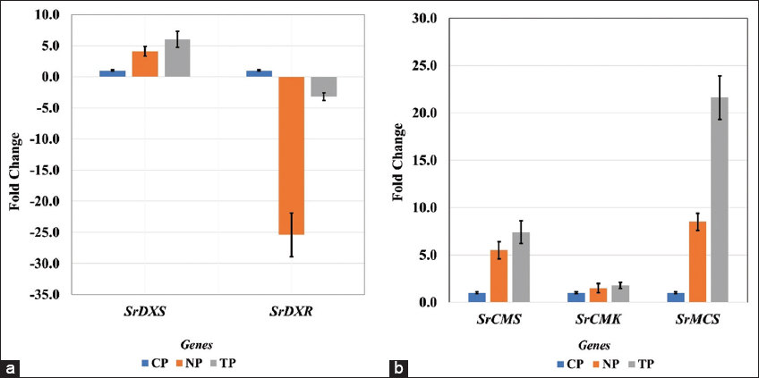
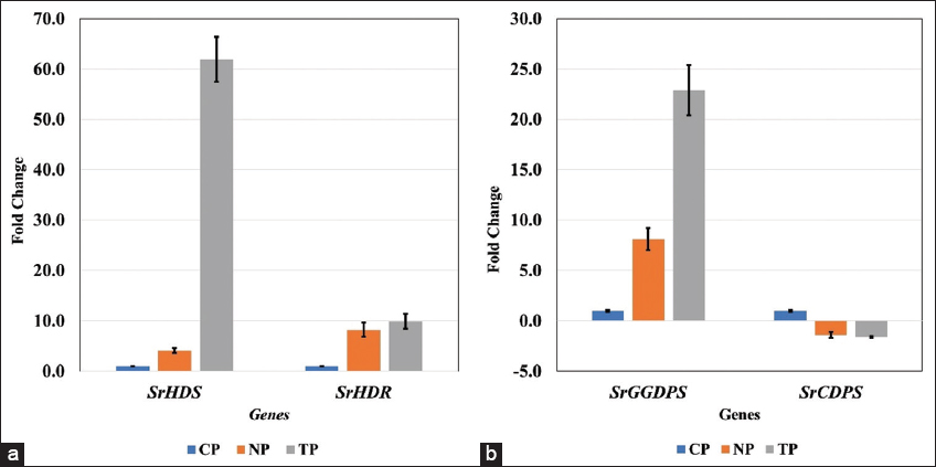
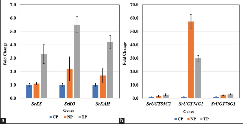
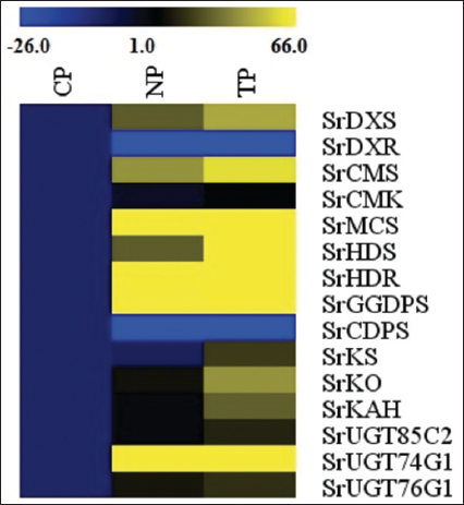
In the current study, two types of transcript accumulation patterns, up-regulation and subsequently varying rates of down-regulation, were seen in the genes of the SG biosynthesis pathway in S. rebaudiana Bertoni, both in transformed and non-transformed lines. The highest transcript levels among the up-regulated genes were found in the leaves of the TPs, indicating that, out of the 15 pathway genes investigated in this study, R. rhizogenes-infected plants had superior transcript level, in general. The present findings are quite similar to the findings of Sarmiento-López, in 2020; Libik-Konieczny et al., in 2020, and Sanchéz-Cordova et al., in 2019 [23-25]. However, specifically, 13 genes were upregulated, and two of them were downregulated in the current study.

In our findings, higher transcript abundance (i.e., up-regulation) in three genes, SrKS (KS), SrKO (KO), and SrKAH (KAH) were recorded in TP and NP as compared to CP, which showed enhanced SGs content in leaves, corroborated with the earlier finding in Stevia by Zheng et al., 2019, and Nasrullah et al., 2023 [26,27]. The genes SrKO and SrUGT74G1 show high expression, leading to an increase in Stevioside level. This finding correlates with studies by Nasrullah et al., 2023 in S. rebaudiana, demonstrating their significant role in enhancing SG content [28].

In the present research, all NP and TP lines were propagated using the same combination of plant hormones in MS media and maintained under the same growth conditions [15], however, a higher SrUGT74G1 expression level was noted in NP when compared with transformed lines (TP). Expression increase of SrUGT74G1 in NP could be due to metabolic reprogramming during micropropagation stress. The lack of comparable expression in TP, even with the same culture conditions, may indicate that in the secondary metabolism of plants, the feedback regulation mechanisms responsive to the complete metabolites tend to inhibit the expression of biosynthetic pathways. For instance, Gachon et al., in 2005 and Tiwari et al., in 2016, discuss how glycosylation, mediated by glycosyltransferases, plays a crucial role in regulating hormone homeostasis and secondary metabolite biosynthesis [28,29].

In TP lines, higher flux through the steviol biosynthetic pathway might activate such feedback loops, leading to the downregulation of SrUGT74G1. Conversely, NP plants, with comparatively lower precursor flux, may not trigger these feedback mechanisms, resulting in higher expression levels of SrUGT74G. Another reason might be the choice of explants [30]. For the NP plants, nodal explants were used for direct shoot regeneration while, in case of TP plants, micro-shoots with hairy roots were used as explants for TP regeneration [15]. In addition, post-transcriptional regulation, including mechanisms mediated by microRNAs (miRNAs), can influence gene expression levels. Kajla et al, in 2023 highlight the role of miRNAs in fine-tuning the expression of genes involved in secondary metabolite biosynthesis. Such regulatory processes can lead to variations in gene expression independent of genetic transformation [31].

In the present study, two genes, SrDXS and SrCDPS indicated a downregulation, and despite that, in in vitro generated plants, stevioside levels were still high, as confirmed by HPLC analysis. This could be achievable due to such genes being part of a complex regulatory network in the SG pathway, which may involve a feedback mechanism, post-transcriptional regulation, and compensatory upregulation of other genes to sustain or even elevate overall SG production. The increased flux through the SG pathway in in vitro plantlets may trigger feedback regulation mechanisms. These mechanisms, which respond to the total metabolites, prefer to suppress the expression of biosynthetic pathways. On the other hand, in vivo plants, with relatively reduced precursor flux, might not activate these feedback mechanisms, resulting in varying expression levels. Moreover, the upregulation of major downstream UGT genes, specifically SrUGT76G1, could counteract the downregulation of upstream genes such as SrCDPS and SrDXR, resulting in net increase in SG accumulation. Similar trends have been reported in Stevia transformation studies, where enhanced glycosylation contributed to higher SG yields despite variations in early pathway gene expression [32,33]. Lower expression of these genes can also act as positive regulators of the pathway. The correlation between gene expression and SG quantification highlights the complex regulation of the biosynthetic pathway. While some genes exhibit downregulation, the overall metabolic flux toward stevioside biosynthesis appears to be driven by the enhanced expression of key glycosyltransferases. This underscores the potential for targeted genetic modifications to optimize SG production in Stevia [34,35].

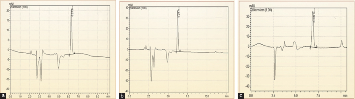
HPLC data of the current research showed that transformed Stevia plants had higher stevioside content compared to micropropagated plants (NP), which in turn accumulated more stevioside than the in vivo, grown CP. This trend largely coincides with the gene expression data obtained through real-time PCR analysis in the current study. Similar findings regarding enhanced SG accumulation in R. rhizogenes-mediated transformed S. rebaudiana plantlets were reported by Sánchez-Córdova and co-workers in (2019) [25]. The rise in stevioside content in NP as compared to the CPs might be attributed to tissue culture-induced metabolic reprogramming [36], where the controlled in vitro environment and synchronized developmental stage of regenerated plantlets can enhance secondary metabolite biosynthesis.

The present work mainly focused on HPLC analysis to quantify stevioside, one of the most predominant and important SGs found in S. rebaudiana. Stevioside is a central intermediate in the biosynthetic pathway and is frequently used as a reliable metabolic marker owing to its high accumulation in the leaf tissues. If other glycosides such as rebaudioside A were to be measured, a better picture of the metabolite spectrum would have emerged; however, the targeted analysis of stevioside alone provides a strong and representative estimate of the biosynthetic activity. This holds true for the main objective of correlating gene expression patterns with the core output of the SG biosynthesis pathway. In addition, focusing on stevioside has allowed for an accurate assessment of the impact of transformation and regeneration conditions on its accumulation.

5. CONCLUSION

In the present study, 13 out of the 15 analyzed genes involved in the SG biosynthesis pathway exhibited higher expression in the leaves of in vitro transformed Stevia plants (TP) compared to in vitro regenerated NP and CP. This upregulation suggests a positive correlation between gene expression and enhanced biosynthesis and accumulation of SGs in leaves. Furthermore, HPLC analysis confirmed that TPs demonstrated a higher stevioside content, indicating their potential reliability for improved SG production. These TP lines could serve as an alternative approach for large-scale production of SGs, facilitating their evaluation in animal trials for prospective commercial applications as a natural sweetener. SGs, being a plant-derived, low-calorie sugar substitute, could be particularly advantageous for individuals managing metabolic disorders, including high blood sugar, cardiovascular issues, and excessive weight gain. Their widespread application in the food industry may contribute to the development of healthier dietary alternatives, promoting overall well-being. This study highlights the potential for molecular-level manipulation of the SG biosynthetic pathway to enhance SG production. However, further molecular studies and pathway exploitation are essential to fully harness the biotechnological potential of S. rebaudiana for commercial applications.

6. ACKNOWLEDGEMENTS

Authors are grateful to Dr. D. Y. Patil Biotechnology and Bioinformatics Institute, Dr. D. Y. Patil Vidyapeeth (Deemed to be University), Pune, for providing the laboratory facilities and financial support. We are thankful to DST, NCL, and Dr. D. Y. Patil Pharmacy College, Pune, for the instruments they provided for the current research.

7. AUTHORS’ CONTRIBUTIONS

All authors made substantial contributions to conception and design, acquisition of data, or analysis and interpretation of data; took part in drafting the article or revising it critically for important intellectual content; agreed to submit to the current journal; gave final approval of the version to be published; and agree to be accountable for all aspects of the work. All the authors are eligible to be an author as per the International Committee of Medical Journal Editors (ICMJE) requirements/guidelines.

8. CONFLICT OF INTEREST

The authors report no financial or any other conflicts of interest in this work.

9. ETHICAL APPROVALS

This study does not involve experiments on animals or human subjects.

10. AVAILABILITY OF DATA AND MATERIALS

All the data pertaining to this study, are in the possession of the authors and will be supplied upon request.

11. PUBLISHER’S NOTE

All claims expressed in this article are solely those of the authors and do not necessarily represent those of the publisher, the editors and the reviewers. This journal remains neutral with regard to jurisdictional claims in published institutional affiliation.

12. USE OF ARTIFICIAL INTELLIGENCE (AI)-ASSISTED TECHNOLOGY

The authors declare that they have not used artificial intelligence (AI)-tools for writing and editing of the manuscript, and no images were manipulated using AI.


REFERENCES

1.  Sharma S, Gupta S, Kumari D, Kothari SL, Jain R, Kachhwaha S. Exploring plant tissue culture and steviol glycosides production in Stevia rebaudiana (Bert.) Bertoni:A review. Agriculture. 2023;13(2):475.[CrossRef]

2.  Hajihashemi S, Geuns JM, Ehsanpour AA. Gene transcription of steviol glycoside biosynthesis in Stevia rebaudiana Bertoni under polyethylene glycol, paclobutrazol and gibberellic acid treatments in vitro. Acta Physiol Plantarum. 2013;35:2009-14.[CrossRef]

3.  Brandle JE, Telmer PG. Steviol glycoside biosynthesis. Phytochemistry. 2007;68(14):1855-63.[CrossRef]

4.  Thakur K, Ashrita, Sood A, Kumar P, Kumar D, Warghat AR. Steviol glycoside accumulation and expression profiling of biosynthetic pathway genes in elicited in vitro cultures of Stevia rebaudiana. In Vitro Cell Dev Biol Plant. 2021;57:214-24.[CrossRef]

5.  Zhou X, Gong M, Lv X, Liu Y, Li J, Du G, et al. Metabolic engineering for the synthesis of steviol glycosides:Current status and future prospects. Appl Microbiol Biotechnol. 2021;105(13):5367-81.[CrossRef]

6.  Ceunen S, Werbrouck S, Geuns JM. Stimulation of steviol glycoside accumulation in Stevia rebaudiana by red LED light. J Plant Physiol. 2012;169(7):749-52.[CrossRef]

7.  Eslami-Firouzabadi A, Karimi M, Abbasi-Surki A, Shafeinia A, Derikvand-Moghadam F. Optimising the rate and stages of application of nitrogen fertiliser for stevia under greenhouse conditions. South African Journal of Plant and Soil 2023;40:58-63.[CrossRef]

8.  Olas B. Stevia rebaudiana Bertoni and its secondary metabolites:Their effects on cardiovascular risk factors. Nutrition. 2022;99:111655.[CrossRef]

9.  Abdel-Aal RA, Abdel-Rahman MS, Al Bayoumi S, Ali LA. Effect of stevia aqueous extract on the antidiabetic activity of saxagliptin in diabetic rats. J Ethnopharmacol. 2021;265:113188.[CrossRef]

10.  Bugliani M, Tavarini S, Grano F, Tondi S, Lacerenza S, Giusti L, et al. Protective effects of Stevia rebaudiana extracts on beta cells in lipotoxic conditions. Acta Diabetol. 2022;59:113-126.[CrossRef]

11.  Peteliuk V, Rybchuk L, Bayliak M, Storey KB, Lushchak O. Natural sweetener Stevia rebaudiana:Functionalities, health benefits and potential risks. EXCLI J. 2021;20:1412.[CrossRef]

12.  Abdullah S, Mohamad Fauzi NY, Khalid AK, Osman M. Effect of gamma rays on seed germination, survival rate and morphology of Stevia rebaudiana hybrid. Malays J Fundam Appl Sci. 2021;17(5):543-9.[CrossRef]

13.  Álvarez-Robles MJ, López-Orenes A, Ferrer MA, Calderón AA. Methanol elicits the accumulation of bioactive steviol glycosides and phenolics in Stevia rebaudiana shoot cultures. Ind Crops Prod. 2016;87:273-9.[CrossRef]

14.  Kahrizi D, Ghaheri M, Yari Z, Yari K, Bahraminejad S. Investigation of different concentrations of MS media effects on gene expression and steviol glycosides accumulation in Stevia rebaudiana Bertoni. Cell Mol Biol. 2018;64(2):23-7.[CrossRef]

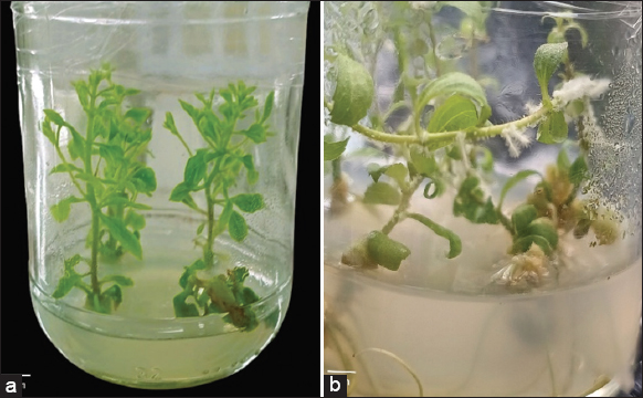
15.  Singh P, Labade D, Chote M, Deshmukh P, Panchal B, Deshpande J, et al. Efficient regeneration of Stevia rebaudiana Bertoni transformants through hairy root culture technique. J Appl Bot Food Qual. 2025;98:22-8. [CrossRef]

16.  Pan H, Xiao L, Tang K, Xia H, Li Y, Jia H, et al. Screening UDP-glycosyltransferases for effectively transforming stevia glycosides:Enzymatic synthesis of glucosylated derivatives of rubusoside. J Agric Food Chem. 2022;70(48):15178-88.[CrossRef]

17.  Yu J, Tao Y, Pan H, Lin L, Sun J, Ma R, et al. Mutation of Stevia glycosyltransferase UGT76G1 for efficient biotransformation of rebaudioside E into rebaudioside M. J Funct Foods. 2022;92:105033.[CrossRef]

18.  Ghaheri M, Kahrizi D, Bahrami G, Mohammadi-Motlagh HR. Study of gene expression and steviol glycosides accumulation in Stevia rebaudiana Bertoni under various mannitol concentrations. Mol Biol Rep. 2019;46(1):7-16.[CrossRef]

19.  Livak KJ, Schmittgen TD. Analysis of relative gene expression data using real-time quantitative PCR and the 2- ΔΔCT method. Methods. 2001;25(4):402-8.[CrossRef]

20.  Herath V, Gayral M, Adhikari N, Miller R, Verchot J. Genome-wide identification and characterization of Solanum tuberosum BiP genes reveal the role of the promoter architecture in BiP gene diversity. Sci Rep. 2020;10(1):11327.[CrossRef]

21.  Phule AS, Barbadikar KM, Maganti SM, Seguttuvel P, Subrahmanyam D, Babu MP, et al. RNA-seq reveals the involvement of key genes for aerobic adaptation in rice. Sci Rep. 2019;9(1):5235.[CrossRef]

22.  Ye J, Jin CF, Li N, Liu MH, Fei ZX, Dong LZ, et al. Selection of suitable reference genes for qRT-PCR normalisation under different experimental conditions in Eucommia ulmoides Oliv. Sci Rep. 2018;8(1):15043.[CrossRef]

23.  Sarmiento-López LG, López-Meyer M, Sepúlveda-Jiménez G, Cárdenas L, Rodríguez-Monroy M. Photosynthetic performance and stevioside concentration are improved by the arbuscular mycorrhizal symbiosis in Stevia rebaudiana under different phosphate concentrations. PeerJ. 2020;8:10173.[CrossRef]

24.  Libik-Konieczny M, Michalec-Warzecha ?, Dziurka M, Zastawny O, Konieczny R, Rozp?dek P, et al. Steviol glycosides profile in Stevia rebaudiana Bertoni hairy roots cultured under oxidative stress-inducing conditions. Appl Microbiol Biotechnol. 2020;104:5929-41.[CrossRef]

25.  Sanchéz-Cordova ÁD, Capataz-Tafur J, Barrera-Figueroa BE, López-Torres A, Sanchez-Ocampo PM, García-López E, et al. Rhizobium rhizogenes-mediated transformation enhances steviol glycosides production and growth in Stevia rebaudiana plantlets. Sugar Tech. 2019;21(3):398-406.[CrossRef]

26.  Zheng J, Zhuang Y, Mao HZ, Jang IC. Overexpression of SrDXS1and SrKAH enhances steviol glycosides content in transgenic Stevia plants. BMC Plant Biol. 2019;19:1-6.[CrossRef]

27.  Nasrullah N, Ahmad J, Saifi M, Shah IG, Nissar U, Quadri SN, et al. Enhancement of diterpenoid steviol glycosides by co-overexpressing SrKO and SrUGT76G1 genes in Stevia rebaudiana Bertoni. PLoS One. 2023;18(2):0260085.[CrossRef]

28.  Gachon CM, Langlois-Meurinne M, Saindrenan P. Plant secondary metabolism glycosyltransferases:The emerging functional analysis. Trends Plant Sci. 2005;10(11):542-9.[CrossRef]

29.  Tiwari P, Sangwan RS, Sangwan NS. Plant secondary metabolism linked glycosyltransferases:An update on expanding knowledge and scopes. Biotechnol Adv. 2016;34(5):714-39.[CrossRef]

30.  Bednarek PT, Or?owska R. Plant tissue culture environment as a switch-key of (EPI) genetic changes. Plant Cell Tissue Organ Cult. 2020;140(2):245-57.[CrossRef]

31.  Kajla M, Roy A, Singh IK, Singh A. Regulation of the regulators:Transcription factors controlling biosynthesis of plant secondary metabolites during biotic stresses and their regulation by miRNAs. Front Plant Sci. 2023;14:1126567.[CrossRef]

32.  Kim MJ, Zheng J, Liao MH, Jang IC. Overexpression of Sr UGT 76G1 in Stevia alters major steviol glycosides composition towards improved quality. Plant Biotechnol J. 2019;17(6):1037-47.[CrossRef]

33.  Abdelsalam NR, Botros WA, Khaled AE, Ghonema MA, Hussein SG, Ali HM, et al. Comparison of uridine diphosphate-glycosyltransferase UGT76G1 genes from some varieties of Stevia rebaudiana Bertoni. Sci Rep. 2019;9(1):8559.[CrossRef]

34.  Singh S, Murmu S, Das AB, Haider ZA, Banerjee M. Establishment of root-to-root culture and evaluation of phytochemicals in Rhizobium rhizogenes transformed roots of Stevia rebaudiana. J Pharmacogn Phytochem. 2017;6(6S):49-54.

35.  Bayraktar M, Naziri E, Karabey F, Akgun IH, Bedir E, Röck-Okuyucu B, et al. Enhancement of stevioside production by using biotechnological approach in in vitro culture of Stevia rebaudiana. Int J Secondary Metab. 2018;5(4):362-74.[CrossRef]

36.  Fazili MA, Bashir I, Ahmad M, Yaqoob U, Geelani SN. In vitro strategies for the enhancement of secondary metabolite production in plants:A review. Bull Natl Res Centre. 2022;46(1):35. https://doi.org/10.1186/s42269-022-00717-z[CrossRef]

Reference

Article Metrics
82 Views 30 Downloads 112 Total

Year

Month

Related Search

By author names

Similar Articles

Enzymes and qualitative phytochemical screening of endophytic fungi isolated from Lantana camara Linn. Leaves

Mbouobda Hermann Desire , Fotso Bernard , Muyang Rosaline Forsah , Chiatoh Thaddeus Assang, Omokolo Ndoumou Denis

Antibacterial activity of Ferula asafoetida: a comparison of red and white type

Richa Bhatnager, Reena Rani, Amita Suneja Dang

Antimicrobial Activity Screening of Marine Bacteria Isolated from the Machilipatnam Sea Coast of Andhra Pradesh, India

K. Bala Chandra, V. Umamaheswara Rao, Subhaswaraj Pattnaik, Siddhardha Busi

Microbial biotechnology for bio-prospecting of microbial bioactive compounds and secondary metabolites

Ajar Nath Yadav

Differential metabolic responses associated with drought tolerance in Egyptian rice

Amira Hassanein, Eman Ibrahim, Rania Abou Ali, Hanan Hashem

Agrobacterium rhizogenes as molecular tool for the production of hairy roots in Withania somnifera

Manali Singh,, Deep Chandra Suyal, Nisha Dinkar, Soniya Joshi, Nishtha Srivastava, Vineet Kumar Maurya, Abhiruchi Agnihotri, Sanjeev Agrawal

Genome mining and AntiSMASH analysis of an Endophytic Talaromyces sp. reveal biosynthetic pathway gene clusters for novel bioactive compounds

Priyanka N. Shenoy, Sneha Bhaskar, M. Manu, M. P. Likitha, N. Geetha, Shailasree Sekhar, K Ramachandra Kini

Secondary metabolite profiles, antimicrobial and antioxidant activities of callus, and leaves extract of Piper sarmentosum Roxb.

Junairiah Junairiah, Listijani Suhargo, Tri Nurhariyati, Nabilah Istighfari Zuraidassanaaz

Antifungal potential of entomopathogenic bacteria, Photorhabdus, and Xenorhabdus (Morganellaceae) against pathogenic fungi

Mary Lalramchuani,, Lal Ramliana, Hrang Chal Lalramnghaki, Albana L. Chawngthu, Van Ramliana, Esther Lalhmingliani

The role of plant growth regulators in modulating secondary metabolite production in nampu (Homalomena rostrata Griff)

Fahrauk Faramayuda, Demia Pratiwi, I. Gusti Ngurah Dwi Wiryawan, Elfahmi Elfahmi

Genomic and functional characterization of Bacillus sp. B.PNR2 from extinct volcanic soil in Buriram province, Thailand

Praphat Kawicha, Kusavadee Sangdee, Thanwanit Thanyasiriwat, Rattana Pengproh, Khanitta Somtrakoon, Aphidech Sangdee

Gene expression study of Saccharomyces cerivisae GPH1 gene in response to chemical modulators

Prasad M. P.

Transcriptional expression of three putative pathogenesis-related proteins in leaves of rubber tree (Hevea brasiliensis) inoculated with Neofusicoccum ribis

A. I. C. Nyaka Ngobisa , Godswill Ntsomboh-Ntsefong , Wong Mui Yun , M. Z. Dzarifah, P. A. Owona Ndongo

Genome-wide identification and expression analysis along the leaf developmental gradient of the sigma factor gene family in foxtail millet (Setaria italica)

Hongyun Liu, Jinjin Cheng, Siyuan Cheng, Hui Fan , Bo Wen , Zheng Liu

Progress in understanding the regulation and expression of genes during plant somatic embryogenesis: A review

Vikrant, Prajisha Janardhanan

Comparative transcriptome analysis to identify common genes involved in the progression of Sjögren’s syndrome and rheumatoid arthritis

Khushboo Choudhury, Navjyoti Chakraborty, Monika Gandhi, Sayan Chatterjee, Ram Singh Purty

Evaluation of seven different wheat cultivars for their resistance to drought in terms of growth indicators and yield

Zeyad H. AL-Fatlawi, Ali Nadhim Farhood, Saleh Abed Alwahed Mahdi, Auday Hamid Taha Al-Tmime

In silico characterization of Melittin from Apis cerana indica and evaluation of melittin intron for transgene expression in mammalian cells

Kevin Kumar Vijayakumar, Abisheik Rajandran, Sandhya Lumumba, Shakila Harshavardhan

Molecular characterization and expression profiling of arsenic mediated stress-responsive genes in Dawkinsia tambraparniei (Silas, 1954)

Selvakumar Sakthivel,, Anand Raj Dhanapal,, Venkataa Suresh Munisamy, Mohammed Parvez Nasirudeen, Varatharaji Selvaraj, Vijay Velu, Annadurai Gurusamy

Computational analysis of differential gene expression in rice during abiotic stress

Shivani Devi, Yogeeta Goyal, Mansi Malik, Navjot Kaur, Yamini Sangar, Kashmir Singh, Ruchi Sachdeva

The comparative anti-obesity potential of Lagerstroemia speciosa (L.) leaf extracts and their synthesized gold nanoparticles by downregulation of PPAR-γ, C/EBP-α, and FABP4/aP2 gene expression

Tarsem Nain, Mahendra Bishnoi, Navpreet Kaur, Santosh Kumar Tiwari, Jaya Parkash Yadav

Genome-wide analysis and gene expression studies revealed putative homeotic genes with a role in flower formation in sesame (Sesamum indicum L.)

H. N. Annapurna, Arya Ramachandran, N. S. Ronald Reagan, Injangbuanang Pamei, K. T. Ramya, Ragiba Makandar