
1. INTRODUCTION
Black pepper (Piper nigrum L.), commonly referred to as the "king" of spices, is extensively used worldwide [1]. Currently, approximately 70 countries cultivate black pepper, with India maintaining the largest cultivation area of 195,000 hectares across its territory, followed by Indonesia with 116,000 hectares, and Brazil with 45,000 hectares. In Vietnam, black pepper cultivation began in the 17th century in regions such as Phu Quoc district, Kien Giang province. By 2022, the total black pepper cultivation area in Vietnam will reach 119,600 hectares, with the Central Highlands region accounting for 60% (71,760 hectares), making it the largest black pepper production region in the country [2]. However, the annual yield of black pepper is significantly impacted by various diseases, particularly root rot caused by Phytophthora capsici, a pathogenic oomycete [3,4]. The widespread presence of this disease in many regions has made black pepper cultivation increasingly challenging [3–6]. Chemical plant protection measures have achieved only partial success, there is a growing demand for spices free from chemical residues. Currently, all available commercial pepper varieties are susceptible to this disease, and continuous efforts over the past 2 decades have only identified a few resistant to P. capsici [5,7,8]. One of the strategies to enhance black pepper resistance against foot rot disease is by grafting, for instance, using Piper colubrinum as a disease-resistant rootstock [9].
Currently, the rootstock source of disease resistance is wild pepper species, such as P. colubrinum [9], P. hancei [10], or P. flaviflorum [11]. Moreover, black pepper is a perennial plant, and conventional breeding programs face numerous difficulties due to its slow growth rate. Therefore, the development of new alternative strategies is essential to identify and produce disease-resistant pepper varieties that meet market demands without chemical residues. In previous reports, we successfully selected Piper hancei varieties resistant to diseases caused by Phytophthora capsici and Meloidogyne incognita [10,12]. Piper hancei belongs to the genus Piper of the family Piperaceae and is mainly distributed in southwest China. Its aerial parts have been widely used in Chinese herbal medicinal prescriptions for the treatment of rheumatism, asthma, and arthritic conditions [13–15]. P. hancei growth habit was climbing and had a polymorphic branching type with the production of many runners. P. hancei produced multiple runner shoots and lots of adventitious roots. Pubescence on the stem was absent. Leaf lamina shapes were ovate-elliptic with acrodromous venation. The flower spike was erect type. The spike shape was cylindrical. P. hancei produced only pistillate flowers. The fruit shape was not round [12].
Biotechnological tools like tissue culture are important for the selection, multiplication, and conservation of medicinal plant genotypes [16,17]. In vitro regeneration plays a great role in the production of high-quality and disease-free plantlets for crop programs [18–20]. However, the study on in vitro propagation of Piper hancei was not well-studied. This study describes the in vitro propagation process of Piper hancei, aimed at mass-producing plantlets to serve as rootstocks in the production of disease-resistant pepper.
2. MATERIALS AND METHODS
2.1. Media and Cultural Conditions
The Piper hancei variety, known for its resistance to Phytophthora capsici and Meloidogyne incognita, have been cultivated at the Institute of Biotechnology, Hue University. The plants, around 2.5 m tall and over 2 years old, robust and disease-free, were selected as initial materials. Healthy stem segments with large leaves, containing 6–7 nodes from the entire plant, were used for sterilizing in sample inoculation. The culture medium used was MS basal medium [21] containing 3.0% sucrose and 0.8% agar, supplemented with different concentrations of plant growth regulators (PGRs). The pH of the medium was adjusted to 5.8. The medium was autoclaved at 121°C, 1 atm for 15 minutes. Experiments were conducted under conditions of 25°C, light intensity of 2,000–3,000 lux, and a photoperiod of 10–12 hours/day [18].
2.2. Explant Sterilization
Explants from the second to fifth nodes counting from the apex of pepper vines cut into 3 cm segments for sterilization. These segments were initially shaken in 1% dilution of Lifebouy soap for 15 minutes. Next, samples were treated in a 2% solution of Ridomil Gold fungicide (Syngenta) for 30 minutes, followed by immersion in the bactericide Antisuper 80wp (2%, Greenfarm) for 60 minutes. After each treatment, the explants were thoroughly rinsed with sterile distilled water [18]. The sterilized segments were transferred to autoclaved Duran bottles within a laminar flow hood. The explants were further treated by shaking in 70% ethanol for 60 seconds, followed by rinsing in sterile distilled water. Additional sterilization with 10% NaClO for 6–12 minutes, followed by rinses in sterile distilled water (5–6 times) [22]. Finally, the explants were cut into 2 cm lengths containing dormant buds and cultured on MS medium supplemented with 3.0% sucrose, 0.8% agar, and 0.5 mg/l [6-Benzylaminopurine (BAP)] [18]. The efficacy of sterilization after 3 weeks of culture was evaluated through the proportions of explants that were non-contaminated, contaminated, and dead.
2.3. Effects of PGRs on In Vitro Shoot Regeneration
In vitro stem explants were cultured on a basal MS medium containing 0.8% agar and 3% sucrose, supplemented with either BAP or kinetin (KIN) (1–2 mg/l) [23], or a combination of BAP (1 mg/l) with 0.1–0.5 mg/l indole butyric acid (IBA) to evaluate the shoot regeneration ability [24]. After four weeks of culture, the following parameters were recorded: percentage of explants forming shoots, shoot numbers per explant, and average shoot height (cm).
2.4. Effects of PGRs on In Vitro Shoot Multiplication
To evaluate rapid shoot multiplication, in vitro shoots were cultured on a basal MS medium containing 0.8% agar and 3% sucrose, supplemented with BAP at concentrations of 1.0–1.5 mg/l in combination with KIN at concentrations of 0.5–2.5 mg/l [23] and IBA at 0.5 mg/l [25]. After eight weeks of culture, the following parameters were recorded: number of shoots per explant and average shoot height (cm).
2.5. In vitro Root Formation
To investigate the root formation and growth potential of in vitro shoots, shoots with an average height of 2 cm, 2–4 leaves, and robust growth were individually separated and cultured on MS basal medium containing 0.8% agar and 3% sucrose, supplemented with IBA (0–2.0 mg/l) [23]. The results were evaluated after a 4-week culture period based on several criteria: percentage of explants forming roots, root number per shoot, root length (cm), and average height of shoots (cm).
2.6. Transplantation
The nursery, covering an area of 200 m², was equipped with a 50% shade net and utilizes natural light and temperature. In vitro healthy plantlets (3–5 leaves, 3–4 cm in height, and 6–11 roots) were selected for transplantation into the nursery. The in vitro culture bags containing the plants were acclimatized to external conditions over a period of 3–5 days. Subsequently, the bags were opened and left undisturbed for 1–2 days to allow the plants to adjust to the natural environment [26]. Post acclimatization, the in vitro plants were removed from their culture medium. The roots were thoroughly washed to remove any adhering medium, and then soaked in a 1% Ridomil Gold (Syngenta) solution for 5 minutes. The treated plants were then transplanted into seedling trays filled with a mixture of fertilizer, sand, and soil in a ratio of 1:1:1. To facilitate acclimatization, the plants were covered with transparent polyethylene bags for 2 weeks. The polyethylene covers were gradually removed over a period of 2 weeks. The plants were then transferred to a greenhouse for four weeks as described by Hussain et al. [26]. After 2–6 weeks of transplantation, the growth of the in vitro plantlets was evaluated by measuring survival rate (%), leaf number/plantlet, and plant height (from the soil surface to the shoot tip). The experiments were conducted in a nursery with ambient temperatures ranging from 28°C –32°C.
2.7. Statistical Analysis
Each experiment was repeated 3 times, and 30 samples were observed in each condition. The data were analyzed in SPSS 19.0 software, using Duncan’s test (p ≤ 0.05).
3. RESULTS
3.1. Effect of Sterilization Time
To enhance the sterilization efficiency of the explants, we conducted preliminary sterilization using commercially available fungicides and bactericides, followed by treatment with 10% NaClO. The sterilization results after three weeks of culture indicated a significant influence of the sterilization time with 10% NaClO on the survival rate, infected rate, and death rate of the explants. When the explants were sterilized for 6 minutes, the number of survival rate was the lowest for uninfected explants, at 4.45%, while the infected rate was very high, at 91.1%. Increasing the sterilization duration from 6 minutes to 8–10 minutes resulted in an increase in the number of surviving samples from 4.45% to 37.78%, and a significant reduction in the infected rate from 91.1% to 51.11%. However, extended sterilize/exposure times to 12 minutes did not significantly change the infected rate, but led to a considerable increase in the death rate (11.11%–24.45%) and reduced the survival rate (Table 1). Hence, the 10% NaClO treatment in 12 minutes duration has been found to be excessively long and caused significant damage to the stem nodes while failing to completely eliminate all microbial sources present in samples. In summary, treating samples with 10% NaClO for 10 minutes yielded the best sterilization results, achieving 37.78% of infection-free survival rates.
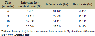 | Table 1. Effect of 10% NaClO sterilization duration on living sample viability. [Click here to view] |
3.2. Effect of PGRs on Shoot Regeneration
Nodal segments were initially sterilized before being cultured on MS basal medium containing 3.0% sucrose and 0.8% agar and supplemented with different concentrations of BAP or KIN or both combined to assess the shoot regeneration capacity. After 2 weeks of cultivation, reproductive buds were formed on the nodes in certain culture media. The regeneration rates of shoots varied across different MS media by the fourth week. The MS medium without PGRs supplementation resulted in the lowest regeneration rate at 70%, while all PGR-supplemented media achieved 100% shoot regeneration (Table 2; Fig. 1).
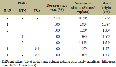 | Table 2. Effect of PGRs on shoot regeneration after 4 weeks. [Click here to view] |
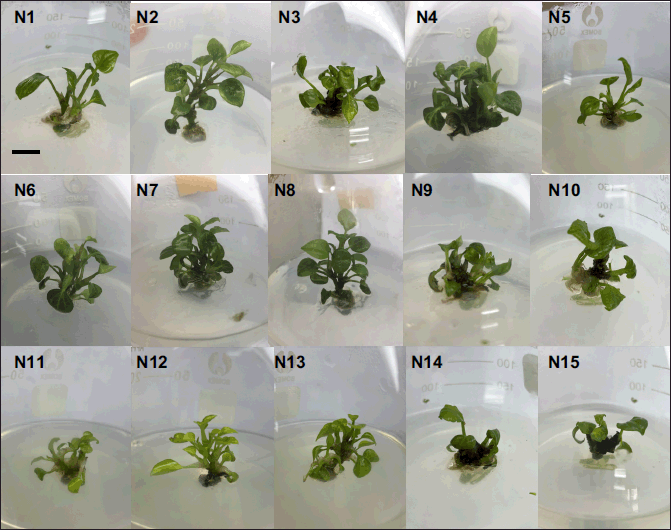
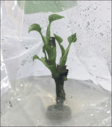 | Figure 1. Regenerated shoots from stem node explants on MS medium supplemented with 1.0 mg/l BAP after 4 weeks. [Click here to view] |
The results illustrate that the MS medium supplemented with 1 mg/l BAP yielded the highest shoot regeneration efficiency, producing robust shoots with a light green color and an average of 1.83 shoots per explant. However, increasing the BAP concentration to 2 mg/l resulted in a decrease in shoot regeneration efficiency, with an average of 1.2 shoots per explant, and the growth of these shoots was slower. In media supplemented solely with KIN and those supplemented with a combination of BAP and IBA, the average number of shoots obtained was nearly equivalent (1.03–1.27 shoots/explant).
3.3. The Effects of PGRs on Shoot Multiplication
BAP stimulates in vitro shoot formation, while KIN prolongs shoot and promotes leaf development. Therefore, we combined these 2 regulators, either alone or in combination with IBA, to investigate the potential for shoot multiplication, aiming to find the optimal conditions for high-quality shoot production. The results were evaluated after 8 weeks of culture.
In comparison to natural shoot regeneration from node explants, in vitro shoot multiplied relatively slowly. Thus, shoot multiplication efficiency was calculated after 8 weeks of cultivation. The data revealed that the number of shoots increased with a medium supplemented with 1 mg/l BAP and increasing KIN concentrations from 0.5 to 2 mg/l, but declined when KIN increased to 2.5 mg/l. In a medium with 1.5 mg/l BAP and KIN ranging from 0.5 to 1.5 mg/l, the shoot number increased, then decreased when KIN was further increased to 2–2.5 mg/l. Shoots formed in BAP and KIN-supplemented media displayed vigorous growth, with large size and dark green foliage (Table 3; Fig. 2).
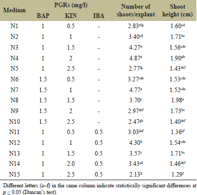 | Table 3. Effect of PGRs on shoot multiplication after 8 weeks. [Click here to view] |
 | Figure 2. Formation of shoot clusters on different media after 8 weeks (bar = 1 cm). Among all experimental media, MS medium supplemented with 1 mg/l BAP and 2 mg/l KIN, and MS medium supplemented with 1.5 mg/l BAP and 1.0 mg/l KIN, yielded the highest and comparable shoot number, at 4.87 and 4.77 shoots/explant, respectively. However, shoots grown in MS medium supplemented with 1.0 mg/l BAP and 2.0 mg/l KIN exhibited stronger growth and larger size than those in the other medium. Therefore, MS medium supplemented with 1.0 mg/l BAP and 2.0 mg/l KIN was selected as the optimal medium for shoot multiplication process. [Click here to view] |
Among all experimental media, MS medium supplemented with 1 mg/l BAP and 2 mg/l KIN, and MS medium supplemented with 1.5 mg/l BAP and 1.0 mg/l KIN, yielded the highest and comparable shoot numbers, at 4.87 and 4.77 shoots/explant, respectively. However, shoots grown in MS medium supplemented with 1.0 mg/l BAP and 2.0 mg/l KIN exhibited stronger growth and larger size than those in the other medium. Therefore, MS medium supplemented with 1.0 mg/l BAP and 2.0 mg/l KIN was selected as the optimal medium for shoot multiplication process.
3.4. Root Formation
In this study, the effect of IBA for in vitro rooting was investigated, and the results after 4 weeks of cultivation were summarized (Table 4). Shoots cultured on IBA-free MS medium showed no rooting ability, with slow growth, underdeveloped leaves with yellowing, and an average shoot height of 2.36 cm.
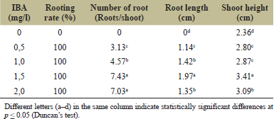 | Table 4. Effect of PGRs on rooting ability of in vitro pepper shoots. [Click here to view] |
On the MS media supplemented with 0.5–2 mg/l IBA, all shoots achieved a rooting percentage of 100%. However, the rooting intensity and shoot development varied with different IBA concentrations. The medium containing 0.5 mg/l IBA yielded the lowest average number of roots per shoot (3.13) with a root length of 1.14 cm and no root hairs. Despite slow growth and pale green leaves, the presence of a developed root system enhanced better growth compared to non-IBA supplemented media, with an average shoot height of 2.8 cm. Increasing the IBA concentration to 1.0 mg/l resulted in an increase in root formation with an average of 4.57 roots/shoot and a root length of 1.42 cm, without root hairs. Shoots exhibited slow growth, reaching an average height of 2.87 cm, with larger green leaves. Further increases in IBA to 1.5–2.0 mg/l significantly enhanced the rooting ability, the highest root numbers ranged from 7.03 to 7.43 roots/shoot (non-significant difference between both media), characterized by abundant root hairs. However, shoots on 1.5 mg/l IBA exhibited better growth and root development compared to those supplemented with 2.0 mg/l IBA. In 1.5 mg/l IBA-supplemented medium, roots formed early and rapidly, averaging 1.97 cm in length and 3.41 cm in height, whereas shoot formation on 2.0 mg/l IBA was slower, with an average root length of 1.35 cm and shoot height of 3.09 cm (Fig. 3). IBA concentrations beyond 1.5 mg/l may inhibit shoot and root formation, so, the higher IBA concentrations were not investigated. The optimal medium for rooting in vitro pepper shoots was found to be MS medium supplemented with 0.8% agar, 3% sucrose, and 1.5 mg/l IBA.
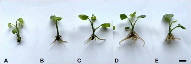 | Figure 3. In vitro shoots after 4 weeks of culture on MS medium supplemented with different concentrations of IBA. (A) 0 mg/l IBA; (B) 0.5 mg/l IBA; (C) 1.0 mg/l IBA; (D) 1.5 mg/l IBA; (E) 2.0 mg/l IBA. (bar = 1 cm). [Click here to view] |
3.5. Acclimatization and Growth of In Vitro Plantlets in Nursery
In vitro plantlets were transplanted to nursery beds containing a mixture of soil, sand, and organic fertilizer in a ratio of 1:1:1. Due to the high susceptibility of in vitro plants to dehydration and mortality upon transfer, transparent plastic sheets were used to cover the trays during the initial 2 weeks, ensuring adequate ventilation while minimizing water loss. In the last 3–5 days of the second week, the plastic coverings were gradually removed to help the plants acclimate to natural growing conditions. Assessment of plant survival and growth parameters including survival rate, number of leaves/plantlet, and plant height were conducted after 2 and 4 weeks of transplantation (Table 5).
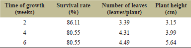 | Table 5. The growth of in vitro plantlets in a nursery. [Click here to view] |
In the first 2 weeks, the plantlets exhibited minimal growth, primarily adapting to external conditions and developing a root system suitable to the new substrate. The survival rate during this period was 86.11%. Some leaves showed slight yellowing and underdevelopment, with an average of 3.39 leaves/plantlet and a plant height of 3.15 cm. These plantlets were exposed to natural environmental conditions after 2 weeks. Beginning from 2 to 4 weeks, the plantlets adapted well to the external environment, although some plants did not survive, reducing the survival rate to 80.55% after 4 weeks. Survived plantlets displayed vigorous growth, with shoot height increasing from 3.15 to 3.99 cm. Most plantlets produced new leaves, with older leaves turning a dark green and robust, while new leaves were light green; some older leaves yellowed and fell off, at around 4.31 leaves/plantlet. By week 6, the plantlets had fully adapted to the external conditions, and no further plant losses occurred between weeks 4 and 6. The plants grew strong, with large robust leaves and an average height of 5.64 cm and 4.49 leaves/plantlet (Fig. 4).
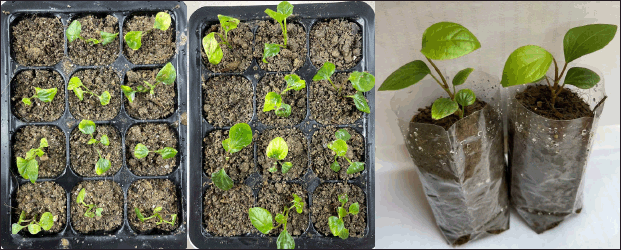 | Figure 4. The growth of in vitro plantlets after 2–6 weeks in nursery. [Click here to view] |
4. DISCUSSION
Traditional methods typically involve pepper propagation through cutting, which can reduce the yield and quality of pepper due to fungal, bacterial, or viral infections. Additionally, plants propagated by cutting increased genetic uniformity but lack uniformity in both morphology and quality [27–29]. With in vitro plantlets, disease-free propagation is ensured, and vigorously grown plantlets can be selected before being transferred to the field [30,31]. In the Piperaceace family, the in vitro propagation of Piper nigrum was well-studied [24,29,32], however, lack of publications on P. hancei micropropagation. Some studies have demonstrated that BAP alone or the combination of BAP and KIN yields the best shoot multiplication results for pepper [33–35]. Betel vine (Piper betle L.) produced the highest number of shoots on Woody Plant Medium (WPM) medium supplemented with 3.0 mg/l BAP and 1.0 mg/l KIN [36]. Long pepper (Piper longum) showed the best shoot formation on MS medium supplemented with 8.9 M BAP and 4.64 M KIN [37]. However, in this study, shoots grown in KIN-supplemented media exhibited faster growth, larger leaves, and greater height compared to those grown in media with a BAP and IBA combination. In comparison to the medium without IBA, the medium supplemented with IBA showed a reduction in the number of shoots formed.
The combination of BAP with KIN and IBA indicated that IBA negatively affected shoot multiplication and growth, resulting in slow, weak shoots with small, pale green leaves. Callus formation was observed at the shoot base, with the outer part being soft, pale yellow, and the firm inner part. High KIN concentrations (1.5–2.5 mg/l) caused the leaf petioles near the shoot base to swell and form soft white callus that later turned black. These results differ from previously published research in root formation of in vitro shoots in various species within the genus Piper. Divakaran et al. [36] reported that Piper betle L. developed a robust root system and grew consistently on WPM medium without PGR supplementation. Similarly, in vitro shoots of Piper aduncum and Piper hispidinervum showed optimal root formation on MS medium without PGR supplementation [38]. Piper auritum shoots successfully rooted (100%) on ½ MS medium supplemented with 2 mg/l IAA [39]. Strong root development from shoots of P. longum was achieved on MS medium supplemented with 2.46 µM IBA [37]. These findings indicated that root development from in vitro shoots varies significantly among different species within the Piper genus.
IBA, a synthetic auxin, is known for its role in stimulating root formation from in vitro shoots of various plant species, including black pepper [18,33,34]. In this study, the effect of IBA concentration for in vitro rooting was investigated, and the optimal medium was found to be 1.5 mg/l IBA. In this medium, roots formed early and rapidly, averaging 1.97 cm in length and 3.41 cm in height. Previous studies on in vitro propagation of some species within the genus Piper have demonstrated high survival rates and vigorous growth when tissue-cultured plantlets were transferred to the nursery [26,40]. P. betle L. was successfully propagated by tissue culture methods, reaching an 80% survival rate when planted in soil [36]. Anand and Srinivasa Rao [41] reported a survival rate of 98% when transplanted in vitro P. barberi plantlets into the nursery, with a subsequent survival rate of 75% in the field [41]. In this study, the survival ratio value of 80.55% after 6 weeks, similar to other reports.
5. CONCLUSION
We have succeeded in developing a procedure to propagate Piper hancei in vitro. Shoots were regenerated from the stem on basal MS medium supplemented with 1.0 mg/l BAP after 4 weeks of culture (1.83 shoots/sample). The MS medium supplemented with 1.0 mg/l BAP and 2.0 mg/l KIN was optimal for shoot multiplication (4.87 shoots/cluster) whereas 1.5 mg/l IBA addition was optimal for rooting (7.43 roots/shoot). The plantlets were successfully transplanted to the nursery with a survival rate of 80.55% after 6 weeks, averaging 4.49 leaves and 5.64 cm in height. The plantlets can be served as rootstocks in the production of disease-resistant pepper.
6. ACKNOWLEDGMENTS
The study was supported by the Ministry of Science and Technology of Vietnam (Grant No. ?T?L.CN08/20). We acknowledge partial financial support from the Core research program (NCTB.DHH.2024.03), which covers article processing charges.
7. CONFLICTS OF INTEREST
The authors report no financial or any other conflicts of interest in this work.
8. AUTHORS’ CONTRIBUTIONS
All authors made substantial contributions to conception and design, acquisition of data, or analysis and interpretation of data; took part in drafting the article or revising it critically for important intellectual content; agreed to submit to the current journal; gave final approval of the version to be published; and agree to be accountable for all aspects of the work. All the authors are eligible to be an author as per the International Committee of Medical Journal Editors (ICMJE) requirements/guidelines.
9. ETHICAL APPROVALS
This study does not involve experiments on animals or human subjects.
10. DATA AVAILABILITY
The data are available from the corresponding author upon appropriate request.
11. PUBLISHER’S NOTE
All claims expressed in this article are solely those of the authors and do not necessarily represent those of the publisher, the editors and the reviewers. This journal remains neutral with regard to jurisdictional claims in published institutional affiliation.
12. USE OF ARTIFICIAL INTELLIGENCE (AI)-ASSISTED TECHNOLOGY
The authors declares that they have not used artificial intelligence (AI)-tools for writing and editing of the manuscript, and no images were manipulated using AI.
REFERENCES
1. Spence C. The king of spices: on pepper's pungent pleasure. Int J Gast Food Sci 2024;35:100900. CrossRef
2. Chung DV, Hoa NX, Mai PLT, Nguyet NTM, Hang HTT, Thanh DD, et al. Assessment of the growth, disease resistance of tissue cultured black pepper plants (Piper nigrum L.) on the farm in Dak Nong province. Tay Nguyen J Sci 2023;62:61–8.
3. Truong NV, Liew EC, Burgess LW. Characterisation of Phytophthora capsici isolates from black pepper in Vietnam. Fungal Biol 2010;114(2-3):160–70. CrossRef
4. Nazeem P, Raghavamenon A, Devassy BT, Parab G, Devaki G, Keshavachandran R, et al. Expression of pathogenesis related proteins in black pepper (Piper nigrum L.) in relation to Phytophthora foot rot disease. J Trop Agri 2008;46:49–51.
5. Vandana VV, Bhai RS, Azeez S. Biochemical defense responses of black pepper (Piper nigrum L.) lines to Phytophthora capsici. Physiol Mol Plant Pathol 2014;88:18–27. CrossRef
6. Long VN. Spread of Phytophthora capsici in Black Pepper (Piper nigrum) in Vietnam. Engineering 2015;7:506–13. CrossRef
7. Bhai RS, Eapen SJ, Anandaraj M, Saji KV. Identification of Phytophthora and nematode-resistant source-from opens pollinated progenies of black pepper (Piper nigrum) using a modified protocol. Ind J Agri Sci 2010;80(10):43–7.
8. Eapen SJ, Ramana KV, Saji KV. Host resistance in black pepper (Piper nigrum L.) against root-knot and burrowing nematode. J Plant Crop 2011;39(3):341–6.
9. Helina S, Suharjo R, Latifa N, Dwiatmini K, Cempaka I, Khodijah S, et al. The role of grafting in the resistance of foot rot disease in black pepper (Piper nigrum). Pak J Phytopathol 2024;36:225–30. CrossRef
10. Truong HTH, Rasphone S, Nguyen BLQ, Nguyen NQ, Nguyen TM, Ho HN, et al. Piper grafts resistant to both pathogenic Phytophthora capsici and Meloidogyne incognita. Ind J Agri Res 2024;AF-894:1–9. CrossRef
11. Hao Y, Fan R, Zhao Y, Nie K, Wang L, Zhao T, et al. Intra species dissection of Phytophthora capsici resistance in black pepper. J Adv Res [In press] 2024. CrossRef
12. Truong HTH, Rasphone S, Nguyen BLQ, Ho HN, Nguyen CQ, Tran TT, et al. Identification of Piper species that are resistant to Phytophthora capsici, Meloidogyne incognita, and waterlogging in Vietnam. Plant Pathol 2023;72(9):1615–25; CrossRef
13. Zhang L, Hu X, Liu M. Complete chloroplast genome sequences of the medicinal plant Piper hancei. Mitochondrial DNA B Resour 2021;6(9):2775–6. CrossRef
14. Yang F, Li H, Yang YQ, Hou Y, Liang D. Lignanamides from the stems of Piper hancei maxim. and their anti-inflammatory and cytotoxic activities. Fitoterapia 2022;161:105231. CrossRef
15. Jiang S, Jianhua X, Li X. A study on the RAPD and SCAR molecular markers of Piper species. J Agri Rural Dev Trop Subtrop 2009;110(2):127–35.
16. Verma A, Janani H, Nishteswar K. Biotechnological tools in the cultivation of medicinal plants. J Ind Syst Med 2016;4:42–5.
17. Debnath M, Malik CP, Bisen PS. Micropropagation: a tool for the production of high quality plant-based medicines. Curr Pharm Biotechnol 2006;7(1):33–49. CrossRef
18. Thi PTD, Man NTN, Quynh NTK, Hoa TT, Thuy PMT, Quang HT. Micropropagation of long-leaved paperbark (Melaleuca leucadendra (L.) L.). Propag Ornam Plants 2023;23(4):91–8.
19. Lemma DT, Banjaw DT, Megersa H. Micropropagation of medicinal plants: review. Int J Plant Breed Crop Sci 2020;7(2):796-802.
20. Podwyszy?ska M, Orlikowska T, Trojak-Goluch A, Wojtania A. Application and improvement of in vitro culture systems for commercial production of ornamental, fruit, and industrial plants in Poland. Acta Soc Bot Pol 2022;91:914. CrossRef
21. Murashige T, Skoog F. A revised medium for rapid growth and bio assays with tobacco tissue cultures. Physiol Plant 1962;15(3):473–97. CrossRef
22. Gunawardhana WPDS, Perera PIP, Muhandiram HMAP, Swarnathilaka DBR, Priyadarshani KDN. Surface sterilization protocols of leaf and bud explants for initiating in vitro cultures of Piper nigrum L. (Pepper). Trop Agri Res Ext 2022;25(2):132–42; CrossRef
23. Sakpere AM, Ezenu VN. Vegetative and micropropagation potential of Piper guineense (Schumach and Thonn). Int J Horti Sci 2023;29(1):29–36. CrossRef
24. Thuyen DA, Du TX, Giap DD, Ton NT. Preliminary study on the micropropagation in vitro of black pepper (Piper nigrum L.). Acad J Biol 2014;27(3):39–45. CrossRef
25. Raju MRI, Junaki M, Hossain MT. Establishment of a suitable regeneration protocol for rapid propagation of Piper nigrum L. through in vitro culture. Jahangirnagar Univ J Biol Sci 2022;11(1&2):31–40. CrossRef
26. Hussain A, Naz S, Nazir H, Shinwari Z. Tissue culture of black pepper (Piper nigrum L.) in Pakistan. Pak J Bot 2011;43:1069–78.
27. Currey CJ, Hutchinson VA, Lopez RG. Growth, morphology, and quality of rooted cuttings of several herbaceous annual bedding plants are influenced by photosynthetic daily light integral during root development. HortSci 2012;47(1):25–30; CrossRef
28. Bamidele Akinrinola T, Abidemi Onadeko R. Performance of black pepper cuttings as influenced by growing media. Chi J Agri Ani Sci 2023;39:256–65.
29. Dat NB, Anh TTH, Hoa BT, Diep PH, Minh TV. Effects of plant growth regulators on micropropagation of black pepper (Piper nigrum) in commercial-scales. Asian J Biotechnol Biores Technol 2024;10(2):45–59. CrossRef
30. Hasnain A, Naqvi SAH, Ayesha SI, Khalid F, Ellahi M, Iqbal S, et al. Plants in vitro propagation with its applications in food, pharmaceuticals and cosmetic industries; current scenario and future approaches. Front Plant Sci 2022;13:1009395. CrossRef
31. Kumar M, Sirohi U, Malik S, Kumar S, Ahirwar GK, Chaudhary V, et al. Methods and factors influencing in vitro propagation efficiency of ornamental Tuberose (Polianthes species): a systematic review of recent developments and future prospects. Horticulturae 2022;8(11):998. CrossRef
32. Kadam S, Rasam D, Joshi K, Jadhav A. Micropropagation of black pepper, cv. panniyur-1: standardization of sterilization protocol and media composition. Glob J Bio-Sci Biotechnol 2020;9:45–9.
33. Deepak DA, Vaidya E, Muthusamy S, Agrawal A. Improved micropropagation protocol and molecular marker based genetic stability assessment of black pepper (Piper nigrum L.). Ind J Plant Genet Res 2022;35:264–74. CrossRef
34. Afroz T, Huq H, Hoque ME. In vitro regeneration of black pepper (Piper nigrum L.). Ban J Agri 2023;48(2):75–85. CrossRef
35. Philip VJ, Joseph D, Triggs GS, Dickinson NM. Micropropagation of black pepper (Piper nigrum Linn.) through shoot tip cultures. Plant Cell Reports 1992;12(1):41–4. CrossRef
36. Divakaran M, Ravindran PN, Pete KV, Babu KN, Rema J, Pillai GS. Micropropagation of betel vine (Piper betle L.). J Spi Aroma Crop 1992;(2):160–2.
37. Soniya EV, Das MR. In vitro micropropagation of Piper longum – an important medicinal plant. Plant Cell Tiss Org Cult 2002;70(3):325–7. CrossRef
38. da Silva Tatiane L, Balzon Talita A, Schewinski-Pereira J. A rapid in vitro protocol for propagation of Piper aduncum and Piper hispidinervum, two species from Amazon region with multipurpose uses. Afri J Biotechnol 2012;11(89):15539–46. CrossRef
39. Dominguez F, Lozoya X, Simon J. Tissue culture regeneration of a medicinal plant from Mexico: Piper auritum Kunth. HortSci 2006;41(1):207–9.
40. Joseph B, Joseph D, Philip VJ. Plant regeneration from somatic embryos in black pepper. Plant Cell Tiss Org Cult 1996;47(1):87–90. CrossRef
41. Anand A, Srinivasa Rao C. A rapid in vitro propagation protocol for Piper barberi Gamble, a critically endangered plant. In Vitro Cell Dev Biol - Plant 2000;36(1):61–4. CrossRef