Research Article | Volume 14, Issue 1, January, 2026

In vitro micropropagation and gas chromatography-mass spectrometry profiling of callus culture in Pulicaria jaubertii for conservation and metabolite production

Fathia Mohamed Noman Salam Fatima Ahmed Alhadi Ebraheem Ali Al-nawd Enas Jabir Al-sanabani Esam Mohammed Aqlan Majed Ahmed Al-mansoub   

Open Access   

Published:  Nov 25, 2025

DOI: 10.7324/JABB.2025.250540
Abstract

Pulicaria jaubertii is an aromatic and medicinal plant endemic to Yemen, currently facing habitat decline. This study aimed to evaluate its in vitro response in full-strength Murashige and Skoog (medium supplemented with different types and concentrations of plant growth regulators. Among the tested plant parts, only seed explants successfully initiated callus formation. Calli were subsequently subcultured in media containing 0.1 mg/L 1-naphthaleneacetic acid (NAA) with kinetin (Kin) at 0, 0.25, 0.5, or 1 mg/L. Additional experiments tested media with 0.1 mg/L 6-benzylaminopurine and indole-3-acetic acid (IAA) (0–1 mg/L), as well as 0.1 mg/L Kin with 2,4-dichlorophenoxyacetic acid (2,4-D) (0–1 mg/L). Growth parameters related to callus induction, root, shoot, and leaf production were assessed. Findings revealed that Kin had no significant effect on most growth parameters except callus colour (P = 0.012), with the best growth at 0.25 mg/L. Similarly, IAA significantly influenced callus induction (P = 0.009), with optimal results at 1.0 mg/L. In contrast, 2,4-D had no significant effect, but its highest concentration (1.0 mg/L) supported optimal growth. Gas chromatography-mass spectrometry (GC-MS) analysis identified 46 compounds in the ethanolic callus extract compared to 25 in the mother plant, which indicates a richer phytochemical profile in the callus. The 2-Ethoxyethylamine (85.60%) and Stigmasterol (58.79%) were most abundant in ethanolic and n-hexane extracts. In conclusion, P. jaubertii seeds are the most responsive explants for micropropagation, forming callus as an initial step. Interestingly, GC-MS profiling identified bioactive compounds with medicinal properties. Further studies should refine auxin and cytokinin ratios to enhance propagation efficiency.


Keyword:     Pulicaria jaubertii Micropropagation Cytokinins Auxins Callus Gas chromatography-mass spectrometry


Citation:

Salam FMN, Alhadi FA, Al-nawd EA, Al-sanabani EJ, Aqlan EM, Al-mansoub MA. In vitro micropropagation and gas chromatography-mass spectrometry profiling of callus culture in Pulicaria jaubertii for conservation and metabolite production. J Appl Biol Biotech 2026;14(1):201-215. http://doi.org/10.7324/JABB.2025.250540

Copyright: Author(s). This is an Open Access article distributed under the terms of the Creative Commons Attribution-NonCommercial-ShareAlike license.

HTML Full Text

1. INTRODUCTION

Pulicaria jaubertii Gamal Eldin, also known as Pulicaria orientalis Jaub., which is a member of the family Asteraceae, is one of the Pulicaria species endogenous to the Arabian Peninsula, particularly in Yemen. It is a perennial aromatic plant with erect branches that may reach 50 cm in height. In Arabic, it is officially called “Eter Elraee” [1,2]. However, it is known locally in Yemen as “Anssif” or “Alkhaoah” and is used as a spicing herb for making some food, such as laban (fermented milk) and soup. Traditionally, it is used as an insecticide, antipyretic, diuretic, and for treating urogenital system disorders, colds, malaria, inflammation, and microbial infections [3-6].

P. jaubertii is a perennial aromatic herb with erect, tomentose, grey-green branches reaching 30–50 cm in height. The plant is strongly fragrant and partially woody at the base. Leaves are sessile, oblong to oblanceolate, measuring 1.5–4.5 cm in length and 0.4–2 cm in width, with toothed margins and an obtuse apex. The inflorescence consists of a few capitula (flower heads), each 1–2 cm in diameter, with yellow florets. Involucral bracts are oblanceolate and densely villous. The ray florets are 3–5 mm long. The fruit is an achene, setulose, and obscurely ribbed, containing small seeds adapted for wind dispersal [2,7].

Several studies revealed that Pulicaria species, including P. jaubertii, have been found to have various bioactive characteristics such as cytotoxic and anticancer [2,8-11], antioxidant [3,5,12-14], antibacterial [2,13,15], antifungal and immuno-regulatory [16], antiinflammatory and antihistaminic [9,10,17], antidiabetic [9], antihypertensive [8,18], and antispasmodic [19] activities.

Detailed agricultural statistics on this species are rare due to its large-scale harvesting rather than cultivation. Ethnobotanical surveys and herbarium records in Yemen indicate that P. jaubertii is seasonally collected because of its high demand for culinary and medicinal uses in rural areas [20,21]. In addition, it is highly sold in local markets; thus, formal data on annual harvest volumes are also lacking. Therefore, the emergence and worsening of habitats have increased concern for the stability of wild populations, which outlines the need for protective measures and further research [22].

P. jaubertii is not yet recorded on the International Union for Conservation of Nature Red List of endangered species. However, many factors like drought, overexploitation, uncontrolled overcutting, and overgrazing in Yemen and Saudi Arabia may put many native species, including P. jaubertii, at high risk and worsen this decline [7]. Therefore, conservation efforts should consist of protecting their natural habitats and preserving them in ex-situ environments through seed banks and botanical gardens [16]. However, some recent techniques, including the micropropagation technique, could be used to improve their production. This technique is an important in vitro tissue culture method that may be used to conserve a great number of threatened and rare crops, medicinal, and economically important plants [23-27]. It could be used to provide several benefits for the plant, such as producing a large amount of pathogen-free and healthy plants, as well as genetically improved crops within a relatively short time and small space [28-32]. Interestingly, the rate of proliferation and other variables could be affected by the use of diverse types and concentrations of plant growth regulators (PGRs), chiefly auxins and cytokinins [33,34]. These types of PGRs are commonly used in combination with plant tissue culture [35,36], and the research area for their action is still broad. Phytochemical composition in plants varies due to genetic, environmental, and methodological factors, which could influence their biological properties and therapeutic potential [37].

P. jaubertii has been widely investigated for its essential oils and secondary metabolites, which vary significantly with geographic origin, extraction method, and plant part used [2,5,13,38]. However, in vitro techniques such as callus culture remain unexplored for this species. Callus cultures offer a controlled environment for enhancing the production of bioactive compounds [16]; and understanding the biochemical and genetic factors influencing such variation may facilitate the synthesis of pharmacologically and industrially valuable phytochemicals [39]. To date, no study has reported a micropropagation protocol or gas chromatography-mass spectrometry (GC-MS)-based phytochemical profiling for P. jaubertii. This study, therefore, presents the first successful micropropagation strategy for the species, employing seed-derived callus as the primary explant source. Although the approach follows standard protocols used in related Asteraceae species such as Pulicaria incisa and Achillea spp., its application to P. jaubertii addresses a critical gap. The developed protocol not only supports conservation but also establishes a reproducible platform for secondary metabolite production under controlled conditions, which aims to assess growth responses in Murashige and Skoog (MS) media supplemented with different types and concentrations of PGRs, specifically cytokinins and auxins.


2. MATERIALS AND METHODS

2.1. Chemicals

Cytokinins (kinetin [Kin] and 6-benzylaminopurine [BAP]) and Auxins (1-naphthaleneacetic acid [NAA] and 2,4-dichlorophenoxyacetic acid [2,4-D]), plant hormones, were purchased from Sigma-Aldrich (St. Louis, MO, USA). Ethanol, n-hexane, and other chemicals used in the study were of the highest quality available.

2.2. Plant Material and Study Design

P. jaubertii [Figure 1a] was collected from Ibb City, Yemen, from March to April 2021 at GPS coordinates of 13º 57’ 46.44” N and 44º 10’ 23.88” E. The taxonomist, Dr. Esam Aqlan, identified the plant at the Biology Department, Faculty of Science, Ibb University. The experimental study was conducted in the Tissue Culture Laboratory, Department of Biology, Faculty of Sciences, Ibb University, in 2021.

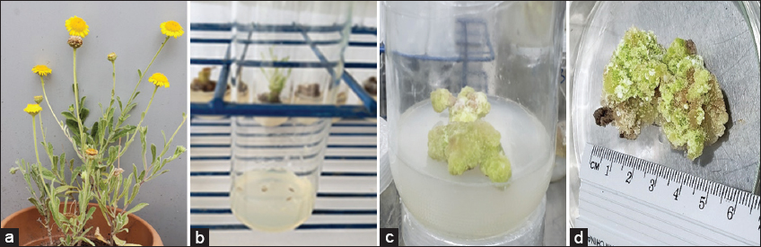
Figure 1: (a) Morphology of Pulicaria jaubertii; (b) Seeds; (c) Callus used as explants; (d) Harvested callus for extraction.


[Click here to view]

2.3. Medium and Cultural Conditions

Several types of plant parts (leaves, nodes, and seeds) were surface-sterilised and inoculated in a full Murashige and Skoog [40] medium as performed by Salam et al. [41] [Figure 1b]. Preliminary trials also tested leaf and stem explants under various hormonal regimes, including high cytokinin and low auxin combinations; however, these explants failed to produce callus or shoots and often exhibited necrosis. Seeds, used with their coats intact, germinated successfully and induced callus without the need for decoating. The MS medium contains specific concentrations of nutrients necessary for most plant growth, but in hormone-free conditions, only seeds produce roots and shoots.

For micropropagation, seeds were supplemented with 0.5 mg/L Kin and 0.1 mg/L 1-naphthalene acetic acid (NAA) in MS medium. Callus was formed within 1–2 weeks [Figure 1c and d], followed by shoot formation and then root production after 3–4 weeks. Explants obtained from these calli were then subcultured with various types and concentrations of PGRs.

2.4. Hormonal Effects on Shoot, Root and Callus Regeneration

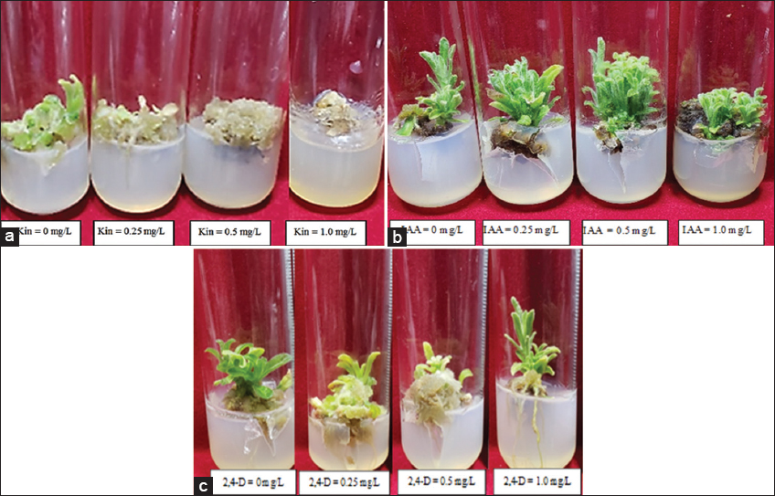
2.4.1. Shoot induction and multiplication

The explants’ response to growth was analysed in full MS media with different concentrations of the cytokinin Kin (0, 0.25, 0.5, and 1 mg/L) and a fixed concentration of the auxin NAA (0.1 mg/L).

2.4.2. Root induction

The explants’ response to growth was investigated in MS media supplemented with various concentrations (0, 0.25, 0.5, and 1 mg/L) of the auxin indole-3-acetic acid (IAA) and a fixed concentration of the cytokinin 6-benzylamino purine (BAP) (0.1 mg/L).

2.4.3. Callus induction

Likewise, explants’ response was tested with different concentrations of growth regulators 2,4-dichlorophenoxyacetic acid (2,4-D) (0, 0.25, 0.5, and 1 mg/L) and a fixed concentration of Kin (0.1 mg/L).

The response included the regeneration of shoots, roots, and callus, as well as their related growth parameters. Cultures were kept at 25 ± 2°C in an air-conditioned environment under a 16-h light/8-h dark photoperiod. Since no previous micropropagation study has focused on P. jaubertii, a stepwise method was used to understand the basic hormonal needs for shoot and callus development. Each experiment changed one growth regulator (cytokinin or auxin) while keeping the concentration of the other constant to find effective baseline conditions.

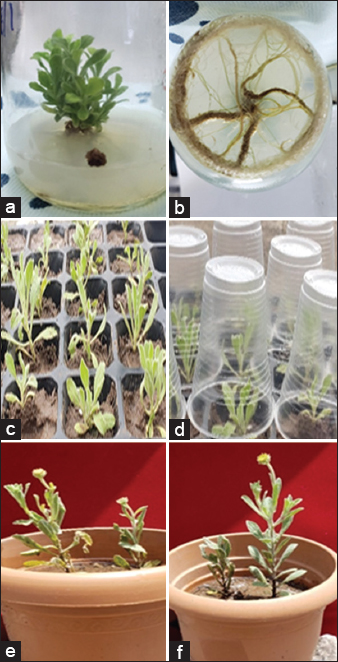
2.4.4. Acclimatisation of in vitro derived plantlets

Acclimatisation was carried out on 50 rooted shoots from 35-day-old in vitro plantlets. These plantlets were moved to plastic pots filled with a soil-sand mixture (70:30, v/v) and kept under laboratory conditions (25 ± 2°C, 16-h photoperiod) for 4 weeks. The survival rate was noted, and 45 plantlets (90%) successfully acclimatised. Among these, 20 plantlets were later transplanted into larger pots and grown under outdoor garden conditions. All 20 plantlets survived and showed healthy growth (100%) after another 4 weeks.

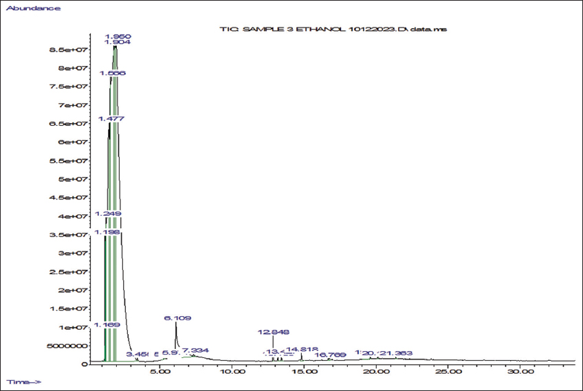
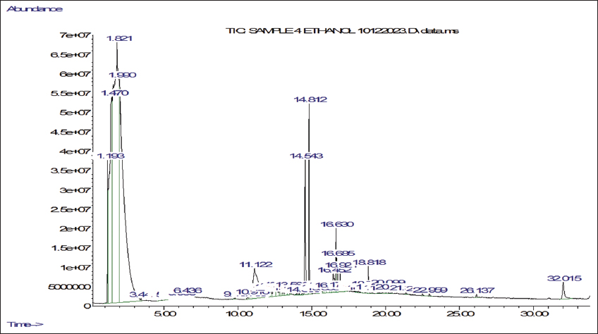
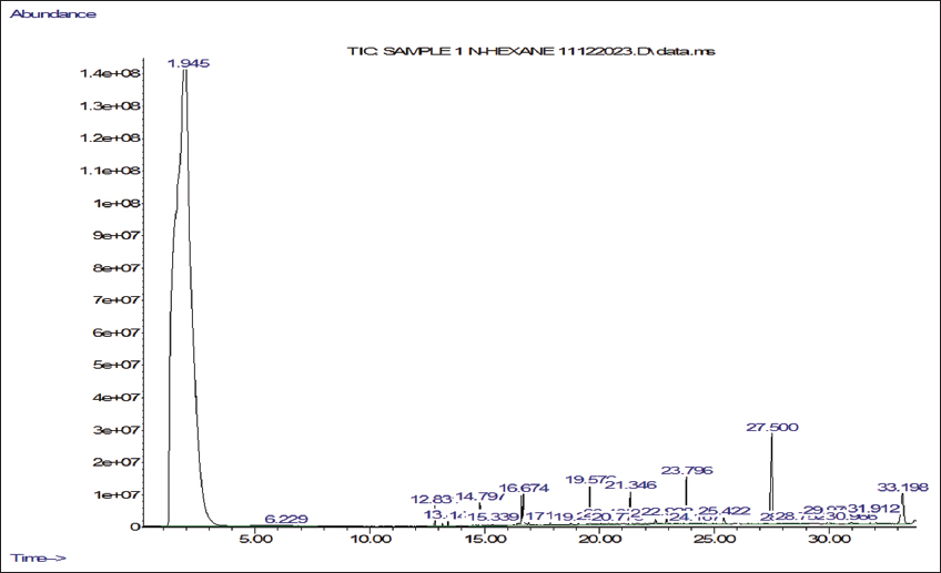
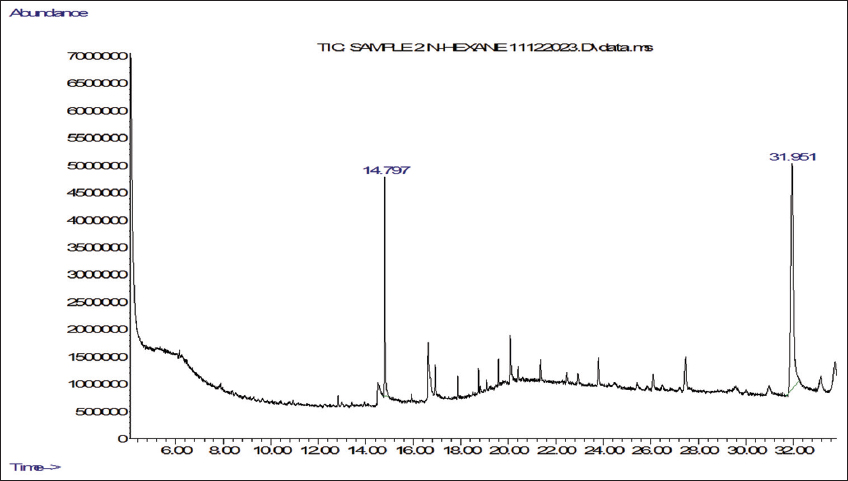
2.5. Gas Chromatography-Mass Spectrometry (GC-MS) Analyses

Ethanol and n-hexane extracts were made from shade-dried powder obtained from the mother and callus derived from micropropagated explants of P. jaubertii. The four extracts were prepared using the cold maceration method and then concentrated at 45°C using a rotary evaporator (Büchi, Switzerland). We determined the volatile phytochemicals following the procedure by Yisak et al. [42] with some modifications. GC-MS analysis was done using an Agilent 7890A gas chromatograph coupled with a 5975C mass spectrometer (Agilent, USA) equipped with electron ionisation (EI) for phytochemical analysis. Chromatographic separations were carried out using an HP-5MS (19091S-433) capillary column that is 30 m long, has a 0.25 mm internal diameter, and a 0.25-µm column phase film thickness. The injection mode was split-less, Helium served as the carrier gas, and we injected 1 µL of the sample at a consistent flow rate of 1 mL/min into an inlet heated to 275°C. The initial oven temperature was 60°C with a 2-minute hold time, then increased to 200°C with a ramp of 10°C/min and 3°C/min to 240°C. The ion source temperature was set to 230°C for the mass spectrometer settings, and the quadrupole temperature was 150°C. The system ran in positive electron impact mode at 70 eV, scanning from 40 to 650 m/z. The total run time was 45 minutes. Phytochemical components were qualitatively identified by comparing their retention times and mass spectral data with those in the NIST98 library.

2.6. Statistical Analysis

Data were summarized in tables as frequencies and proportions. Crosstabulation and Fisher’s exact test were used to study the effect of different concentrations of the hormones on the non-parametric markers of plant growth. One-way analysis of variance, followed by Duncan’s Multiple Range test, was used to compare the means of the parametric values produced at various concentrations of the hormones. Data analysis was performed using the statistical software Statistical Package for the Social Sciences version 20.0, and P ≤ 0.05 was taken as significant.


3. RESULTS

3.1. Hormonal Effects on Callus, Shoot, and Root Regeneration

Seeds were the only plant part that responded to growing in the media, whether supplemented or not with the tested PGRs. Shoot proliferation was first achieved from callus induced on MS medium supplemented with 0.5 mg/L Kin and 0.1 mg/L NAA. This medium served as the baseline for subsequent experiments in which Kin, IAA, and 2,4-D concentrations were varied. As shown in Table 1A and Figure 2a, adding Kin at any tested concentration (0, 0.25, 0.5, or 1 mg/L) to the MS medium containing 0.1 mg/dL of NAA induced rooting in some tubes (n = 14, 38.9%) and shooting and callus formation were noticed in most tubes (n = 26, 72.2%, and n = 28, 77.8%, respectively). Although the highest callus induction (n = 9, 100%) occurred at 1.0 mg/L kin, no significant differences were noticed between concentrations for callus induction, shooting, or rooting. In contrast, callus color varied significantly (P = 0.012) between treatments, with yellowish-brown calli being most frequent (n = 12, 33.3%), followed by greenish-brown calli (n = 8, 22.2%).

Table 1: Effect of different types and concentrations of cytokinins and/or auxins on the induction of roots, shoots, and callus and the colour and morphology of Pulicaria jaubertii callus.

Comparisons variablesABC


Concentration of the cytokinin: Kinetina (n=36)Fisher’s exact test (P-value))Concentration of the auxin: IAAb (n=36)Fisher’s exact test (P-value))Concentration of auxin: 2,4-Dc (n=36)Fisher’s exact test (P-value)
Total n (%)0 ml/L n (%)0.25 mg/L n (%)0.5 mg/L n (%)1 mg/L n (%)Total n (%)0 ml/L n (%)0.25 ml/L n (%)0.5 ml/L n (%)1 ml/L n (%)Total n (%)0 ml/L n (%)0.25 ml/L n (%)0.5 ml/L n (%)1.0 ml/L n (%)
Root inductionNS
 No22 (61.1)4 (44.4)7 (77.8)5 (55.6)6 (55.7)2.333 (0.668)13 (36.1)8 (88.9)6 (66.7)7 (77.8)6 (66.7)1.739 (0.833)0 (0)0 (0.0)0 (0.0)0 (0.0)0 (0.0)
 Yes14 (38.9)5 (55.6)2 (22.2)4 (44.4)3 (44.4)23 (63.9)1 (11.1)3 (33.3)2 (22.2)3 (33.3)36 (100)9 (100.0)9 (100.0)9 (100.0)9 (100.0)
Shoot induction
 No10 (27.8)2 (22.2)2 (22.2)4 (44.4)2 (22.2)1.634 (0.784)27 (75)4 (44.4)3 (33.3)5 (55.6)1 (11.1)4.201 (0.317)19 (52.8)4 (44.4)6 (66.7)7 (77.8)2 (22.2)6.330 (0.117)
 Yes26 (72.2)7 (77.8)7 (77.8)5 (55.6)7 (77.8)9 (25)5 (55.6)6 (66.7)4 (44.4)8 (88.9)17 (47.2)5 (55.6)3 (33.3)2 (22.2)7 (77.8)
Callus induction
 No8 (22.2)3 (33.3)2 (22.2)3 (33.3)0 (0.0)4.108 (0.297)4 (11.1)4 (44.4)0 (0)0 (0)0 (0)8.521 (0.009*)4 (11.1)0 (0.0)3 (33.3)0 (0.0)1 (11.1)4.937 (0.163)
 Yes28 (77.8)6 (66.7)7 (77.8)6 (66.7)9 (100.0)32 (88.9)5 (55.6)9 (100)9 (100)9 (100)32 (88.9)9 (100.0)6 (66.7)9 (100.0)8 (88.9)
Colour of callus
 No callus produced8 (22.2)3 (33.3)2 (22.2)3 (33.3)0 (0.0)20.388 (0.012**)4 (11.1)4 (44.4)0 (0)0 (0)0 (0)14.277 (0.139)4 (11.1)0 (0.0)3 (33.3)0 (0.0)1 (11.1)14.422 (0.138)
 Green6 (16.7)3 (33.3)0 (0.0)2 (22.2)1 (11.1)17 (47.2)3 (33.3)4 (44.4)6 (66.6)4 (44.4)14 (38.9)6 (66.7)2 (22.2)3 (33.3)3 (33.3)
 Yellowish Brown12 (33.3)2 (22.2)6 (66.7)3 (33.3)1 (11.1)1 (2.8)0 (0)0 (0)1 (11.1)0 (0)16 (44.4)2 (22.2)3 (33.3)6 (66.7)5 (55.6)
 Dark Brown2 (5.6)0 (0.0)1 (11.1)0 (0.0)1 (11.1)12 (33.3)2 (22.2)4 (44.4)2 (22.2)4 (44.4)1 (2.8)1 (11.1)0 (0.0)0 (0.0)0 (0.0)
 Greenish brown8 (22.2)1 (11.1)0 (0.0)1 (11.1)6 (66.7)2 (5.6)0 (0)1 (11.1)0 (0)1 (11.1)1 (2.8)0 (0.0)1 (11.1)0 (0.0)0 (0.0)

Values are expressed as n (%); (n=36 for each hormonal mixture and n=9 for each concentration). P≤0.01,
P≤0.05. aWith a fixed concentration of the cytokinin, Kin. bWith a fixed concentration of the auxin, NAA. cWith a fixed concentration of the cytokinin, BAP. NS: No statistics were computed because root induction is constant. IAA: Indole-3-acetic acid, 2,4-D: 2,4-dichlorophenoxyacetic acid.

Figure 2: Growth of Pulicaria jaubertii in Murashige and Skoog media: (a) Growth at different concentrations of kinetin (Kin) and 0.1 mg/L of 1-naphthaleneacetic acid; (b) Growth at different concentrations of indole-3-acetic acid and 0.1 mg/L of 6-benzylaminopurine; (c) Growth at different concentrations of 2,4-dichlorophenoxyacetic acid and 0.1 mg/L of Kin (n=36 for each hormonal mixture and n=9 for each concentration).


[Click here to view]

Furthermore, as shown in Table 1B and Figure 2b, it was observed that the addition of the auxin IAA at various concentrations (0, 0.25, 0.5, or 1 mg/L) to the MS medium containing a fixed concentration of BAP (0.1 mg/L) did not produce any significant difference in the shooting and rooting induction (P > 0.05). In addition, as revealed in Table 1C and Figure 2c, it was observed that the addition of auxin 2,4-D at various concentrations (0, 0.25, 0.5, or 1 mg/L) to the MS medium that contained a fixed concentration of Kin (0.1 mg/L) had no significant effect (P > 0.05) on shooting, rooting, and callus induction. However, the highest 2,4-D concentration (1.0 mg/L) resulted in the best shoot induction (n = 7, 77.8%). Root growth was observed in all tubes, regardless of the 2,4-D concentration. The best multiplication and growth performance were achieved at 1.0 mg/L 2,4-D.

As shown in Table 2A, the means of the number and length of shoots, length of roots, and the number of leaves formed on the calli showed no significant difference (P > 0.05) across various concentrations of Kin for each one of these parameters. However, the best multiplication and overall growth performance were observed at the lowest Kin concentration (0.25 mg/L). However, the highest concentration of IAA (1 mg/L) caused the highest shooting rate (88.89%). On the other hand, a significant difference in the callus formation (P = 0.009) was observed between tubes containing BAP alone without IAA and those supplemented with IAA. Callus formation was achieved 100% in all tubes with IAA, while only 55.56% of the tubes without IAA produced calli. Although calli exhibited different colours, no significant differences (P > 0.05) were detected across IAA concentrations. Similarly, as shown in Table 2B, the number and length of shoots, root length, and number of leaves did not significantly differ (P > 0.05) across IAA concentrations. These findings were confirmed by Duncan’s test, which showed no significant differences in growth means across hormone treatments. However, the best overall induction and growth performance was achieved at the highest IAA concentration (1.0 mg/L).

Table 2: The effect of various concentrations of the cytokinin Kin and the auxin NAA on the number and lengths of shoots, length of roots, and number of leaves of Pulicaria jaubertii.

Growth parameterAF-testP-valueDuncan P-valueBF- testP-valueDuncan P-value


Concentration of the cytokinin: Kinitin (n=36)Concentration of auxin: IAA (n=36)


0 mg/L (n=9)0.25 mg/L (n=9)0.5 mg/L (n=9)1 mg/L (n=9)0 mg/L (n=9)0.25 mg/L (n=9)0.5 mg/L (n=9)1 mg/L (n=9)
Number of shoots1.89±0.542.56±0.681.44±0.562.11±0.650.5690.6390.2521.56±0.411.44±0.441.22±0.491.56±0.340.1360.9380.620
Length of shoots2.06±0.582.06±0.471.06±0.371.34±0.421.1810.3330.1771.22±0.381.28±0.400.89±0.301.06±0.380.2300.8750.501
Length of roots1.00±0.350.39±0.260.72±0.300.61±0.320.6680.5780.2140.56±0.561.11±0.590.78±0.521.22±0.620.2860.8350.460
Number of leaves15.33±5.6019.11±5.737.78±3.1910.89±3.651.1290.3520.1279.33±4.157.78±2.933.33±1.156.67±2.690.7530.5290.196

Values are expressed as mean±standard error of the mean. (n=36 for each hormonal mixture and n=9 for each concentration). IAA: Indole-3-acetic acid, NAA: 1-naphthaleneacetic acid, Kin: kinetin.

3.2. Acclimatisation of In vitro Derived Plantlets

A total of 50 plantlets were acclimatised under optimal laboratory conditions in a soil-sand mixture (70:30, v/v) following successful shoot and root inductions. After 4 weeks, 45 plantlets survived, showing a 90% survival rate. Twenty of these acclimatised plantlets were then transferred to larger pots and grown under outdoor garden conditions, where all survived and exhibited healthy growth after an additional 4 weeks (100% survival), as shown in Figure 3. These results confirm the feasibility of acclimatising regenerated P. jaubertii plantlets for ex vitro establishment.

Figure 3: Acclimatisation of Pulicaria jaubertii plantlets: (a) Rooted shoots; (b) Root initiation; (c) Plantlets grown in soil-rite mixture; (d) Vessels covered with transparent containers; (e and f) Successful growth In vitro-derived plantlets in the garden after 4 weeks of acclimatisation.


[Click here to view]

3.3. GC-MS Analyses

The GC-MS analysis allows the identification of the main secondary metabolites and provides insights into their composition and variability. This study investigates the phytochemical differences between the ethanolic and n-hexane extracts of the mother plant and in vitro-derived callus of P. jaubertii to assess their metabolic range and potential pharmaceutical significance. The GC-MS analysis of ethanolic extracts identified 25 compounds [Table 3 and Figure 4] in the mother plant extract and 46 in the callus extract [Table 4 and Figure 5], which indicates that the callus contains more phytochemicals. The most abundant volatile components found in the ethanolic extract of the mother plant are Formic acid, 1-methylethyl ester (39.95%), 2-Ethoxyethylamine (26.5%), Formic acid (13.11%), Boron, trihydro(N-methylmethanamine)-, (T-4)- (6.55%), 2-Decanol (3.46%), and 2-Cyclohexen-1-one, 2-methyl-5-(1- methylethyl)-, (S)- (2.95%). However, the analysis showed the presence of many compounds in small amounts. Similarly, the ethanolic extract of the callus exhibited the presence of high quantities of 2-Ethoxyethylamine (85.60%), Methyl serine (4.39%), n-Hexadecanoic acid (2.83%), and Pentadecanoic acid, ethyl ester (2.22%).

Table 3: Volatile compositions of ethanolic extract of Pulicaria jaubertii mother plant.

No.CompoundsRetention timeRelative area (%)Molecular formulaMolecular weight (g/mol)
12-Decanol1.2493.46C10H22O158.167
2Formic acid1.47613.11CH2O248.005
3Boron, trihydro (N-methylmethanamine-, (T-4)-1.5646.55C2H7BN46.022
42-Ethoxyethylamine1.83226.50C4H11NO89.084
5Formic acid, 1-methylethyl ester1.95439.95C4H8O288.052
6Propanamide, 2-hydroxy-3.4520.07C3H7NO289.048
7Phenol, 4-(2-aminopropyl)-5.2590.14C9H13NO151.100
82-Octanol, (R)-5.4050.01C8H18O130.136
92-Formylhistamine5.9700.12C6H9N3O139.075
10Xanthatin, 8-[4-[[(isopropylamino) carbonyl] methoxy] phenyl]-1,3-dipropyl6.0630.01UnknownUnknown
112-Cyclohexen-1-one, 2-methyl-5-(1- methylethyl)-, (S)-6.1102.95C10H18O152.120
12Pyrazole[4,5-b] imidazole, 1-formyl-3-ethyl-6-.beta.-d-ribofuranosyl7.4740.01C12H16N4O5296.279
13(R)-(-)-14-Methyl-8-hexadecyn-1-ol12.8480.28C17H32O252.245
141,2-Benzenediol, 4-[2-(methylamino) ethyl]-13.1860.05C9H13NO2167.095
151-(3,5-Dimethyl-1-adamantanoyl) semicarbazide13.4250.06C3H4N2O4265.179
161,2-Ethanediamine, N-(2-aminoethyl)14.6720.03C4H13N3103.166
17Hexadecanoic acid, ethyl ester14.8180.12C18H36O2284.272
18Benzeneethanamine, N-methyl-16.6710.05C10H15N135.105
19Benzenemethanol, 3-hydroxy-.alpha. [(methylamino) methyl]-16.7710.07C9H13NO2167.095
20Cyanacetyl urea16.9450.03C4H5N3O2194.044
21Methylpent-4-enylamine19.5920.03C6H13N99.105
22Cyclobutanol20.1160.02C4H8O72.058
231-Octadecanamine, N-methyl-21.3630.02C19H41N283.324
242-Hexanamine, 4-methyl23.8170.02C7H17N115.136
25Phenethylamine p,α-dimethyl31.9720.05C10H15N149.12

All reported compounds were identified with reference compounds in the NIST 98 library.

Figure 4: Gas chromatography-mass spectrometry chromatogram of ethanolic extract of Pulicaria jaubertii mother plant.


[Click here to view]

Table 4: Volatile compositions of ethanolic extract of Pulicaria jaubertii callus.

No.CompoundsRetention timeRelative area (%)Molecular formulaMolecular weight (g/mol)
12-Ethoxyethylamine1.82685.60C4H11NO89.084
2Methoxymethyl isothiocyanate3.4400.06C3H5NOS103.009
32-Formylhistamine4.9440.09C6H9N3O139.075
4Acetic acid, (aminooxy)-5.2590.07C2H5NO391.027
5Propanoic acid, 2-(aminooxy)5.6840.17C3H7NO3105.043
62-O-Mesyl arabinose5.7540.07C6H12O7S228.03
7N-Methoxy-1-ribofuranosyl-4-carboxylic amide5.8300.04C7H12N2O6273.096
8Phenol, 4-(2-aminopropyl)-5.9470.10C9H13NO151.100
92-Cyclohexen-1-one, 2-methyl-5-(1- methylethyl)-, (S)-6.2440.11C10H18O152.12
10Azetidin-2-one 3,3-dimethyl-4-(ethyl-1-amino)-6.4360.10C7H14N2O142.111
11Benzenemethanol, 3-hydroxy-.alpha. [(methylamino) methyl]-10.7670.07C10H15NO3167.095
12Benzeneethanamine, N-methyl-10.9190.07C10H15N135.105
13Methyl serine11.1234.39C30H30O8119.058
14Allantoic acid11.9330.16C4H8N4O4176.055
15l-2,4-Diaminobutyric acid12.2420.07C4H10N2O2118.074
16Benzocycloheptano[2,3,4-I, j] isoquinoline, 4,5,6,6a-tetrahydro-1,9-dihydroxy-2,10-dimethoxy-5-methyl-12.5330.21C20H23NO4341.163
17Imidazole-5-carboxylic acid, 2-amino-12.5850.36C5H5N3O2127.038
18p-Hydroxynorephedrine12.8480.04C9H13NO2167.095
192H-Pyran-2,6 (3H)-dione12.9640.06C5H6N2O2112.016
20Tetraacetyl-d-xylonic nitrile13.1040.06C14H17NO9343.09
214-Amino-1-pentanol13.4250.20C5H13NO103.100
22Octanoic acid, ethyl ester13.5880.07C10H20O2172.146
233,4-Furandiol, tetrahydro-, trans-14.1360.05C4H8O3104.047
24n-Hexadecanoic acid14.5442.83C16H32O2256.24
25Hexadecanoic acid, ethyl ester14.7940.20C18H36O2284.272
26Pentadecanoic acid, ethyl ester14.8122.22C17H34O2270.256
27Benzeneethanamine, 2-fluoro-.beta.,5-dihydroxy-N-methyl-15.3770.07C9H12FNO2185.085
28l-Threitol15.7800.04C4H10O4122.058
291,7-Diaminoheptane16.1530.15C7H18N2130.147
303,3-Dimethyl-4-methylamino-butan-2-one16.2690.08C7H15NO129.115
312-Amino-4-hydroxypteridine-6-carboxylic acid16.4500.77C7H5N5O3207.039
32Benzyl alcohol, .alpha.-(1-aminoethyl)-m-hydroxy-, (-)-16.4500.77C9H13NO2167.095
33Linoleic acid ethyl ester16.6310.72C20H36O2308.272
34Oleic Acid16.6830.95C18H34O2282.256
35Octadecanoic acid, ethyl ester16.9220.33C20H40O2312.303
36o-Veratramide17.9250.03C9H11N3S181.074
374-Fluorohistamine18.4840.08C9H8FN2129.07
383-Piperidinol18.7460.06C5H11NO101.084
39Hexanedioic acid, bis (2-ethylhexyl) ester18.8160.32C22H42O4370.308
40bicyclo[2.2.1], heptane-5-(ethyl-1-amine)19.4810.07C20H27N139.136
41Benzenepropanamine, N-(1,1-dimethylethyl)-.alpha.-methyl-.gamma.-phenyl19.8890.10C9H17N281.214
421-[a-(1-Adamantyl) benzylidene] thiosemicarbazide19.9880.06C18H23N3O313.161
43Bis (2-ethylhexyl) phthalate20.0990.11C24H38O4390.277
44Benzenemethanol, .alpha.-(1-aminoethyl)-, (R*, S*)-(.+/-.)-21.3690.06C9H13NO151.1
45Methylpent-4-enylamine22.4880.03C6H13N99.105
46Cathinone32.0120.82C9H11NO149.084

All reported compounds were identified with reference compounds in the NIST 98 library. The asterisk indicates undefined sterochemistery; (R*, S*) denotes mixed stereoisomers; (+/-) indicates a racemic mixture.

Figure 5: Gas chromatography-mass spectrometry chromatogram of ethanolic extract of Pulicaria jaubertii callus.


[Click here to view]

In contrast, the n-hexane extracts contained 25 compounds in the mother plant and 14 in the callus. The n-hexane of this mother extract contained Tetracosane (2.04%), Hexatriacontane (1.05%), Stigmasterol (1.05%), and Docosane, 9-butyl- (1.05%) as the most abundant ones [Table 5 and Figure 6]. The n-hexane extract of callus [Table 6 and Figure 7] has the lowest list of compounds, although it contains high amounts of Stigmasterol (58.79%), Hexadecanoic acid, ethyl ester (16.20%), Phenethylamine, p.,alpha.-dimethyl (4.77%), and 1,2-benzenedicarboxylic acid diisooctyl ester (4.32%). Furthermore, some volatile components have been shown in both ethanolic extracts (e.g., 2-Ethoxyethylamine, Phenol, 4-(2-aminopropyl)-, 2-Formylhistamine, and 2-Cyclohexen-1-one, 2-methyl-5-(1 1-methylethyl)-, (S)-. Hexadecanoic acid, ethyl ester was found in all extracts, while Benzeneethanamine, N-methyl-, and Benzenemethanol, 3-hydroxy-.alpha. [(methylamino)methyl]- were not found in the n-hexane extract of the callus. Overall, these findings suggest that the ethanolic extract of callus possesses the richest phytochemical profile among the tested extracts and highlight it as a promising source for bioactive compound production. These compounds may serve as starting materials, intermediates, or derivatives in pharmaceutical production, thereby contributing to drug development and therapeutic applications.

Table 5: Volatile compositions of n-hexane extract of Pulicaria jaubertii mother plant.

No.CompoundsRetention timeRelative area (%)Molecular formulaMolecular weight (g/mol)
11,4-Eicosadiene12.8300.16C20H38278.297
23,7,11,15-Tetramethyl-2-hexadecen-1-ol13.4070.10C20H40O296.308
3n-Hexadecanoic acid14.5260.20C16H32O2256.424
4Hexadecanoic acid, ethyl ester14.7940.10C18H36O2284.272
5Linoleic acid, ethyl ester16.6130.20C20H36O2306.483
69,12,15-Octadecatrienoic acid, ethyl ester, (Z, Z, Z)-16.6770.21C20H34O2306.256
79,12,15-Octadecatrien-1-ol, (Z, Z, Z)-16.6940.34C18H32O264.245
89,12,15-Octadecatrienoic acid, methyl ester, (Z, Z, Z)-16.6940.34C19H32O2292.240
9Heptadecanoic acid, 15-methyl-, ethyl ester16.9100.04C20H40O2298.504
10Tridecane, 7-hexyl-19.5740.25C19H40268.313
11Eicosane, 10-methyl-19.5790.25C21H44296.344
12Octadecane20.4010.04C18H38254.297
13Pentatriacontane21.3450.31C35H27492.563
14Octacosane22.4530.07C28H58394.454
15Trichothec-9-en-4-ol, 7,8:12,13-diepoxy-, 2-butenoate, [4.beta.(Z),7.beta.,8.beta.]-22.9250.07C19H24O5332.162
16Eicosane, 9-octyl-23.7990.64C28H58394.454
17Eicosane25.4200.11C20H42282.329
18Heptadecane25.4200.11C17H36240.471
19Tetracosane27.5122.04C24H50338.391
20Hexadecane29.9780.17C16H34226.266
21Stigmastan-6,22-dien, 3,5-dedihydro-31.9070.29C29H46394.360
22Hexatriacontane33.2131.05C36H74506.579
23Stigmasterol33.2131.05C29H48O412.371
24Docosane, 9-butyl-33.2131.05C26H54366.423
255-Methyl-2-phenylindolizine33.7550.01C15H13N207.105

All reported compounds were identified with reference compounds in the NIST 98 library.

Figure 6: Gas chromatography-mass spectrometry chromatogram of n-hexane extract of Pulicaria jaubertii mother plant.


[Click here to view]

Table 6: Volatile compositions of n-hexane extract of Pulicaria jaubertii callus.

No.CompoundsRetention timeRelative area (%)Molecular formulaMolecular weight (g/mol)
1Methylpent-4-enylamine14.5031.48C6H13N99.105
2Hexadecanoic acid, ethyl ester14.79416.20C18H36O2284.272
3p-Hydroxynorephedrine16.6302.21C9H13NO2167.095
4Benzeneethanamine, N-methyl-16.6650.52C9H13N135.105
5Amphetamine16.9271.82C9H13N135.105
62-Aminononadecane17.8661.34C19H41N283.324
7Benzenepropanamine, .alpha.-methyl18.7461.27C14H14N4O4149.12
8Imidazole-5-carboxylic acid, 2-amino-19.5791.57C4H5N3O2127.038
91,2-benzenedicarboxylic acid diisooctyl ester20.0874.30C24H38O4390.556
10l-Alanine, N-(3-methyl-1-oxobutyl)-, methyl ester21.3511.77C34H46N2O8187.121
11Benzenemethanol, 3-hydroxy-.alpha. [(methylamino) methyl]-23.7942.92C6H13N99.105
12Phenethylamine, p,.alpha.-dimethyl27.4664.77C10H15N149.12
13Stigmasterol31.95458.79C29H48O412.371
14Benzenemethanol, .alpha.-(1-aminoethyl)-, (R*, S*)-(.+/-.)-33.7661.03C9H13NO151.100

All reported compounds were identified with reference compounds in the NIST 98 library. The asterisk indicates undefined sterochemistery; (R*, S*) denotes mixed stereoisomers; (±) indicates a racemic mixture.

Figure 7: Gas chromatography-mass spectrometry chromatogram of n-hexane extract of Pulicaria jaubertii callus.


[Click here to view]

4. DISCUSSION

Our study showed that the plant seeds successfully responded to growth on the media, and callus induction was greater for plant multiplication with all the studied hormones. This was consistent with those of other studies. For instance, Ghareb [43], in her research on another species of Pulicaria named P. incisa, reported that a combination of various concentrations of the auxin NAA and the cytokinin 2-isopentenyladenine produced a large amount of callus and the lowest number of shoots per explant. The study also showed that similar observations were made with several plants belonging to other genera and/or families, e.g., Acacia chundra [27], Acacia mangium [44], Acacia auriculiformis [45], Pogostemon cablin [46], and Pterocarpus santalinus [47]. The combination of cytokinins and auxins was hypothesised to play an important role in callus induction and increasing its percentage [48].

In this study, auxin IAA significantly enhanced callus induction in all tested concentrations. Consistent with this, previous studies have highlighted the important role of auxins in callus induction [36,49]. Our findings revealed that the highest concentrations of Kin produced callus in all the tubes. This observation is comparable with results from Younes et al. [50], who declared that Achillea fragrantissima (Family: Asteraceae) had increased callus formation at higher kin concentrations. Similarly, Sivanesan and Jeong [51] observed callus production in Pentanema indicum (Family: Asteraceae) with high cytokinin and low auxin levels, which is also seen in Vicoa indica (Asteraceae) [52].

This current study observed successful callus formation and plant proliferation using NAA and Kin. Kamili et al. [53] reported comparable findings on Artemisia annua L. (Family: Asteraceae). The callus was successfully induced from leaf explants on MS medium with NAA. In addition, NAA alone promoted root regeneration, while a mixture of NAA and Kin was required for shoot regeneration. Equally, Juan et al. [54] noted that callus induction and plant proliferation in Atractylodes macrocephala Koidz (Family: Asteraceae) were achieved using leaf explants cultured on MS medium supplemented with Kin and NAA. Sánchez-Ramos et al. [55] reported that a combination of NAA and Kin could significantly increase callus production in Ageratina pichinchensis (Family: Asteraceae) within 15 days.

Similar findings extend to other plant families. Manasa et al. [56] reported that callus induction in Mussaenda frondosa L. (Family: Rubiaceae) was achieved on MS medium supplemented with 2 mg/L NAA and 4 mg/L Kin, whereas the highest callus formation rate (81.7%) was achieved with 1.0 mg/L NAA and 0.1 mg/L Kin. Our results indicate an inverse relationship between Kin concentration and growth parameters. Under a fixed NAA concentration, optimal plant proliferation was achieved at the lowest Kin concentration. Consistently, Mehta and Subramanian [57] noted that Asparagus adscendens Roxb. (Family: Asparagoideae) produced multiple shoots after 3 weeks on MS medium with 0.46 µM NAA and 0.27 µM Kin, though higher concentrations of both hormones did not yield better results.

In addition, BAP combined with IAA significantly induced callus, followed by plant multiplication, with higher IAA concentrations yielding the best results. Sivanesan and Jeong [51] noted that P. indicum Ling. (Family: Asteraceae) callus produced the highest number of shoots on MS medium with 4.0 mg/L BAP and 1.0 mg/L IAA. Pramanik et al. [58] reported that Pluchea indica (Family: Asteraceae) produced the most leaves at 0.087 µM IAA, with a count of 55%. However, higher concentrations at 1.312 µM caused a slight decrease to 49%. Despite this reduction, they still encouraged callus formation at 275 mg/L.

Noticeably, our results showed that increasing the 2,4-D concentration did not significantly affect plant growth parameters, including callus formation. Still, the highest concentration produced the best results for most parameters. In contrast, Rouane et al. [59] noted that P. incisa capitula performed better in a medium with 2,4-D and Kin. Similarly, numerous studies pointed out the positive effects of 2,4-D and Kin on callus induction [60-63]. However, consistent with our findings, Ali and Afrasiab [64] and Dangash et al. [65] observed reduced callogenic responses at higher 2,4-D concentrations across various plant species. Uddin et al. [66] reported that Stevia rebaudiana (Family: Asteraceae) produced maximum callus at 3.0 mg/L 2,4-D, while higher concentrations resulted in poor callus formation.

Callus colour and shape variations observed in this study were also noted by Rouane et al. [59], who reported similar changes in P. incisa calli over time and with different PGR concentrations. Sari and Kusuma [67] similarly observed friable callus formation in Myrmecodia tuberosa (Family: Rubiaceae). According to Elias et al. [68], these variations are derived from interactions between endogenous and exogenous PGRs, the type of explant, and environmental conditions.

Our results showed that different hormone concentrations had no significant positive effect on shoot and root induction, as new shoots only developed on pre-existing callus. Similar outcomes were found by Sivaram and Mukundan [69]. In their study, Stevia rebaudiana leaf explants did not produce shoots when using Kin along with IAA or NAA. Tamura et al. [70] also observed slow shoot proliferation in Stevia rebaudiana on a high-Kin medium. Likewise, Jin et al. [46] reported that the addition of NAA to Pogostemon cablin (Family: Lamiaceae) cultures induced callus formation but slowed shoot growth compared to cytokinin alone. Singh et al. [71] further reported that NAA with Kin promoted callus formation but did not induce shoot formation. Ghareb [43] suggested that lower shoot multiplication might happen because of too much callus formation at the base of shoot buds.

The high survival rates of plantlets were achieved during acclimatisation in this study, which evidences the strong adaptability of regenerated P. jaubertii plantlets to ex vitro conditions. These results are similar to, or even better than, those seen in other micropropagated Asteraceae species. Survival during acclimatisation is often a major challenge in large-scale propagation [41,50]. Successful acclimatisation is an important procedure of any micropropagation protocol, as it determines whether in vitro plantlets can transition to soil-based growth and maintain healthy development under ambient conditions. The current findings show that our protocol works well for inducing callus and regenerating shoots. It also helps produce strong plantlets that can survive in uncontrolled environments. This supports both conservation and possible cultivation efforts for this species.

Organic solvents used in this study facilitated the extraction of a broad spectrum of phytochemicals from P. jaubertii. Ethanol solvent could extract both polar and moderately lipophilic compounds, such as phenolics and alkaloids, while n-hexane targets non-polar lipophilic constituents, such as terpenes and sterols. The use of different solvents allows for a more comprehensive phytochemical analysis by capturing a wide range of compounds with different solubilities in both the mother plant and in vitro-derived callus samples. GC-MS analysis revealed unique volatile components in the callus. It also identified compounds that have been reported in the same species [2,5,12,13] or other Pulicaria species [72-74]. This supports our observation that the in vitro-derived callus showed a richer phytochemical profile than the mother plant.

The current study found many volatile components in the ethanolic extract of P. jaubertii callus compared to the extract from the mother plant, which could be attributed to the dedifferentiated nature and metabolic plasticity of callus culture. In a lab setting, PGRs like auxins and cytokinins could stimulate biosynthetic pathways of the plant. This results in producing a wider range of secondary metabolites that are not usually found in mature cultures [75,76]. Furthermore, the simple structure and loose cells of callus allow solvents to penetrate more easily and improve the extraction of metabolites. Unlike the mother plant, the callus lacks specialised structures like oil glands and cuticular wax layers that compartmentalise lipophilic constituents, allowing free diffusion of intracellular compounds. The ethanolic extract of the callus produced several polar and semi-polar compounds, including nitrogen-containing molecules, amino alcohols, alkaloid derivatives, and sugar alcohols, not detected in the mother plant [77,78]. In contrast, the n-hexane extract from callus had fewer lipophilic compounds compared to the extract from the mother plant. This is likely due to the limited accumulation of non-polar metabolites such as long-chain hydrocarbons, terpenoids, and triterpenoids in undifferentiated tissues. As a non-polar solvent, n-hexane primarily extracts terpenoids, fatty acids, and sterols. In the structurally mature mother plant, these compounds accumulate in specialised tissues such as glandular trichomes and cuticular layers, which are either absent or poorly developed in callus. This anatomical and biochemical disparity explains the reduced abundance of lipophilic constituents in the callus-derived hexane extract [76].

Importantly, GC-MS analysis revealed that the ethanolic callus extract yielded several compounds reported for the first time in P. jaubertii, including Methoxymethyl isothiocyanate, 2-O-Mesyl arabinose, N-Methoxy-1-ribofuranosyl-4-carboxylic amide, Azetidin-2-one 3,3-dimethyl-4-(ethyl-1-amino)-, Allantoic acid, 1,7-Diaminoheptane, l-2,4-Diaminobutyric acid, 2-Amino-4-hydroxypteridine-6-carboxylic acid, Benzocycloheptano [2,3,4-I,j]isoquinoline derivative, p-Hydroxynorephedrine, 2H-Pyran-2,6(3H)-dione, Tetraacetyl-d-xylonic nitrile, l-Threitol, and Cathinone. In the n-hexane callus extract, new components included 2-Aminononadecane, Amphetamine, l-Alanine, N-(3-methyl-1-oxobutyl)-, methyl ester, and 1,2-Benzenedicarboxylic acid diisooctyl ester. These compounds were not previously reported in previous phytochemical studies of P. jaubertii [2,5,9,13,16,18,38], which primarily examined essential oils and solvent extracts from aerial parts, roots, or flowers. Their detection in callus culture shows the importance of tissue culture systems as alternative ways to discover new plant-based molecules that could have medical uses.

In our analysis, 2-Ethoxyethylamine was identified for the first time, while Stigmasterol was abundant in the current n-hexane extracts [5,16] and has been previously reported in this species. Some identified compounds were also found in other plant families, including Dipterygium glaucum (Capparidaceae) [79] and Urginea maritima (L.) (Asparagaceae) [80]. These findings suggested possible biochemical convergence across other plant families and highlighted the diverse secondary metabolite production in callus cultures.

Phytochemical variation among P. jaubertii samples has been consistently reported. The oxygenated monoterpene named carvotanacetone (2-Cyclohexen-1-one, 2-methyl-5-(1- methylethyl)-, (S)-) was found in low amounts (2.95%) in the ethanolic extract of the mother plant but reached 98.34% in plants from Sana’a, Yemen [12], 63.957% in Hajja Province, Yemen [5], 93.5% in Lahj Province, Yemen [13], and 98.59% in Jazan Province, Saudi Arabia [2]. However, these studies looked at essential oils taken through hydrodistillation from leaves or flowers. In contrast, this research examined ethanolic and n-hexane extracts of the whole plant and callus extracted by maceration. In line with our findings, Alharthi et al. [16] did not detect carvotanacetone in the methanolic extract of P. jaubertii from Ibb, Yemen. Phytochemical content variation can result from many factors, such as plant genotype, geographical location, climate, season, reproductive stage, and extraction methods [37].

These findings underscore the potential metabolic differences between the callus and mother plant, which provide new insights into the biosynthetic potential of callus cultures and their relevance for pharmacological and biotechnological applications. PGRs can stimulate the production of secondary metabolites by changing the activity of important regulatory genes in metabolic pathways. Cytokinins and auxins, in particular, have been shown to influence the expression of genes like phenylalanine ammonia-lyase and chalcone synthase. These genes play a crucial role in the biosynthesis of flavonoids, terpenoids, and phenolics. Several identified compounds possess therapeutic properties. Carvotanacetone and other oxygenated monoterpenes exhibit antimicrobial activity [2,13], n-Hexadecanoic acid has antioxidant, antiandrogenic, antimicrobial, and hypocholesterolemic properties, while phytol (3,7,11,15-Tetramethyl-2-hexadecen-1-ol) is known for its antioxidant, antimicrobial, anti-inflammatory, neuroprotective, anticancer, and diuretic effects [81]. While these results highlight the phytochemical richness and morphogenetic potential of P. jaubertii callus cultures, the study presents several limitations that warrant further investigation.

4.1. Study Limitations and Future Directions

A primary limitation of this study is its non-factorial experimental design. While the stepwise approach allowed the initial assessment of hormonal effects on callus induction and organogenesis, it did not fully explore interactive effects among PGRs. Future studies should assume factorial designs to evaluate hormone combinations and optimise regeneration efficiency systematically. Another limitation concerns the explant type. Although we tested leaf and stem explants with high cytokinin and low auxin levels, they did not start callus or morphogenesis. This response likely happened because of their higher differentiation status, the buildup of inhibitory secondary metabolites, or differences in hormonal response specific to the genotype. The study, therefore, focused on seeds, the only explant type that consistently responded. Additional investigations using pre-treatments or rejuvenation techniques may enhance regeneration from mature tissues in P. jaubertii. Although gene expression profiling was not investigated in the present study, the observed increase in metabolite diversity in callus culture may be partially attributed to PGR-mediated activation of these pathways [75]. Future studies are recommended to include molecular tools such as reverse transcription quantitative polymerase chain reaction to explore the correlation between hormonal treatments and the expression of biosynthetic genes responsible for secondary metabolite accumulation.

GC-MS identified volatile compounds qualitatively, but no quantification was done with authentic standards. This limits the pharmacological relevance of the findings. Future efforts should include validated quantification techniques, e.g., high-performance liquid chromatography or GC-MS with calibration curves, to determine absolute concentrations of key metabolites like Stigmasterol or 2-Ethoxyethylamine [79]. In addition, this study successfully achieved callus induction and shoot/root emergence, as well as plantlet acclimatisation, with high survival rates under both laboratory (90%) and garden (100%) conditions. These inductions are critical components of any propagation protocol designed for conservation or commercial application. However, a genetic fidelity assessment using molecular markers such as random amplified polymorphic DNA or inter-simple sequence repeat was not conducted, and this remains a limitation. Future studies should focus on this part to support the conservation and commercial growth potential of P. jaubertii. The current study serves as a pilot framework toward establishing a complete and genetically validated micropropagation protocol [53,64].

The callus culture protocol offers a useful platform for both conservation and biotechnological exploitation of P. jaubertii. Scaled-up in vitro propagation could support the sustainable supply of plant material, reducing pressure on wild populations affected by overharvesting and habitat degradation. Furthermore, callus culture can serve as a renewable source of bioactive compounds under controlled conditions. Future research should focus on enhancing metabolite production via elicitation, precursor feeding, or metabolic engineering. Gene expression studies linked to secondary metabolite pathways could clarify how PGRs influence biosynthesis. Finally, it is recommended to use bioactivity-guided fractionation and pharmacological screening to confirm the therapeutic potential of high quantities of identified metabolites, especially from hormonally enriched callus extracts.


5. CONCLUSION

To conclude. P. jaubertii seeds were the only plant part that successfully grew in MS medium, first inducing callus formation before plant vegetation. Since 2,4-D showed no significant effect on callus induction or growth parameters, while IAA achieved optimal growth at its highest concentration and Kin was most effective at its lowest concentration, we propose an optimised micropropagation protocol. This protocol supplements the growth medium with 0.25 mg/L Kin and 1.0 mg/L IAA to ensure efficient callus induction and optimal growth. Further studies are needed to establish a standardised protocol based on these findings.


6. ACKNOWLEDGMENTS

The authors thank the Department of Biology, Faculty of Sciences, Ibb University, for providing the materials and equipment necessary to conduct this research. We also acknowledge the Faculty of Medicine and Health Sciences, Universiti Malaysia Sarawak (UNIMAS) for providing academic and institutional support. Special thanks to Dr. Mohammed Alsamei, Faculty of Agriculture, Sana’a University, and Mr. Mohammed Alnabhan, Faculty of Sciences, Sana’a University, for their helpful support and assistance.


7. AUTHOR’S CONTRIBUTIONS

All authors made substantial contributions to conception and design, acquisition of data, or analysis and interpretation of data; took part in drafting the article or revising it critically for important intellectual content; agreed to submit to the current journal; gave final approval of the version to be published; and agree to be accountable for all aspects of the work. All the authors are eligible to be an author as per the International Committee of Medical Journal Editors (ICMJE) requirements/guidelines.


8. CONFLICTS OF INTEREST

The authors report no financial or any other conflicts of interest in this work.


9. ETHICAL APPROVALS

This study does not involve any experiments on animals or human participants.


10. FUNDING

There is no funding to report.


11. DATA AVAILABILITY

All data supporting this study are available from the authors and could be provided upon request.


12. PUBLISHER’S NOTE

All claims expressed in this article are solely those of the authors and do not necessarily reflect the views of the publisher, editors, or reviewers. The journal remains neutral regarding jurisdictional claims in institutional affiliations.


13. USE OF ARTIFICIAL INTELLIGENCE (AI)-ASSISTED TECHNOLOGY

The authors confirm that no artificial intelligence (AI) tools were used for writing or editing this manuscript, and no images were generated or manipulated using AI.


REFERENCES

1.  Chaudhary SA, Al-Jowaid AA. Vegetation of the Kingdom of Saudi Arabia. Riyadh (Saudi Arabia):Ministry of Agriculture and Water;1999.

2.  Fawzy G, Ati HA, Gamal AE. Chemical composition and biological evaluation of essential oils of Pulicaria jaubertii. Pharmacogn Mag. 2013;9(33):28-32.[CrossRef]

3.  Muneer A, Shamsan A, Mohammed Y, Alsanea E, Alfardi T, Alhaidari S, et al. Nutritional, health-promoting properties and antioxidant activity of Yemeni fermented milk (laban) and a laban-Pulicaria jaubertii mixture. Turk J Agric Food Sci Technol. 2020;8:2049-58.[CrossRef]

4.  Dubaie AS, El-Khulaidi AA. Medicinal and Aromatic Plants in Yemen, Deployment-Components of Effective-Uses. Sana'a-Yemen:Ebadi Center for Studies and Publishing;2005.

5.  Algabr MN, Ameddah S, Menad A, Mekkiou R, Chalchat JC, Benayache S, et al. Essential oil composition of Pulicaria jaubertii from Yemen. Int J Med Aromat Plant. 2012;2(4):688-90.

6.  Patni B, Chandra H. Identification of phytochemical contents and antimicrobial activity of Saraca asoca leaves extract. Environ Conserv J. 2016;17(3):19-24.[CrossRef]

7.  Ghazal EM. Floristic study on the vegetation of Hajjah government, West of Yemen. J Med Plants Stud. 2019;7(2):12-7.

8.  Ragab EA, Raafat M. A new monoterpene glucoside and complete assignments of dihydroflavonols of Pulicaria jaubertii:Potential cytotoxic and blood pressure lowering activity. Nat Prod Res. 2016;30(11):1280-8.[CrossRef]

9.  Mohammed HA, Abdelwahab MF, El-Ghaly EM, Ragab EA. Phytochemical characterization, in vitro anti-inflammatory, anti-diabetic, and cytotoxic activities of the edible aromatic plant;Pulicaria jaubertii. Molecules. 2021;26(1):203.[CrossRef]

10.  Al-Yahya MA, Khafagy S, Shihata A, Kozlowski JF, Antoun MD, Cassady JM. Phytochemical and biological screening of Saudi medicinal plants, part 6. Isolation of 2 alpha-hydroxyalantolactone the antileukemic principle of Francoeuria crispa. J Nat Prod. 1984;47(6):1013-7.[CrossRef]

11.  Al-Yahya MA, El-Sayed AM, Mossa JS, Kozlowski JF, Antoun MD, Ferin M, et al. Potential cancer chemopreventive and cytotoxic agents from Pulicaria crispa. J Nat Prod. 1988;51(3):621-4.[CrossRef]

12.  Hussein SR, Marzouk MM, Soltan MM, Ahmed EK, Said MM, Hamed AR. Phenolic constituents of Pulicaria undulata (L.) C.A. Mey. Sub sp. Undulata (Asteraceae):Antioxidant protective effects and chemosystematic significances. J Food Drug Anal. 2017;25(2):333-9.[CrossRef]

13.  Al-Fatimi M, Awadh Ali NA, Wurster M, Al-Sokari SS, Lindequist U, Setzer WN. Chemical composition, antimicrobial and antioxidant activity of the essential oil of Pulicaria jaubertii from South Yemen. World J Pharm Res. 2015;4(12):1-9.

14.  Algabr MN, Mekkiou R, Ameddah S, Menad A, Boumaza O, Seghiri R, et al. Antioxydant activities from the aerial parts of Pulicaria jaubertii. Adv Nat Appl Sci. 2010;4(1):63-70.

15.  Al-Naqeb G. Antioxidant and antibacterial activities of some Yemeni medicinal plants. Int J Herb Med. 2015;3(3):6-11.

16.  Alharthi AS, Alruwaili NW, Al-Baadani HH, Al-Garadi MA, Shamlan G, Alhidary IA. Investigating the effect of Pulicaria jaubertii as a natural feed additive on the growth performance, blood biochemistry, immunological response, and cecal microbiota of broiler chickens. Animals (Basel). 2023;13(6):1116.[CrossRef]

17.  Mahfouz M, Ghazal A, El-Dakhakhny M, Ghoneim M. Pharmacological studies on the active principle isolated from Pulicaria dysenterica. J Drug Res. 1973;5(2):151-72.

18.  El-Ghaly ES, Shaheen U, Ragab E, El-Hila AA, Abd-Allah MR. Bioactive constituents of Pulicaria jaubertii:A promising antihypertensive activity. Phcog J. 2016;8(1):81-6.

19.  Tanira MO, Ali BH, Bashir AK, Wasfi IA, Chandranath I. Evaluation of the relaxant activity of some United Arab Emirates plants on intestinal smooth muscle. J Pharm Pharmacol. 1996;48(5):545-50.[CrossRef]

20.  Al-Fatimi M. Ethnobotanical survey of medicinal plants in central Abyan governorate, Yemen. J Ethnopharmacol. 2019;241:111973.[CrossRef]

21.  Chhetri BK, Awadh Ali NA, Setzer WN. A survey of chemical compositions and biological activities of Yemeni aromatic medicinal plants. Medicines (Basel). 2015;2(2):67-92.[CrossRef]

22.  Al-Fatimi M. Traditional knowledge of wild plants on traditional tools, materials, products and economic practices in Southern Yemen. J Ethnobiol Ethnomed. 2024;20(1):62.[CrossRef]

23.  Delgado-Paredes GE, Vásquez-Díaz C, Esquerre-Ibañez B, Bazán-SernaquéP, Rojas-Idrogo C. In vitro tissue culture in plants propagation and germplasm conservation of economically important species in Peru. Sci Agropecu. 2021;12(3):337-49.[CrossRef]

24.  Gulzar B, Mujib A, Malik MQ, Mamgain J, Syeed R, Zafar N. Plant tissue culture:Agriculture and industrial applications. In:Transgenic Technology Based Value Addition in Plant Biotechnology. Netherlands:Elsevier;2020. 25-49. [CrossRef]

25.  Holobiuc M, Blindu R, Mitoi M, Cristea V. The establishment of an in vitro gene bank in Dianthus spiculifolius Schur and D. Glacialis ssp. Gelidus (Schott Nym. Et Kotschy) Tutin:I. The initiation of a tissue collection and the characterization of the cultures in minimal growth conditions. Ann For Res. 2009;52:117-28.[CrossRef]

26.  Rajasekharan PE, Ambika SR, Ganeshan S. In-vitro conservation of Tylophora indica:A threatened medicinal plant. Icfai Univ J Gen Evol. 2009;11:26-35.

27.  Rout GR, Mohapatra A, Jain SM. Tissue culture of ornamental pot plant:A critical review on present scenario and future prospects. Biotechnol Adv. 2006;24(6):531-60.[CrossRef]

28.  Debnath M, Malik CP, Bisen PS. Micropropagation:A tool for the production of high quality plant-based medicines. Curr Pharm Biotechnol. 2006;7(1):33-49.[CrossRef]

29.  Espinosa-Leal CA, Puente-Garza CA, García-Lara S. In vitro plant tissue culture:Means for production of biological active compounds. Planta. 2018;248:1-18.[CrossRef]

30.  Podwyszynska M, Orlikowska T, Trojak-Goluch A, Wojtania A. Application and improvement of in vitro culture systems for commercial production of ornamental, fruit, and industrial plants in Poland. Acta Soc Bot Pol. 2022;91(1):914.[CrossRef]

31.  Rani V, Raina SN. Genetic fidelity of organized meristem-derived micropropagated plants:A critical reappraisal. In Vitro Cell Dev Biol Plant. 2000;36:319-30.[CrossRef]

32.  Xiao Y, Niu G, Kozai T. Development and application of photoautotrophic micropropagation plant system. Plant Cell Tissue Organ Cult. 2011;105:149-58.[CrossRef]

33.  Hajare ST, Chauhan NM, Kassa G. Effect of growth regulators on in vitro micropropagation of potato (Solanum tuberosum L.) Gudiene and belete varieties from Ethiopia. Sci World J. 2021;2021(1):5928769.[CrossRef]

34.  Werbrouck S. Merits and drawbacks of new aromatic cytokinins in plant tissue culture. In:IV International Symposium on Acclimatization and Establishment of Micropropagated Plants. Belgium:International Society for Horticultural Science;2008.103-7.[CrossRef]

35.  Zebarjadi A, Bourjian L, Ghasempor HR, Kahrizi D. Study on effect of different concentration of plant hormones (BAP, NAA, 2,4-D and kinetin) on callus induction in Brassica napus. J Biotechnol. 2013;136(2):S151.[CrossRef]

36.  Ghasempour H, Dabiri S, Cheraghi M. Composition of the volatile oil, tissue culture and micro-regeneration optimization of wild yarrow (Achillea biebersteinii). Iran J Plant Physiol. 2012;2(4):539-46.

37.  Sifaoui I, López-Arencibia A, Martín-Navarro CM, Reyes-Batlle M, Wagner C, Chiboub O, et al. Programmed cell death in Acanthamoeba castellanii Neff induced by several molecules present in olive leaf extracts. PLoS One. 2017;12(8):e0183795.[CrossRef]

38.  Hussein K, Ahmed AH, Al-Maqtari MA. Composition and radical scavenging activity of edible wild Pulicaria jaubertii (Asteraceae) volatile oil. PSM Biol Res. 2017;2(1):21-9.

39.  Hassanpour H. Optimized medium composition in Physalis alkekengi callus culture altered nitric oxide level for inducing antioxidant enzyme activities and secondary metabolites. Sci Rep. 2024;14(1):16425.[CrossRef]

40.  Murashige T, Skoog F. A revised medium for rapid growth and bio assays with tobacco tissue cultures. Physiol Plant. 1962;15(3):473-97.[CrossRef]

41.  Salam FM, Alhadi FA, Al-Thobhani MA. Effect of full and half ms-salt strength on some commercial cultivars of carnation (Dianthus caryophyllus L.). PSM Biol Res. 2021;6(1):5-12.

42.  Yisak H, Yaya EE, Chandravanshi BS, Redi-Abshiro M. Volatile compounds in two varieties of teff (Eragrostis tef (Zuccagni) Trotter) cultivated in Ethiopia by gas chromatography-mass spectrometry. Int J Food Prop. 2021;24(1):1279-88.[CrossRef]

43.  Ghareb HE. In vitro propagation of the rare Pulicaria incisa DC. J Basic Appl Sci. 2018;8(7):26-33.

44.  Nanda RM, Das P, Rout GR. In vitro clonal propagation of Acacia mangium Willd. and its evaluation of genetic stability through RAPD marker. Ann For Sci. 2004;61(4):381-6.[CrossRef]

45.  Mittal A, Agarwal R, Gupta SC. In vitro development of plantlets from axillary buds of Acacia auriculiformis-a leguminous tree. Plant Cell Tissue Organ Cult. 1989;19:65-70.[CrossRef]

46.  Jin H, Deng ZC, He H. Effect of explant types and plant growth regulators on direct regeneration in medicinal plant 'Pogostemon cablin'. Plant Omics. 2014;7(5):322-7.

47.  Sita GL, Sreenatha K, Sujata S. Plantlet production from shoot tip cultures of red sandalwood (Pterocarpus santalinus L.). Curr Sci. 1992;62(7):532-5.

48.  George EF, Hall MA, De Klerk GJ. Plant Propagation by Tissue Culture:The Background. Vol. 1. Berlin:Springer Science and Business Media;2008.

49.  Baskaran P, Rajeswari B, Jayabalan N. Development of an in vitro regeneration system in sorghum [Sorghum bicolor (L.) Moench] using root transverse thin cell layers (tTCLs). Turk J Bot. 2006;30(1):1-9.

50.  Younes L, Shibli R, Al-Qudah T. In vitro propagation and acclimatization of Achillea fragrantissima frossk sch. Bip. Jordan J Agric Sci. 2015;11(2):339-51.[CrossRef]

51.  Sivanesan I, Jeong BR. Micropropagation and in vitro flowering in Pentanema indicum Ling. Plant Biotechnol. 2007;24(5):527-32.[CrossRef]

52.  Thulaseedharan A, Vaidyanathan C. Induction of callus and plant regeneration in Vicoa indica. Plant Cell Tissue Organ Cult. 1990;23:45-8.[CrossRef]

53.  Kamili AN, Kaloo ZA, Shah AM. Plant regeneration from callus cultures of Artemisia annua Linn. J Res Dev. 2001;1:100-6.

54.  Juan C, Kaiwen P, Bin G, Jinchuang W, Yuehua W, Tao WT. Atractylodes macrocephala rapid propagation by direct shoot and plant regeneration by leaf. Chin Agric Sci Bull. 2006;22(11):65-9.

55.  Sánchez-Ramos M, Bahena SM, Romero-Estrada A, Bernabé-Antonio A, Cruz-Sosa F, Gonzálesssz-Christen J, et al. Establishment and phytochemical analysis of a callus culture from Ageratina pichinchensis (Asteraceae) and its anti-inflammatory activity. Molecules. 2018;23(6):1258.[CrossRef]

56.  Manasa DJ, Chandrashekar KR, Bhagya N. Rapid in vitro callogenesis and phytochemical screening of leaf, stem and leaf callus of Mussaenda frondosa Linn.:A medicinal plant. Asian J Pharm Clin Res. 2017;10(6):81-6. [CrossRef]

57.  Mehta SR, Subramanian R. Direct in vitro propagation of Asparagus adscendens Roxb. Plant Tissue Cult. 2005;15(1):25-32.

58.  Pramanik KC, Biswas R, Mitra A, Bandyopadhyay D, Mishra M, Chatterjee TK. Tissue culture of the plant Pluchea indica (L.) Less. And evaluation of diuretic potential of its leaves. Adv Tradit Med. 2007;7(2):197-204. [CrossRef]

59.  Rouane A, Chabane D, Arab K. Evaluation of acute toxicity and anti-inflammatory activity of callus extracts of Pulicaria incisa (Lam.) DC. Appl Ecol Environ Res. 2019;17(1):1013-25.[CrossRef]

60.  Farvardin A, Ebrahimi A, Hosseinpour B, Khosrowshahli M. Effects of growth regulators on callus induction and secondary metabolite production in Cuminum cyminum. Nat Prod Res. 2017;31(17):1963-70.[CrossRef]

61.  Gaurav N, Singh A, Srivastava A, Kumar A, Gariya HS. In vitro propagation of Withania somnifera l.(dunal) from callus of embryonic cotyledon explants in B5 medium. Indian For. 2018;144(1):36-40.

62.  Gupta M, Kour B, Kaul S, Dhar MK. Mucilage synthesis in callus cultures of Plantago ovata forsk. Natl Acad Sci Lett. 2015;38:103-6.[CrossRef]

63.  Shariff N, Sudarshana M, Umesha S, Hariprasad P. Antimicrobial activity of Rauvolfia tetraphylla and Physalis minima leaf and callus extracts. Afr J Biotechnol. 2006;5(10):946-50.

64.  Ali N, Afrasiab H. Effect of TIBA and other plant growth regulators on callogenic response from different explants of safflower (Carthamus tinctorius). Int J Agric Biol. 2014;16(6):1112-6.

65.  Dangash A, Ram M, Niranjan R, Bharillya A, Misra H, Pandya N, et al. In vitro selection and hormonal regulation in cell culture of Artemisia annua L. Plant. JSM Cell Dev Biol. 2015;3(1):1013.

66.  Uddin MS, Chowdhury MS, Khan MM, Uddin MB, Ahmed R, Baten M. In vitro propagation of Stevia rebaudiana bert in Bangladesh. Afr J Biotechnol. 2006;5(13):1238-40.

67.  Sari Y, Kusuma R. Modification of sucrose concentration in solid and liquid medium for Myrmecodia tuberosa Jack callus growth in vitro. Biowallacea J Penelitian Biol. 2015;1(1):9-13.

68.  Elias H, Taha RM, Hasbullah NA, Mohamed N, Manan AA, Mahmad N, et al. The effects of plant growth regulators on shoot formation, regeneration and coloured callus production in Echinocereus cinerascens in vitro. Plant Cell Tissue Organ Cult. 2015;120:729-39.[CrossRef]

69.  Sivaram L, Mukundan U. In vitro culture studies on Stevia rebaudiana. In Vitro Cell Dev Biol Plant. 2003;39:520-3.[CrossRef]

70.  Tamura Y, Nakamura S, Fukui H, Tabata M. Comparison of Stevia plants grown from seeds, cuttings and stem-tip cultures for growth and sweet diterpene glucosides. Plant Cell Rep. 1984;3:180-2.[CrossRef]

71.  Singh M, Saharan V, Dayma J, Rajpurohit D, Sen Y, Sharma A. In vitro propagation of Stevia rebaudiana (Bertoni):An overview. Int J Curr Microbiol Appl Sci. 2017;6(7):1010-22.[CrossRef]

72.  Boga M, Ertas A, Yesil Y, Hasimi N, Yilmaz MA, Özaslan C. Phytochemical analysis and antioxidant and anticholinesterase activities of Pulicaria dysenterica from Turkey. Dicle Univ Tip Fakul Derg. 2014;3(1):53-60.

73.  Helal NM, Ibrahim N, Khattab H. Phytochemical analysis and antifungal bioactivity of Pulicaria undulata (L.) methanolic extract and essential oil. Egypt J Bot. 2019;59(3):827-44.[CrossRef]

74.  Lougraimzi H, Benhima R, Kholssi R, Fatima E, Achbani E, Fadli M. Chemical composition and insecticidal potential of Pulicaria incisa (Lam) essential oil from Moroccan plant against Sitophilus oryzae (L.) and Tribolium castaneum (Herbst.). Biointerface Res Appl Chem. 2022;12:2262-74.[CrossRef]

75.  Georgiev V, Schumann A, Pavlov A, Bley T. Temporary immersion systems in plant biotechnology. Eng Life Sci. 2014;14(6):607-21.[CrossRef]

76.  Ramachandra Rao S, Ravishankar GA. Plant cell cultures:Chemical factories of secondary metabolites. Biotechnol Adv. 2002;20(2):101-53.[CrossRef]

77.  Garg M, Datta S, Ahmad S. Plant tissue culture:A potential tool for the production of secondary metabolites. In:In Vitro Propagation and Secondary Metabolite Production from Medicinal Plants:Current Trends (Part 2). United Arab:Bentham Science Publishers;2024. 15-63.[CrossRef]

78.  Ozyigit II, Dogan I, Hocaoglu-Ozyigit A, Yalcin B, Erdogan A, Yalcin IE, et al. Production of secondary metabolites using tissue culture-based biotechnological applications. Front Plant Sci. 2023;14:1132555.[CrossRef]

79.  Choudhary D, Shekhawat J, Kataria V. GC-MS analysis of bioactive phytochemicals in methanol extract of aerial part and callus of Dipterygium glaucum Decne. Phcogn J. 2019;11(5):1055-63.[CrossRef]

80.  El-Naggar HM, Shehata AM, Morsi MA. Micropropagation and GC-MS analysis of bioactive compounds in bulbs and callus of white squill. In Vitro Cell Dev Biol Plant. 2023;59(1):154-66.[CrossRef]

81.  Olivia NU, Goodness UC, Obinna OM. Phytochemical profiling and GC-MS analysis of aqueous methanol fraction of Hibiscus asper leaves. Futur J Pharm Sci. 2021;7:59.[CrossRef]

Reference

Article Metrics
93 Views 55 Downloads 148 Total

Year

Month

Related Search

By author names

Similar Articles

In vitro plant regeneration in Capsicum chinense Jacq. (Naga Chili)

Rahul P. Raj,V. D. Glint,K. Nirmal Babu

Evaluation of salt tolerance ability in some fig (Ficus carica L.) cultivars using tissue culture technique

Hemaid Ibrahim Ahemaidan Soliman, Mohamed R. A. Abd Alhady

In vitro propagation by axillary shoot proliferation, assessment of antioxidant activity, and genetic fidelity of micropropagated Paederia foetida L.

Biswaranjan Behera, Priyajeet Sinha, Sushanto Gouda, Sakti K. Rath, Durga P. Barik, Padan K. Jena, Pratap C. Panda, Soumendra K. Naik

Sustainable development of plant tissue culture industry: The Indian scenario

Shashank M. Patil, V. B. Chandana Kumari, K. Sumana, S. Sujay, M. Tejaswini, Prithvi S. Shirahatti, Ramith Ramu

An insight on micro propagation of Myrica species for improvement in cultivation practices of nutraceutically important fruits

Sohan Lal, Amita Kumari, Ishita Guleria, Jyoti Dhatwalia, Shabnam Thakur, Shailja Kumari, Subhash Sharma

Micropropagation strategies in medicinally important turmeric (Curcuma sp): Current research and future challenges

Hemavathi Brijesh, Brijesh Ajjappala

The development of multiplication conditions in vitro of Stevia rebaudiana Bertoni in Morocco

Bouaaza Ghizlane,, Chetto Ouiam, Beniken Lhou, Dziri Laila, Benkirane Rachid, Hamid Benyahia

Improving micropropagation of Moringa oleifera: The use of semi-solid medium for rooting and sucrose-free liquid medium combined with temporary ventilation for hardening

Eufrocinio Dela Cruz Marfori

Optimized protocol for high-efficiency micropropagation of banana varieties G9 and Malbhog

Anjali Kumari, Vinay Bhushan Kumar

Rapid in vitro adventitious rooting and proliferation by leaf and nodal cultures of Momordica cymbalaria Fenzl.

Chaitanya Gopu, Chandra Shekar Chakilam, Pavani Chirumamilla, Suvarchala Vankudoth, Shasthree Taduri

In-Vitro Propagation of Dioscorea alata for Tyrosinase Production

B. Gopal Samy, K. Jegatheesan, C. Infant Francina

Effect of growth regulators on callus morphology of Rice anther culturec

Avinash Sharma, Dalpat Lal and Monoj Sutradhar

Effect of growth hormones in induction of callus, antioxidants, and antibacterial activity in Nerium odorum

Avinash Prakasha, S Umesha

Callus induction, Indirect Organogenesis and Plantlet Regeneration from different explants of Vernonia anthelmintica (L.) Willd

Maya Rajan, Shahena Soororbhavan, Vinaya Chandran, Linu Mathew

Optimization of culture variables for efficient callus induction and rapid plant regeneration in zinc rich rice (Oryza sativa L.) cv. “Chittimuthyalu”

Swapan Kumar Tripathy

Effect of nutrient media enhanced with plant-growth regulators on indirect somatic embryogenesis induction for the tissue culture of Digitalis purpurea

Mohammed Ahmed Al-Oqab, Salim Zaid, Youssef Al-Ammouri

Effect of nutrient media enhanced with plant growth regulators on genetic stability in sub-cultures of Digitalis purpurea callus

Mohammed Ahmed AL-Oqab, Salim Zaid, Youssef Al-Ammouri, Shawqi H. Alawdi

Differential response of oil palm (Elaeis guineensis Jacq.) genotypes on somatic embryogenesis and plantlet regeneration from zygotic embryo

D. S. Sparjanbabu, Naveen Kumar Prathapani, M. S. R. Krishna, D. Ramajayam, B. Susanthi

Thidiazuron-mediated and genotype-independent regeneration system for tomato (Solanum lycopersicum L.)

Arshlyonel C. Velda, Sherry Anne G. Rodriguez, Michelle Lyka V. Descalsota, Olivia P. Damasco

Secondary metabolite profiles, antimicrobial and antioxidant activities of callus, and leaves extract of Piper sarmentosum Roxb.

Junairiah Junairiah, Listijani Suhargo, Tri Nurhariyati, Nabilah Istighfari Zuraidassanaaz

The role of plant growth regulators in modulating secondary metabolite production in nampu (Homalomena rostrata Griff)

Fahrauk Faramayuda, Demia Pratiwi, I. Gusti Ngurah Dwi Wiryawan, Elfahmi Elfahmi