
1. INTRODUCTION
Infectious diseases are majorly caused by microorganisms which lead to high levels of morbidity and mortality across the globe. Approximately 60 million deaths per year are estimated to be caused due to these microbial infections [1]. In developed and developing countries, even with the existing knowledge for management of pathogenic microorganisms, the occurrence and spread of unknown strains and evolution of drug resistance in the known organisms possess great public health concerns [2]. Among the emerging new strains of multidrug resistant microorganisms, bacteria are becoming more prevalent [3]. Most of these infectious diseases affect people of all ages, class, ethnicity, and results in an increased rate of mortality and morbidity [4]. This ever growing situation insists researchers to discover more new and effective antibacterial agents that can stand against newly evolving pathogens and increasing resistance [5].
Developing and implementing a wide range of vaccines for these new and evolving pathogens faces more challenges and hence for immediate and safe remedy, more focus is being diverted on natural and traditional medicine [6]. Plants have stood as an elite contributor in traditional medicine since ancient times and the historical evidence of different civilizations confirm the same [7]. The medicinal functionalities of the plants are due to their phytochemical nature [8]. Different parts of the plant from leaves to root [9-11] were reported to contain these medicinal compounds in different ratios which can be isolated, purified, and used for varied treatment purposes including the antibacterial focus [12,13].
The medicinal constituents of the plants mostly belong to the class of secondary metabolites and this class includes a wide range of bioactive compounds including alkaloids, flavonoids, terpenoids, and steroids. Among the list, flavonoids which belong to the family of phenols are the most potent compounds. It occurs in free state as well as in the bound state with other sugar moieties such as glycosides. The medical potency of flavonoid glycosides has been reported widely to control microbial and viral infections, inhibit or cure inflammation, control cancerous cell growths, etc. Although there are 100 of plants from the traditional medicine usage, which have been scientifically studied and reported over years, still many plants are unexplored.
Glinus oppositifolius (L). Aug. DC. is one such herbal plant belonging to the Molluginaceae family which is distributed in Southeast Asian tropical and subtropical countries. It is present in India, particularly in southern and eastern regions [14]. Plants of the Molluginaceae family have been reported for multiple medicinal benefits [15]. The plant has a great potential to deliver therapeutic effects such as antioxidants which could control the cell damaging free radicals [16], anthelmintic which can kill different worms, hepatoprotective which can cure and manage liver-related diseases [17], analgesic which can control pain, anti-inflammatory which can control different inflammatory disorders [18], and anti-hyperglycemic which can help in diabetic management [19]. Tracing the traditional history of the plant, it is found to be used by the tribes as a tonic to postnatal mothers [20] and also for curing liver disorders [21]. The plant parts are practiced as a good healer for stomachic and antiseptic purposes, lochia, ear ache and dermatitis, pruritus, and other skin diseases [22]. A range of phytochemical compounds of G. oppositifolius were reported in recent years; however, a detailed study on specifically using a purified chemical compound of the plant has not been done so far [23,24]. This current research aimed at isolating flavonoid glycosides of G. oppositifolius and to explore its antibacterial efficacy.
2. MATERIALS AND METHODS
2.1. Chemicals and Reagents
All the chemicals and reagents used in the experiment were purchased from HiMedia, India and Merck, India. Quercetin used in the study was purchased from Sigma-Aldrich, India.
2.2. Plant Collection and Authentication
The leaf and stem parts of G. oppositifolius (L.) Aug. DC. were collected from the lake shore areas of Surapet, Chennai, Tamil Nadu, India. The plant was authenticated (PARC/2017/4765) at Plant Anatomy Research Center, Institute of Herbal Science, Chennai – 600045, Tamil Nadu, India. The plant sample was subjected to shade drying for 15 days and made as fine powder using laboratory crusher. The dried powder was, further, sieved and stored in an airtight container for further use.
2.3. Extraction of Active Compounds using Different Solvents
The active phytochemical compounds of the aerial parts of G. oppositifolius were extracted using maceration technique. Different solvents such as aqueous, methanol, chloroform, and ethyl acetate were used to extract the active compounds subsequently and better solvent for isolating flavonoid glycosides was finalized. The solvent was mixed with the plant sample in 1:10 ratio and the mixture was stirred in a laboratory stirrer under room temperature (27°C) at 100 rpm for 48 h. The extraction mixture was subjected to centrifugation for 20 min at 6000 rpm. Following the centrifugation, the supernatant was collected. The extract was, then, dried in a desiccator and the dried powder was then stored in an airtight container under refrigerated conditions. The same protocol was performed for all the tested solvents.
2.4. Qualitative Analysis of Plant Extract for Selective Phytochemicals
The plant extracts obtained were subjected to selective qualitative phytochemical tests to confirm the presence of flavonoid glycosides. The tests were performed to all the extracts which were made using different solvents.
2.4.1. Test for polyphenols
Ferric chloride (FeCl3) test was performed to detect the presence of phenolic compounds in the plant extract. Briefly, 10 mg of the dried extract was mixed 1 ml of deionized water and warmed in a thermostat water bath for 5 min. 2 ml of 5% FeCl3 was added to the warm extract in a clean test tube. The solution was observed for green or blue color formation which indicates the presence of phenols [25]. The test was performed for all the extracts which were obtained using.
2.4.2. Test for flavonoids
Alkali test was performed to detect the presence of flavonoids in the plant extract. 10 mg of the dried extract was taken and added with 1 ml of deionized water in a clean test tube. To the diluted extract, 1% sodium hydroxide (NaOH) solution was added and the tube was observed for yellow color formation which indicates the presence of flavonoids [25].
2.4.3. Test for glycosides
Keller-Killiani test was performed to detect the presence of glycosides in the plant extract. 10 mg of the dried extract was added to 2 ml of deionized water. 1 ml of 5% glacial acetic acid and 1% FeCl3 was added to the diluted extract, followed by addition of a few drops of concentrated hydrochloric acid. The test mixture was observed for the development of greenish-blue color which indicates the presence of glycosides [25].
2.5. Quantitative Estimation
2.5.1. Estimation of total flavonoids in methanolic extract
A spectrophotometry-based aluminium chloride assay was used to estimate the quantity of flavonoids present in the methanolic extract. 250 ml of 5% Sodium nitrite solution was added to 0.5 ml of the plant sample along with 150 ml of 10% aluminium chloride (AlCl3.H2O) solution and incubated at room temperature for 5 min. Following the incubation, 0.5 ml of 1M NaOH solution was dissolved, and then, the total volume was made up of 2.5 ml with distilled water. The absorbance of the mixture was measured with a visible spectrophotometer at 510 nm [26]. Quercetin was used as standard for flavonoid quantification. Tests were carried out in triplicates and the mean value of the results has been taken into consideration. The total flavonoid content was calculated using the following formula:
C = cV/m
where, C = total flavonoid content in mg Quercetin Equivalent/g of dry extract,
c = concentration of quercetin obtained from calibration curve in mg/ml,
V = volume of extract in ml,
m = mass of extract in gram
2.5.2. Estimation of total carbohydrates in methanolic extract
Phenol sulfuric acid assay was performed to detect the estimation of total carbohydrates (which includes the glycosides) in the methanolic extract. 2 ml of the extract was mixed with 1 ml of 5% aqueous phenol and then added with 1 ml of concentrated sulfuric acid, mixed well, and allowed to stand for 10 min. The mixture was, then, vortexed for 30 s and placed in a thermostat water bath for 20 min and then cooled at room temperature for 15 min. After cooling, the absorption was measured at 490 nm using a spectrophotometer. Readings were tabulated and Glucose was used as standard for carbohydrates quantification [27]. Tests were carried out in triplicates and the mean value of the results has been taken into consideration. The total carbohydrate content was calculated using the following formula:
C = cV/m
where, C = total carbohydrate content in mg Glucose Equivalent/g of dry extract,
c = concentration of glucose obtained from calibration curve in mg/ml,
V = volume of extract in ml,
m = mass of extract in gram
2.6. Isolation of Flavonoid Glycosides from Methanolic Extract using Column Chromatography (CC)
The flavonoid glycosides present in the methanolic extract were separated and partially purified through column chromatographic technique. Silica gel (100 g, 60–120 mesh size) was used as the stationary base for the separation of the flavonoids. A clean glass column was taken and glass wool was placed at the bottom of the column to provide an even base for the stationary phase. One-third of the column was filled with aqueous methanol solvent. The silica gel was dissolved in aqueous methanol and was mixed well to form silica slurry. The slurry was added to the column. Additional solvent was poured into the column to pack the silica gel. 3 ml of the extract was added on the top of the column using pipette. Then, the column was eluted with aqueous methanol as a mobile phase. Totally, seven (F1 to F7) fractions were collected and subjected further for experiments [28].
2.7. Estimation of Flavonoids and Carbohydrate Moiety in Column Fractions
Samples from each fraction were subjected to qualitative as well as quantitative estimation of flavonoids and carbohydrate moieties using the above-mentioned procedure of Mathur and Vijayvergia [26] and Saha and Brewer [27], respectively. Quercetin was used as a standard reference compound for flavonoids and Glucose was used as standard for carbohydrates. The readings were tabulated. The amount of flavonoid and carbohydrates present in the fractions were calculated and the fraction with maximum desired compound content was chosen for further analysis.
2.8. Partial Purification of Flavonoid Glycosides using High-Performance Liquid Chromatography
Fraction 3 (F3) of the CC was subjected to HPLC for further purification of the flavonoid glycosides. Agilent HPLC instrument was used for the purpose. The analysis was performed using a C18 column (reverse phase) of 250 mm in length; 4.5 mm width and 5 micrometer of particle size. Acetonitrile and phosphoric acid were used as mobile phase A and B, respectively. Elution was performed under 5°C and at a rate of 0.5 ml/min. Quercetin is used as the standard flavonoid compound. UV spectra analysis was carried out at 260 nm [29]. The chromatogram of the partially purified flavonoid glycoside was compared with the chromatogram of Quercetin standard and elution parameters such as retention time were analyzed.
2.9. Antibacterial Analysis of Partially Purified Flavonoid Glycosides
Antibacterial activity analysis of the partially purified flavonoid glycosides was performed using the well-diffusion method against Escherichia coli (ATCC 25922), Staphylococcus aureus (MTCC 740), Klebsiella pneumoniae (MTCC 7028), Pseudomonas aeruginosa (MTCC 424), Proteus vulgaris (ATCC 8427), and Salmonella enterica Typhimurium (MTCC 1252). The test cultures were spread evenly on Mueller–Hinton Agar plates using sterile cotton swab. Wells with the diameter of 8 mm were made in the agar plates using sterile cork borer. 50 ml of the partially purified flavonoid glycosides (50 mg/ml conc.) is added in the test well. Sterile deionized water is used as negative control and 50 ml of the following standard antibiotics (2.5 mg/ml conc.) Amoxicillin (E. coli and P. vulgaris); Erythromycin (S. enterica Typhimurium and S. aureus); and Ciprofloxacin (K. pneumonia and P. aeruginosa) which were used as positive controls for the respective test cultures. The plates were incubated for 24 h at 37°C in a bacteriological incubator at upright position and, then, observed for the zone of inhibition [30]. Measurements were tabulated. Well assay was carried out in triplicates and mean value has been analyzed.
2.10. Minimum Inhibitory Concentration (MIC) of Partially Purified Flavonoid Glycosides
MIC analysis of the partially purified flavonoid glycosides was studied using the broth dilution method. The plant compounds was taken in increasing concentration from 0.4 mg/ml, 0.8 mg/ml, 1.2 mg/ml, 1.6 mg/ml, 2.0 mg/ml, 2.4 mg/ml, and 2.8 mg/ml and added to seven different sterile test tubes containing 5 ml of standardized suspension of the test cultures (E. coli [ATCC 25922], S. aureus [MTCC 740], K. pneumonia [MTCC 7028], P. aeruginosa [MTCC 424], P. vulgaris [ATCC 8427], and S. enterica Typhimurium [MTCC 1252]). The tubes were kept in a bacteriological incubator at 37°C for 24 h. The control tube contained only the test culture without the plant compound. The lowest concentrations which did not show any growth of tested organisms were determined as MIC [31].
3. RESULTS
3.1. Extraction and Qualitative Analysis of Active Compounds from G. oppositifolius
Leaf and stem part of G. oppositifolius [Figure 1] were collected, cleaned, air dried, and used in the study. Different solvents such as aqueous, methanol, chloroform, and ethyl acetate were used to extract the active constituents from the plant. All the extracts were subjected to selective qualitative phytochemical screening. All the four extracts were found to contain glycosides, phenols, and flavonoids [Table 1]. Further, the selective phytochemical screening confirms that methanolic extract contains high amounts of glycosides, phenols, and flavonoids (+++). The quantitative analysis of total flavonoid and carbohydrate moiety content were assessed from the methanolic extract of G. oppositifolius through established quantitative assays. Total flavonoid calculation and total carbohydrates calculation were carried out based on the standard curves of Quercetin and Glucose. The mean value of total flavonoids content was found to be 210.83 ± 4.63 mg Quercetin equivalent/g of dry extract, whereas the mean value of total carbohydrates content was found to be 167.77 ± 1.73 mg Glucose equivalent/g of dry extract [Table 2].
 | Figure 1: Leaf and stem parts of Glinus oppositifolius. [Click here to view] |
Table 1: Phytochemical constituents of leaf extract of Glinus oppositifolius.
| Extract | Glycosides | Phenol | Flavonoids |
|---|---|---|---|
| Aqueous | + | + | + |
| Methanol | +++ | +++ | +++ |
| Chloroform | ++ | ++ | ++ |
| Ethyl acetate | ++ | ++ | ++ |
+: Limitedly present, ++: Moderately present, +++: Strongly present
Table 2: Total flavonoid and carbohydrate contents of methanol extract of Glinus oppositifolius.
| Compounds | Concentration in mg (Equivalent to standard) |
|---|---|
| Total flavonoids | 210.83±4.63 |
| Total carbohydrates | 167.77±1.73 |
3.2. Isolation of Flavonoid Glycosides using CC and Qualitative Confirmation
Seven column fractions (F1 to F7) were collected from methanolic extract of G. oppositifolius and the fractions were subjected to qualitative as well as quantitative analysis. The qualitative test results given in Table 3 show presence of varying amounts of flavonoid and glycosides across the fractions. Among the seven fractions, F3 fraction has registered the strongest responses for flavonoid and glycoside tests. There is no compound detection found in F7 fraction.
Table 3: Qualitative tests of column fractions.
| Fraction | Qualitative observation value for presence of flavonoid | Qualitative observation value for presence of glycosides |
|---|---|---|
| F1 | + | + |
| F2 | +++ | +++ |
| F3 | ++++ | ++++ |
| F4 | ++ | ++ |
| F5 | + | + |
| F6 | + | + |
| F7 | - | − |
+: Limited presence, ++: Moderate presence, +++: High presence, ++++: Very high presence, −: Absence
3.3. Quantitative Estimation of CC Fractions
The total flavonoids and total carbohydrates content of the CC fractions were calculated and tabulated [Table 4]. Fraction 3 (F3) was found to contain relatively high amount of flavonoids (67.92 ± 0.58 mg Quercetin Equivalent/g of dry fraction) as well as high amount of carbohydrates (38.54 ± 1.75 mg GC/g of dry fraction) [Figure 2]. Next to F3, F2 contains a high amount of flavonoids and carbohydrates. F7 does not contain any quantifiable amount of flavonoids and carbohydrates.
Table 4: Quantitative tests of column fractions.
| Sample label | TFC (mg QE/g of dry extract) | TFC (mg GE/g of dry extract) |
|---|---|---|
| F1 | 0.28±0.058 | 2.01±0.058 |
| F2 | 11.63±0.064 | 10.35±0.064 |
| F3 | 67.85±0.064 | 38.58±0.064 |
| F4 | 5.82±0.081 | 5.66±0.058 |
| F5 | 0.49±0.058 | 1.61±0.055 |
| F6 | 0.17±0.064 | 0.19±0.029 |
| F7 | 0.00 | 0.00 |
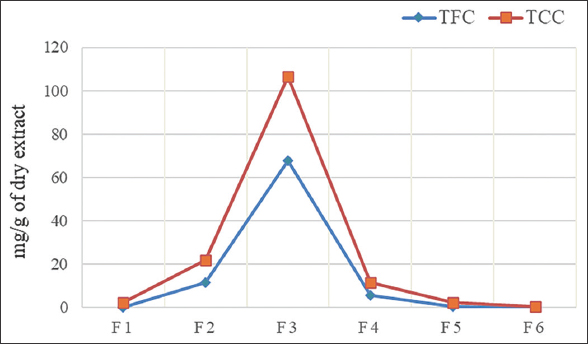 | Figure 2: Quantitative estimation of column chromatography fractions. [Click here to view] |
3.4. Partial Purification of Flavonoid Glycosides using High-Performance Liquid Chromatography
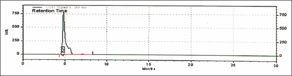
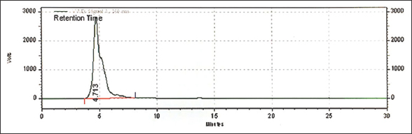
The column F3 which was shown to have better concentration of flavonoids and carbohydrate content was subjected to HPLC analysis along with Quercetin as a standard reference compound. Both the test fraction and reference standard have shown similar peaks in the HPLC graph around the retention time of 4.6 to 4.7 min [Figures 3 and 4]. The fraction which has shown a high peak of flavonoid glycosides was eluted and stored for further experiments.
 | Figure 3: HPLC Chromatogram of Quercetin standard. [Click here to view] |
 | Figure 4: HPLC Chromatogram of chromatographic fraction 3. [Click here to view] |
3.5. Antibacterial Activity of Isolated Flavonoids Glycosides of G. oppositifolius
Antibacterial efficiency of flavonoid glycoside of G. oppositifolius was tested against six pathogenic bacteria through well diffusion assay [Figure 5]. The compound has shown bactericidal ability against all the tested pathogens. However, the zone of inhibition exhibited by the compound varied greatly for different organisms [Figure 6]. The maximum zone of inhibition 28.3 mm was found against S. enterica Typhimurium.
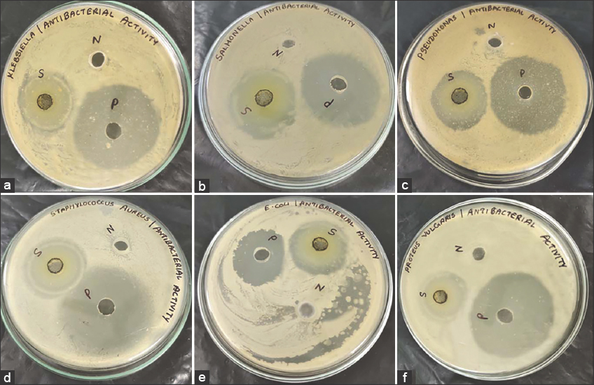 | Figure 5: Antibacterial activity of flavonoid glycosides using well-diffusion assay on (a) Klebsiella pneumonia, (b) Salmonella enterica Typhimurium, (c) Pseudomonas aeruginosa, (d) Staphylococcus aureus, (e) Escherichia coli, and (f) Proteus vulgaris. [Click here to view] |
 | Figure 6: Zone of inhibition of flavonoid glycosides against different pathogens. [Click here to view] |
3.6. MIC of Isolated Flavonoids Glycosides of G. oppositifolius
Inhibitory activity was observed in all the tested concentrations against all the pathogenic bacteria [Figure 7]. Among the different concentrations tested, the 2800 μg/ml has registered the complete growth inhibition percentage (100%) followed by 2400 μg/ml has shown the 83.4%, 100%, 93.67%, 100%, 87.5%, and 87.5% of inhibition against K. pneumoniae, S. enterica Typhimurium, P. aeruginosa, S. aureus, E. coli, and P. vulgaris, respectively. The least inhibition activity was observed in 400 μg/ml against all bacteria tested.
 | Figure 7: Minimum inhibitory concentration analysis of flavonoid glycosides. [Click here to view] |
4. DISCUSSION
Finding and utilizing plant compounds effectively against pathogenic bacteria is one of the good alternative approaches in the health care sector and it helps us greatly prepare to withstand and fight against newly emerging pathogens and also to combat evolving multi-drug resistance among pathogens worldwide [32]. The current research work was focused to isolate and partially purify flavonoid glycosides from G. oppositifolius [Figure 1] and to experiment its antibacterial efficiency against a range of pathogens.
The majority of flavonoids exist in combination with glycosides under natural conditions [33]. Flavonoids with its derivatives were reported to be an effective class of compounds to act against pathogenic bacteria [34]. Flavonoids were found to possess high ability to perform as agents for antioxidant, anticancer, antitumor, hepatoprotective, anti-inflammatory, antidiabetic, antiviral activity, antifungal, etc., [35]. Hence, we have chosen flavonoid as our target molecule among the different compounds reported in G. oppositifolius, and proceeded with isolation, partial purification, and antibacterial analysis on the same. Extraction experiments were carried out using different solvents chosen from different polarity domains. Water (polarity value 1), methanol (polarity value 0.762), chloroform (polarity value 0.259), and ethyl acetate (polarity value 0.228) were employed in the present study [36]. Extraction was carried out using maceration technique, which was reported to be a simpler and better methodology for extraction of flavonoids [37]. Among the different solvent tested, methanol extract was found to contain more expected compounds such as flavonoids, phenols and glycosides through our qualitative experiments [Table 1]. The previous literature evidence also confirms the extraction of a good amount of flavonoid in methanolic extract of G. oppositifolius leaves [18,38]. Hence, further study was carried out using methanolic extract.
Quantitative estimation in the methanolic extract has shown presence of good amount of flavonoids (210.83 ± 4.63 mg Quercetin equivalent/g of dry extract) and carbohydrate contents (167.77 ± 1.73 mg Glucose equivalent/g of dry extract) [Table 2]. A total flavonoid content of 168 mg quercetin equivalent/g of dry ethanolic extract of G. oppositifolius was previously reported by Asok Kumar et al. [16]. Isolation and partial purification of the flavonoid glycosides were carried out using CC with silica gel as the standard phase. Silica gel CC was reported to be one of the good methods for isolation of flavonoid derivatives [39]; hence, the method has been employed in the study. Seven fractions were collected from the chromatography and were subjected for qualitative and quantitative tests. The obtained results [Table 3 and Figure 2] have shown that fraction 3 (F3) contains more amounts of flavonoid glycosides relatively. Fraction 7 (F7) does not contain any of the expected compounds, and hence, further fractions were not collected. Further, purification of the F3 fraction to separate the flavonoid fractions was carried out using high-performance liquid chromatography. HPLC was reported to be a good method for isolation and partial purification of the flavonoid compounds across literatures [40]. The partially purified flavonoid glycosides were eluted almost at similar retention time relative to the Quercetin which was used as the reference compound [Figures 3 and 4].
The partially purified flavonoid glycosides were subjected to antibacterial analysis and found to be effective to inhibit the growth of all pathogens tested through well diffusion assay [Figures 5 and 6]. Known effective antibiotics against the test organisms were used as positive control in all the antibacterial assays to compare the efficiency of the isolated flavonoid glycosides with existing best growth controllers. Several investigations stated that the antibacterial mechanisms of flavonoids include inhibition of nucleic acid synthesis, functionality of cytoplasmic membrane, and due to the interactions with important enzymes [41-43]. Further, the MIC analysis was carried out [Figure 7]. A minimum concentration of 2400 mg/ml was found sufficient to inhibit the visible growth of S. enterica Typhimurium and S. aureus. All other tested pathogens were attained 100% inhibition at the concentration of 2800 mg/ml.
MIC by flavonoid glycosides is in accordance with existing published reports of, Reda et al., Tagousop et al., who have reported the antibacterial activity of flavonoids glycosides from different plants [44,45]. In accordance with the present results, Adamczak et al. examined the MIC of flavonoids and organic acids against clinical pathogens, in which the growth of tested bacteria (S. aureus, Enterococcus faecalis, E. coli, and P. aeruginosa) was shown to be inhibited by salicylic acid at the MIC of 250–500 mg/mL [46]. Zhang et al. reported that the MIC of flavonoid glycosides of Castanea mollissima Blume was 0.00625 g/mL and 0.0125 g/mL against Proteus and Micrococcus genus, respectively [47].
5. CONCLUSION
The current research results showed that methanol as an effective solvent for extraction of flavonoids glycosides of G. oppositifolius using simple maceration technique, silica gel CC, and HPLC as a better channel for isolating and purifying the flavonoid glycosides from G. oppositifolius. The study concludes that the partially purified flavonoid glycosides possess potential inhibitory effect on the growth of a range of pathogenic bacteria. The results open a new way for a special flavonoid glycoside compound from the G. oppositifolius to be tested further against different bacterial genus as well as against other eukaryotic pathogens. Future studies in terms of structural elucidation of this flavonoid glycoside should be carried out to find other related medical potency of the compound as well as to study its drug likeness properties.
6. AUTHORS’ CONTRIBUTIONS
All authors made substantial contributions to conception and design, acquisition of data, or analysis and interpretation of data; took part in drafting the article or revising it critically for important intellectual content; agreed to submit to the current journal; gave final approval of the version to be published; and agreed to be accountable for all aspects of the work. All the authors are eligible to be an author as per the International Committee of Medical Journal Editors (ICMJE) requirements/guidelines.
7. FUNDING
No funding was obtained from any external source.
8. CONFLICTS OF INTEREST
The authors declare no conflicts of interest relevant to this article.
9. ETHICAL APPROVALS
This study does not involve experiments on animals or human subjects.
10. DATA AVAILABILITY
All data supporting this study are available on request.
11. PUBLISHER’S NOTE
This journal remains neutral with regard to jurisdictional claims in published institutional affiliation.
REFERENCES
1. Gulati K, Busari J. Vaccinating a billion people against COVID-19:India's quest for systems leadership in exceptional times. Leadersh Health Serv (Bradf Engl) 2022;35:137-48. [CrossRef]
2. Iwu MW, Duncan AR, Okunji CO. New Antimicrobials of Plant Origin. In:Perspect New Crops New Uses. Alexandria:ASHS Press;1999. 457-62.
3. Fauci AS, Touchette NA, Folkers GK. Emerging infectious diseases:A 10-year perspective from the national institute of allergy and infectious diseases. Emerg Infect Dis 2005;11:519-25. [CrossRef]
4. Pham-Huy LA, He H, Pham-Huy C. Free radicals, antioxidants in disease and health. Int J Biomed Sci 2008;4:89-96.
5. Tacconelli E, Carrara E, Savoldi A, Harbarth S, Mendelson M, Monnet DL, et al. Discovery, research, and development of new antibiotics:The WHO priority list of antibiotic-resistant bacteria and tuberculosis. Lancet Infect Dis 2018;18:318-27. [CrossRef]
6. Abera Z, Degefu H, Gari G, Kidane M. Sero-prevalence of lumpy skin disease in selected districts of West Wollega zone, Ethiopia. BMC Vet Res 2015;11:135. [CrossRef]
7. Malik S. Biotechnology and Production of Anti-Cancer Compounds. Germany:Cham Springer International Publishing;2017. [CrossRef]
8. Geetha K, Kakarla S, Seru G. Screening of crude plant extracts for anti-adipogenesis activity in 3T3-L1 Cells. J Pharm Res 2014;8:81-6.
9. Blumberg J. Introduction to the proceedings of the third international scientific symposium on tea and human Health. J Nutr 2003;133:3244S-6. [CrossRef]
10. Elmasta?M, Gülçin ?, I?ildak Ö, Küfrevio?lu Ö?, ?bao?lu K, Aboul-Enein HY. Radical scavenging activity and antioxidant capacity of bay leaf extracts. J Iran Chem Soc 2006;3:258-66. [CrossRef]
11. Xu ML, Wang L, Hu JH, Lee SK, Wang MH. Antioxidant activities and related polyphenolic constituents of the methanol extract fractions from Broussonetia papyrifera stem bark and wood. Food Sci Biotechnol 2010;19:677-82. [CrossRef]
12. Kumar G, Karthik L, Bhaskara Rao K. A review on medicinal properties of Elaeocarpus ganitrus Roxb. ex G. Don. (Elaeocarpaceae). Res J Pharm Technol 2014;7:1184-6.
13. Sies H, Stahl W, Sundquist AR. Antioxidant functions of vitamins. Vitamins E and C, beta-carotene, and other carotenoids. Ann N Y Acad Sci 1992;669:7-20. [CrossRef]
14. Burkill HM. The Useful Plants of West Tropical Africa. 2nd ed. Kew, UK:Royal Botanic Gardens;1994.
15. Pratap GP, Jyothi B, Husain MK, Nagaraj V, Sudarsanam G. Pharmacognostical and phytochemical studies of Mollugo nudicaulis Lam.:A controversial plant origin ayurvedic drug. Ann Phytomed Int J 2021;10:44-52. [CrossRef]
16. Asok Kumar K, Umamaheswari M, Sivashanmugam AT, Subhadra Devi V, Subhashini N, Ravi TK. Free radical scavenging and antioxidant activities of Glinus oppositifolius (carpet weed) using different in vitro assay systems. Pharm Biol 2009;47:474-82. [CrossRef]
17. Gopinathan S, Nija S. Gastric ulcer curative potential of Mollugo oppositifolia L. extract-a preclinical study. World J Pharma Res 2014;3:929-48.
18. Hoque N, Imam MZ, Akter S, Ehsanul M, Hasan R, Ahmed J, et al. Antioxidant and antihyperglycemic activities of methanolic extract of Glinus oppositifolius leaves. J Appl Pharm Sci 2011;1:50-3.
19. Sahu S, Das D, Tripathy N, Dinda S, Sandeep Kumar H. Evaluation of hypoglycemic activity of Mollugo pentaphylla and Glinus oppositifolius L. Rasayan J Chem 2012;5:57-62.
20. Ramaseshan ST, Pitchaiah P, Bharti V, Ramakrishna KK, Gaddam V, Tewari D, et al. Pharmacognostical, phytochemical and nutritional evaluation of Glinus oppositifolius (L.) Aug. DC. Pharmacogn J 2015;8:31-6. [CrossRef]
21. Khare CP. Indian Medicinal Plants:An Illustrated Dictionary. New York:Springer;2007. [CrossRef]
22. Kirtikar KR, Basu BD. Indian Medicinal Plants. 2nd ed., Vol. 4. Allahabad:Lalit Mohan Basu;1935.
23. Inngjerdingen KT, Debes SC, Inngjerdingen M, Hokputsa S, Harding SE, Rolstad B, et al. Bioactive pectic polysaccharides from Glinus oppositifolius (L.) Aug. DC., a Malian medicinal plant, isolation and partial characterization. J Ethnopharmacol 2005;101:204-14. [CrossRef]
24. Sheu SY, Yao CH, Lei YC, Kuo TF. Recent progress in Glinus oppositifolius research. Pharm Biol 2014;52:1079-84. [CrossRef]
25. Khandelwal K. Practical Pharmacognosy. Pune:Pragati Books Pvt. Ltd;2008.
26. Mathur R, Vijayvergia R. Determination of total flavonoid and phenol content in Mimusops elengi Linn. Int J Pharm Sci Res 2017;8:5282-5.
27. Saha SK, Brewer CF. Determination of the concentrations of oligosaccharides, complex type carbohydrates, and glycoproteins using the phenol-sulfuric acid method. Carbohydr Res 1994;254:157-67. [CrossRef]
28. Arora S, Itankar P. Extraction, isolation and identification of flavonoid from Chenopodium album aerial parts. J Tradit Complement Med 2018;8:476-82. [CrossRef]
29. Lin LZ, Harnly JM. A screening method for the identification of glycosylated flavonoids and other phenolic compounds using a standard analytical approach for all plant materials. J Agric Food Chem 2007;55:1084-96. [CrossRef]
30. Desai SP, Momin YH, Taralekar ST, Dange YD, Jagtap SR, Khade HP. Evaluation of potential in vitro anticancer and antimicrobial activities of synthesized 5-mercapto-4-substituted 1, 2, 4 triazole derivatives. Ann Phytomedicine Int J 2021;10:273-79. [CrossRef]
31. Shekar BR, Nagarajappa R, Jain R, Suma S, Singh R, Thakur R. Minimum inhibitory concentration of the plant extracts'combinations against dental caries and plaque microorganisms:An in vitro study. J Indian Assoc Public Health Dent 2016;14:456. [CrossRef]
32. Subramani R, Narayanasamy M, Feussner KD. Plant-derived antimicrobials to fight against multi-drug-resistant human pathogens. 3 Biotech 2017;7:172. [CrossRef]
33. Martens S, Preuss A, Matern U. Multifunctional flavonoid dioxygenases:Flavonol and anthocyanin biosynthesis in Arabidopsis thaliana L. Phytochemistry 2010;71:1040-9. [CrossRef]
34. Xie Y, Yang W, Tang F, Chen X, Ren L. Antibacterial activities of flavonoids:Structure-activity relationship and mechanism. Curr Med Chem 2014;22:132-49. [CrossRef]
35. Xiao J, Capanoglu E, Jassbi AR, Miron A. Advance on the flavonoid C-glycosides and health benefits. Crit Rev Food Sci Nutr 2016;56:S29-45. [CrossRef]
36. Abubakar AR, Haque M. Preparation of medicinal plants:Basic extraction and fractionation procedures for experimental purposes. J Pharm Bioallied Sci 2020;12:1-10. [CrossRef]
37. Lezoul NE, Belkadi M, Habibi F, Guillén F. Extraction processes with several solvents on total bioactive compounds in different organs of three medicinal plants. Molecules 2020;25:4672. [CrossRef]
38. Martin-Puzon JJ, Rivera WL. Free-radical scavenging activity and bioactive secondary metabolites from various extracts of Glinus oppositifolius (L.) Aug. DC. (Molluginaceae) roots, stems and leaves. Asian Pac J Trop Dis 2015;5:711-5. [CrossRef]
39. Feng W, Hao Z, Li M. Isolation and structure identification of flavonoids. In:Justino GC, editor. Flavonoids from Biosynthesis Human Health. London, UK:In Tech;2017. [CrossRef]
40. Sen AK, Sen DB, Maheshwari RA. Extraction, isolation, and quantitative determination of flavonoids by HPLC. In:Sen S, Chakraborty R, editors. Herbal Medicine in India. Singapore:Springer;2020. 303-36. [CrossRef]
41. Barbieri R, Coppo E, Marchese A, Daglia M, Sobarzo-Sánchez E, Nabavi SF, et al. Phytochemicals for human disease:An update on plant-derived compounds antibacterial activity. Microbiol Res 2017;196:44-68. [CrossRef]
42. Górniak I, Bartoszewski R, Króliczewski J. Comprehensive review of antimicrobial activities of plant flavonoids. Phytochem Rev 2019;18:241-72. [CrossRef]
43. Khameneh B, Iranshahy M, Soheili V, Fazly Bazzaz BS. Review on plant antimicrobials:A mechanistic viewpoint. Antimicrob Resist Infect Control 2019;8:118. [CrossRef]
44. Mohammed R, Souda S, Taie H, Moharam ME, Shaker K. Antioxidant, antimicrobial activities of flavonoids glycoside from Leucaena leucocephala leaves. J Appl Pharm Sci 2015:5:138-47. [CrossRef]
45. Tagousop CN, Tamokou JD, Ekom SE, Ngnokam D, Voutquenne-Nazabadioko L. Antimicrobial activities of flavonoid glycosides from Graptophyllum grandulosum and their mechanism of antibacterial action. BMC Complement Altern Med 2018;18:252. [CrossRef]
46. Adamczak A, O?arowski M, Karpi?ski TM. Antibacterial activity of some flavonoids and organic acids widely distributed in plants. J Clin Med 2019;9:109. [CrossRef]
47. Zhang LQ, Xue HB, Zhu WL, Li YM, Chen KX. Two new flavonoid glycosides isolated from the fruits of Catalpa ovata. Pharmacogn Mag 2020;16:817. [CrossRef]