Mini Review | Volume 13, Issue 2, March, 2025

Monoclonal antibodies for diagnosis of viral diseases: a comprehensive review

Abhishek Kasana Aishwarya Dwivedi Vaishali Verma   

Open Access   

Published:  Jan 25, 2025

DOI: 10.7324/JABB.2025.205679
Abstract

Viruses cause infectious diseases in humans, ranging from mild to life-threatening. Early and accurate diagnosis is essential for timely treatment and prevention of complications. Diagnostic methods typically rely on detecting viral genetic material, proteins, or the host’s immune response, depending on the infection stage. Monoclonal antibodies, known for their specificity and high affinity, are widely used for diagnosing viral infections by enabling precise and sensitive detection of viral proteins. This review explores the applications of monoclonal antibody-based methods for detecting viral infections, emphasizing their pivotal role as sensitive detection reagents. These methods are crucial for developing robust immunodiagnostic assays capable of diagnosing various viral diseases in human, animal, and plant hosts, with special emphasis on SARS-CoV-2. The review also explores various assays that employ monoclonal antibodies for diagnostic purposes and the technologies used for their production. Understanding the principles and applications of monoclonal antibodies in diagnosing viral diseases is essential for implementing effective public health interventions and preventing future pandemics.


Keyword:     Hybridoma Infectious diseases Immunodiagnosis Phage display Point-of-care Virus


Citation:

Kasana A, Dwivedi A, Verma V. Monoclonal antibodies for diagnosis of viral diseases: a comprehensive review. J App Biol Biotech. 2025;13(2):20-31. http://doi.org/10.7324/JABB.2025.205679

Copyright: Author(s). This is an Open Access article distributed under the terms of the Creative Commons Attribution-NonCommercial-ShareAlike license.

HTML Full Text

1. INTRODUCTION

Viral diseases are illnesses caused by viruses that replicate inside the living cells of other organisms (referred to as the host). Viruses cannot replicate independently outside of a host cell. Consequently, they must infect host cells and take over their machinery to generate more virus particles. Viral pathogens have significantly threatened human safety during the past few decades. Viral diseases can cause a range of symptoms depending on the virus, from mild respiratory symptoms to life-threatening conditions. Such diseases are often contagious and have the potential to rapidly disseminate among populations, making prevention and control efforts crucial for public health. Consequently, rapid and precise diagnostic procedures are essential for the timely identification of viruses. This enables the prompt administration of appropriate treatments, thereby improving clinical outcomes and aiding in the control of infection spread [13].An ideal detection method must meet several key criteria: high specificity, quick turnaround time, operational simplicity (no need for time-consuming sampling procedures or specialized equipment), and affordability. Diagnostic approaches for viral diseases can be broadly classified into nucleic acid-based methods and protein-based methods. The nucleic acid-based methods rely on detecting the viral genome, whereas protein-based methods focus on detecting virus-specific proteins (antigen-detection methods) or antibodies produced by the host in response to the virus (serological/antibody-detection methods) [4].Nucleic acid-based methods necessitate the design of specific primer sets to amplify virus-specific sequences, offering high sensitivity. However, these methods require costly chemicals and equipment, such as polymerase chain reaction (PCR) machines, for amplification reactions. In addition, they are susceptible to contamination during sample handling, necessitating skilled personnel. Protein-based antigen detection methods use target-specific antibodies to detect viral proteins [5]. Due to their high specificity towards targets; antibody-based detection tools are a viable diagnostic option for diagnosing a wide range of pathogens . These methods can be highly sensitive, depending on antibody affinity, and can detect proteins during the early stages of infection [6]. Furthermore, antigen detection tests are highly stable and have a longer shelf life [7]. Antibody-based assays can be formatted into point-of-care assays, which are rapid (completed in ~30 min), easy to perform, and generally do not require specialized laboratory equipment or skilled personnel. The COVID-19 pandemic underscored the significance of rapid antigen-based lateral flow assays (LFAs), which proved invaluable for detecting SARS-CoV-2 and implementing containment measures, demonstrating the utility of protein-based assays in viral disease diagnosis [8].

Antibodies are classified into monoclonal and polyclonal types [9]. Monoclonal antibodies are derived from a single clone of plasma B cells [9]. In contrast, polyclonal antibodies are obtained from multiple clones of plasma B cells. Monoclonal antibodies exhibit greater specificity, consistency, and reliability than polyclonal antibodies, with reduced cross-reactivity against other antigens [10]. Various diagnostic techniques utilize monoclonal antibodies, namely enzyme-linked immunosorbent assay (ELISA) [11], Western blotting [12], radioimmunoassay (RIA), and LFA [13]. This review highlights the pivotal role of monoclonal antibodies in developing highly specific, sensitive, and rapid immunodiagnostic tests for detecting viral pathogens. We reviewed existing literature to examine the applications of specific monoclonal antibodies against viral targets for diagnostic tests, with special emphasis on SARS-CoV-2. Additionally, we discussed strategies for their production for diagnostic applications along with the immunoassays that employ them.


2. ASSAYS USED FOR THE DETECTION OF DISEASES USING MONOCLONAL ANTIBODIES

Currently, antibody-based detection of target proteins plays a crucial role in disease diagnostics. These assays rely on non-covalent antibody-antigen interactions, in which antibodies specifically recognize the target antigen, thereby confirming the presence of pathogens. The sensitivity and specificity of these techniques depend on the strength of antibody binding to its antigen [14]. The most widely used antibody-based antigen detection formats include ELISA, LFA, Western Blot, dot blots, and immunohistochemistry (IHC). These assays offer distinct advantages in sensitivity and ease of use, catering to diverse diagnostic needs [Table 1 and Figure 1]. Both polyclonal and monoclonal antibodies are utilized to develop immunodiagnostic tests [15]. The scalable production of recombinant monoclonal antibodies ensures a reliable and cost-effective supply for diagnostic applications, making them highly attractive for commercialization.

Table 1: Summary of the properties of key immunodiagnostic techniques.

Detection MethodSensitivitySpecificityEase of UseAdvantagesDisadvantages
ELISAHigh
(Typically, >10-100 pg; depends on substrate and antibodies used)
Highly specificRequires trained manpower and is labor intensiveHighly sensitive and specific, faster than Western blotTime consuming (at least 2-3 h), requires specialized equipment, expensive
Western blotHigh
(Typically, >100 pg; depends on substrate, membrane, and antibodies used)
Highly specificRequires trained manpower and is labor intensiveHighly sensitive and specificTime consuming (at least 4-6 h), requires specialized equipment, expensive
Lateral flow assayLower as compared to ELISA and Western blot
(Typically, >10 ng; depends on membrane, and antibodies used)
Highly specificEasy to perform and doesn’t require trainingRapid results (15-20 min), easy to use, economical, portableLow sensitivity as compared to ELISA and Western blot
Figure 1: Applications of antibodies specific to viral targets in immunodiagnostic assays.


[Click here to view]

ELISA, a specific and sensitive biochemical test, facilitates analyte detection and quantitative analysis without requiring specialized equipment [16]. It is based on the principle of antigen-antibody binding, where the protein of interest is immobilized, and a specific antibody binds to it. Detection of this binding is achieved using a secondary antibody coupled with an enzyme, resulting in a color change upon reaction with a chromogenic substrate [17]. Fluorogenic or chemiluminescent substrates provide enhanced sensitivity for quantitative measurements [17]. Four major types of ELISA are commonly employed for the detection of proteins: direct, indirect, sandwich, and competition ELISA. In direct ELISA, the surface of the plate is coated with either the antibody or antigen present in the sample. Detection is achieved through an enzyme attached to either the antibody or antigen, facilitating accurate measurement [18,19]. It finds applications in the detection of various pathogens, including viruses, such as SARS-CoV-2 [20,21], Porcine Circovirus 3 (PCV3) [22], Cache Valley virus (CVV) [23], Pseudorabies virus, etc. [24]. Indirect ELISA is a two-step detection assay. Initially, the target-specific primary antibody attaches to the antigen, and subsequently, an enzyme-linked secondary antibody is introduced to detect and bind to the primary antibody [2527]. Indirect ELISA is mostly used for serological testing, which involves the detection of the host’s immune response against the pathogen. It has shown high sensitivity and specificity for detecting IgG and IgM antibodies against Crimean-Congo hemorrhagic fever (CCHF) in acute and convalescent human sera [28]. This assay also finds applications in detecting SARS-CoV-2 antibodies in cats [29]. Sandwich ELISA, by immobilizing antigens between capture and detector antibodies, offers increased sensitivity and specificity in detecting target antigens. It finds applications in the detection of the Ebola virus [30] and Citrus leprosis virus C2 [31]. Competition ELISA assesses antigen-antibody interactions by competitive binding assays [32]. This assay finds various applications in the diagnosis of different viral diseases, such as porcine epidemic diarrhea virus (PEDV) [33] and Goose astrovirus (GAstV) [34].

Western blotting, or immunoblotting, identifies proteins separated by gel electrophoresis using antibody detection [35]. Multiple studies have reported the use of this technique in the characterization of antibodies developed for the detection of viral diseases [21,22,3638]. Immunohistochemistry (IHC) utilizes antibodies to determine the location of proteins or antigens in tissues through visualization of antigen-antibody interactions in tissue samples using colored enzyme substrates or fluorescent dyes [39]. Researchers use this technology to study virus-infected tissue samples [40,41].

Lateral Flow Assays (LFA) first reported in the 1960s, revolutionized diagnostic accessibility by employing monoclonal antibodies for rapid and straightforward antigen detection [42]. LFAs detect viral antigens or antibodies in diverse samples (e.g., blood, saliva) via specific immobilized monoclonal antibodies on a nitrocellulose membrane. Multiple studies have reported the development of LFA-based methods for detecting viral antigens [36,43,44]. Advanced LFAs integrated with artificial intelligence-based reading technologies have been employed to enhance detection sensitivity for viruses such as SARS-CoV-2, achieving up to 86.2% sensitivity compared to 71.4% when assessed by human eye. This advancement underscores the effectiveness of combining traditional diagnostic methods with cutting-edge technology to improve accuracy and reliability in virus detection [45].


3. TECHNOLOGIES FOR THE DEVELOPMENT OF MONOCLONAL ANTIBODIES FOR DIAGNOSTIC APPLICATIONS

As elaborated above, monoclonal antibodies play a crucial role in disease diagnosis. Several technologies are available for developing monoclonal antibodies for diagnostic and therapeutic applications [Figure 2]. These techniques include hybridoma technology, phage display, single B cell cloning technology, the use of humanized animals (such as mice) encoding human antibody genes, next-generation sequencing-based technology, and computational/AI-based technologies.

Figure 2: Technologies used for the production of monoclonal antibodies.


[Click here to view]

The hybridoma method developed by G. Köhler and C. Milstein [46] is a time-tested technique to obtain monoclonal antibodies (2). Once desirable antibody-secreting hybridoma clones are selected, they are cultured on a large scale to produce antibodies in desired quantities [47]. Monoclonal antibodies developed using this technique are mostly preferred among all the available methods because the isolated antibodies are highly specific to the target antigen [9]. Also, this technique is more convenient and cost-effective than other techniques [48]. This technique has expanded the antibody discovery and production scope across various applications. Multiple researchers have reported the development of monoclonal antibodies against viral antigens using hybridoma technology [21,36,49,50].

George Smith invented the phage display technology in 1985 and revolutionized the process of discovery of antibodies for diagnostic, therapeutic, and basic research applications. This technology is based on the ability of bacteriophages to display foreign proteins, such as antibody fragments, on their surface. At the same time, they encode the gene inside the phage particle. Usually, M13 filamentous bacteriophage is used as the viral vector for displaying antibody fragments on its surface. The process involves the construction of a phage display library, which is a collection of millions to billions of different phage particles, each displaying a unique antibody fragment on its surface [Table 2]. Typically, such a library is generated by amplification of genes encoding antibody variable heavy and light chain domains from the B cells isolated from immunized (animals such as mice or convalescent humans) or non-immunized sources, followed by their PCR-based assembly and cloning in frame with the coat proteins (mostly g3 protein) of M13 bacteriophage. The resulting phage library is then subjected to biopanning, to eliminate non-binding or low-affinity phages, and recover target specific phages. After several rounds of biopanning, the individual phage clones displaying the desired antibody fragments are isolated, and the DNA encoding the antibody fragments is sequenced. After determining the sequences, the variable genes of antibodies can be subcloned into different expression systems. Recombinant monoclonal antibodies developed using phage display offer several advantages over traditional hybridoma-derived antibodies [Table 2]. These include improved stability, consistent quality, and the ability to engineer desired properties like higher affinity or specificity [51].

Table 2: Comparison of techniques to produce monoclonal antibodies for diagnostic applications.

TechnologyPrincipleAdvantagesLimitations
Hybridoma technologyAnimals are immunized with the target antigen, followed by the fusion of immortal myeloma cells with B lymphocytes [77].Robust and has been used for the discovery of large number of antibodies for different applications [78].
Once clones are established, monoclonal antibody production becomes straightforward.
Preserves original pairing of antibody chains present in B cells [79].
Hybridoma antibodies can be used directly and can be cryopreserved until the subsequent use for an indefinite time period [80].
Non-availability of fusion partners limits its use and multi-species applicability [81].
Requires a purified antigen for immunization. In some cases, it may be difficult to purify the antigen.
Efficacy depends on animal health and immunization efficiency.
High risk of contamination [82].
Majority of B cells are lost during fusion, which leads to a loss of efficiency [9].
Phage display technologyAntibodies derived from naïve or immunized B cell sources are displayed on the surface of bacteriophages [83]. A library of these phage particles is created, and each displays a unique antibody fragment on its surface. It can be used in the isolation of specific binders.Comparatively faster and more optimized.
There is no necessity to immunize animals for phage display when using naïve libraries.
Sufficient depth of coverage for finding antigen-specific antibodies compared to other methods [84].
Good clones may get missed due to poor RNA recovery or loss of DNA during library construction [84].
Requires high transformation efficiency during preparation of large libraries.

Typically, single B cell cloning technology involves the isolation of B cells from vaccinated or convalescent humans, followed by the sorting and culturing of B cells as single cells, screening of B cell supernatant for antibody specificity against the target, and cloning of antibody genes into mammalian expression vectors to produce full-length monoclonal antibodies [52,53]. This technology retains the inherent pairing of light and heavy chains expressed by a B cell and has been successfully used to isolate fully human antibodies as therapeutics for the treatment of a wide range of viral diseases, including SARS-CoV-2 [54]. Researchers have also developed humanized mice in which the locus encoding mouse antibody genes is replaced with human antibody genes. Consequently, upon immunization, these mice yield human antibodies instead of mouse antibodies, which can be directly employed for therapeutic applications without requiring humanization [55]. Deep sequencing of B cell repertoires using next generation sequencing (NGS) technologies has also enabled the identification of antibodies against desired targets [56]. NGS can also be combined with other antibody discovery technologies, such as phage display, to discover antibody leads [57]. Recently, artificial intelligence (AI)-based methods have also become available to predict antibody sequences specific to a target in silico [58]. AI-based methods are actively being explored for discovering new antibodies and improving the properties of existing antibodies [59].

While all these technologies have been successfully employed for the production of monoclonal antibodies for therapeutic and diagnostic applications, hybridoma technology and phage display technology are the most commonly used techniques for developing monoclonal antibodies for diagnostic applications. Both techniques facilitate the isolation of antibodies with desired characteristics, such as high affinity, specificity, and tailored cross-reactivity.


4. DEVELOPMENT OF MONOCLONAL ANTIBODIES FOR THE DETECTION OF SARS-CoV-2

During the COVID-19 pandemic, there was significant emphasis on developing monoclonal antibodies against SARS-CoV-2 for both diagnostic and therapeutic purposes. SARS-CoV-2, a positive-strand virus, encodes multiple structural and non-structural proteins. Several research groups reported successful efforts in developing monoclonal antibodies aimed at rapid detection of SARS-CoV-2 [Table 3].

Table 3: Studies reporting the development of monoclonal antibodies for the detection of SARS-CoV-2.

S. No.ImmunogenTechnology used for antibody discoveryMethods used for antibody characterizationDetection limitReferences
1.Spike protein ECDPhage DisplayIndirect ELISA, SPR, Competitive ELISA, and Sandwich ELISA43.2 ng/mL with sandwich ELISA[20]
2.Spike protein (Full-length)Hybridoma TechnologyELISA, Dot Blot, and LFA781 PFU with sandwich ELISA[49]
3.Multiple SARS-CoV-2 proteinsPhage DisplayELISA, BLI, Western Blot, and IFANot reported[37]
4.UVC-inactivated SARS-CoV-2QLD02Hybridoma TechnologyIHC, Indirect IFA, ELISA, Western Blot, and LFA100 ng/mL using LFA[36]
5.Nucleocapsid and Spike protein (S1)Hybridoma TechnologyELISA, Flow cytometry, and LFA4.89–9.06 ng/mL with S1 and 0.76–6.95 ng/mL with nucleocapsid-based LFA[43]
6.Nucleocapsid proteinHybridoma TechnologyELISA, BLI, and LFA240 ng/mL using LFA[44]
7.Truncated Nucleocapsid proteinHybridoma TechnologyBLI, Immunoblotting, IFA, IHC, sandwich ELISA and LFA6.3x104 virus copies/mL or 6.25 pg/mL of recombinant protein using LFA[50]
8.Nucleocapsid proteinPhage DisplayELISA, Western blot, Dot blot and BLI2.5x104 PFU per reaction[60]
9.Truncated Nucleocapsid proteinHybridoma TechnologyIndirect ELISA, Immunofluorescence Analysis, Western Blot, Sandwich ELISA5500 PFU/mL using sandwich ELISA[21]

Kim et al., reported a sandwich ELISA-based method for detecting SARS CoV-2 [20]. Using phage display technology, they developed mouse monoclonal antibodies against the extracellular domain of spike protein (SARS-CoV-2 S-ECD). Two monoclonal antibodies (K104.1 and K104.2) were isolated from a synthetic phage display library and were characterized using phage ELISA, surface plasmon resonance (SPR), competition ELISA, and sandwich assay. These were used as capture and detection antibodies to develop sandwich ELISA-based immunoassays. The limit of detection (LOD) was found to be 43.2 ng/mL for the extracellular domain of the spike protein. Since this pair targeted a relatively more conserved region of spike protein, it also allowed detection of the spike proteins of other SARS-CoV-2 variants [20]. Mariotti et al., explored the development of LFA-based methods for detecting SARS-CoV-2 [49]. They developed mouse monoclonal antibodies using hybridoma technology against the full-length spike protein of the SARS-CoV-2 and characterized 13 antibodies using ELISA and Western blot. Epitope mapping revealed that nine antibodies were specific to the S1 domain, while four recognized the S2 domain. The antibodies were also characterized using dot blot and LFA. They were found to be highly specific and could detect viruses produced in the lab and in the biological samples. The best pair of antibodies, S71-S79, allowed the detection of 781 plaque-forming units (PFU) using sandwich ELISA.

In addition to spike protein, other proteins have also been explored for the development of diagnostic tests against SARS-CoV-2. Morgan et al., developed mouse monoclonal antibodies against the UVC-inactivated whole virus (SARS-CoV-2QLD02) using hybridoma technology [36]. Two antibodies, SCV2-6A11 and SCV2-7E9, were used to develop LFA, which allowed the detection of >100 ng of the recombinant spike protein. One antibody, SCV2-1E8, allowed the detection of multiple virus variants and worked efficiently in ELISA, Western blot, and immunofluorescence assay (IFA) applications [36]. Mishra et al., reported the development of 18 monoclonal antibodies against 9 different SARS-CoV-2 proteins using a synthetic Fab-displayed phage library [37]. The antibodies were characterized by ELISA, Biolayer Interferometry (BLI), Western Blot, and IFA, and antibodies 15884 and 15887 were useful for studying subcellular locations of SARS-CoV-2 viral proteins using IFA. The antibodies were found to target different epitopes, allowing their use for the development of multiple assays for detecting SARS-CoV-2. Salcedo et al., developed mouse monoclonal antibodies against both nucleocapsid and spike protein (S1) of SARS-CoV-2 using hybridoma technology [43]. B cells derived from the spleen and lymph nodes of immunized mice were used for spike protein (S1), whereas lymph nodes were used for nucleocapsid protein. Eleven antibody pairs (7 for S1 and 4 for nucleocapsid protein) were tested using LFA, and the LOD ranged from 4.89–9.06 ng/mL for recombinant S1 protein and 0.76–6.95 ng/mL for nucleocapsid proteins. Further, the nucleocapsid-based LFA developed using antibodies 1 and 453 showed 84.6% sensitivity and 100% specificity using patient nasal samples. Xie et al., developed an LFA-based test for detecting SARS-CoV-2 virus using mouse monoclonal antibodies against nucleocapsid protein [44]. The antibodies were characterized using a paired screening by a double sandwich method using BLI, and three antibodies, namely N3, N10, and N15, were selected to develop LFA. The N10 (gold conjugated) and N15 (test line) pair showed the maximum sensitivity and allowed the detection of 240 ng/mL of recombinant nucleocapsid. The LFA also detected the recombinant nucleocapsid produced by the Delta (B.1.617.2) variant of the SARS-CoV-2.

Yamaoka et al., used truncated nucleocapsid protein (121–419 amino acids) for the development of mouse monoclonal antibodies [50]. They established 144 stable hybridomas and characterized them using indirect ELISA, AlphaScreen, and BLI. The affinity of antibodies was found to be in the sub-nanomolar range for three antibody clones. These clones allowed detection of nucleocapsid protein in the lysates of virus infected cells using immunoblot. Antibodies 7 and 98 also worked in IFA, whereas 98 also allowed the detection of virus in the paraffin-embedded lung tissue of an infected patient using IHC. Antigen-capture ELISA using antibodies 9 (capture) and 98 (detector) allowed highly sensitive detection of as low as 3.2 pg/mL of recombinant nucleocapsid protein or 3.3x10⁴ copies/mL of the inactivated virus. LFA developed using the optimized pair was found to be highly specific to SARS-CoV-2 and allowed detection of 6.25 pg/mL of recombinant nucleocapsid protein or 6.3x10⁴ copies/mL of the inactivated virus. Kim et al., developed chicken scFv antibodies against full-length nucleocapsid protein using phage display technology [60]. Purified nucleocapsid protein was panned against a chicken naïve phage library, and during selections, clones reactive to SARS-CoV and MERS-CoV were eliminated. The SARS-CoV-2 specific scFvs were produced in scFv-Fc format for further characterization. The affinity of four clones was found to be in the nanomolar range. The LOD of selected clones was tested using LFA and a combination of 12H8–12H1 antibodies allowed the detection of as low as 2.5×10⁴ virus PFU per reaction with no cross-reactivity to other viruses, namely SARS-Co-V, MERS-CoV, and influenza virus. Terry et al., developed mouse monoclonal antibodies against truncated nucleocapsid protein (133-419 amino acids) of SARS-CoV-2 using hybridoma technology [21]. Two antibodies, mBG17 and mBG86, allowed the detection of virion-derived N protein from 5500 PFU/mL and 55,000 PFU/mL SARS-CoV-2 virions, respectively, with rabbit polyclonal antibody as the capture antibody in sandwich ELISA.

Overall, based on studies describing the development of monoclonal antibodies for detecting SARS-CoV-2, it is evident that hybridoma technology and phage display are the most widely used technologies for producing these antibodies. The primary target proteins for these antibodies are spike protein and nucleocapsid protein [Table 3]. The spike protein is present on the surface of the SARS-CoV-2, while the nucleocapsid protein coats the RNA genome present inside the virus particle. Due to their high copy numbers, both proteins are important targets for developing sensitive detection strategies. Both truncated and full-length versions of these proteins have been employed to generate antibodies. The developed antibodies have been utilized in various assay formats, including ELISA-based methods (such as sandwich and indirect ELISA), LFAs, and IFAs. This variety allows for flexibility in different diagnostic settings and resource availability. The assays have demonstrated high sensitivity, with LOD reported as low as single-digit picogram ranges [50] or detecting as few as 781 plaque-forming units (PFU) of virus [49]. While studies have explored antibodies from human [37] and chicken origin [60] for diagnostic development, the mouse remains the most widely used host for producing monoclonal antibodies. Robust screening procedures have enabled the isolation of antibodies capable of detecting multiple variants of SARS-CoV-2. This versatility underscores their potential application in monitoring viral evolution and the emergence of new variants.


5. DEVELOPMENT OF MONOCLONAL ANTIBODIES FOR THE DETECTION OF VIRUSES OTHER THAN SARS-CoV-2

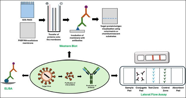
While the importance of monoclonal antibodies in diagnostics became prominently evident during the COVID-19 pandemic, it is noteworthy that even before this global health crisis, monoclonal antibodies were extensively utilized for detecting various viral diseases. Several research groups have reported the development and applications of monoclonal antibodies for detecting viruses other than SARS-CoV-2 [Table 4]. During the selection of targets for antibody development in diagnostic applications, a critical factor is choosing proteins abundant in the virus particle, such as spike and nucleocapsid proteins in the case of SARS-CoV-2, which are also highly immunogenic. These proteins facilitate the production of high-affinity antibodies essential for sensitive detection in immunodiagnostic assays.

Table 4: Studies reporting the development of monoclonal antibodies for detecting viruses other than SARS-CoV-2.

S. No.VirusImmunogenMethod Used for Antibody CharacterizationDetection LimitReferences
1.Porcine Circovirus 3Capsid proteinIndirect ELISA, Western blot, IFA, Dot blot, EB-ELISANot reported[22]
2.Varicella-zoster virus (VZV)Glycoprotein E (gE)IFA, immunoperoxidase monolayer assay, ELISA, Western Blot, LFA30 ng/mL of purified gE using LFA[61]
3.Pseudorabies virusInactivated whole virus proteinIndirect ELISA, IFA, Western BlotNot reported[24]
4.Classical swine fever virusEnvelope protein E2Indirect ELISA, Western blot, competition ELISANot reported[71]
5.Cache Valley virus (CVV)NucleoproteinELISA, Western blot, IFA, MAC-ELISANot reported[23]
6.Hepatitis E virus (HEV)HEV ORF3 proteinIndirect ELISA, Western blot, Competitive ELISANot reported[70]
7.Zika virusEnvelope proteinELISA, LFA33 μg/mL of recombinant E protein and 6.3×10⁶ PFU/mL of ZIKV using LFA[62]
8.H3 influenza A virusInactive H3 virusSandwich ELISA, Western Blot>104 virus dilution using Sandwich ELISA; Amount not reported[64]
9.Chikungunya VirusEnvelope 2 proteinELISA, Immunoblotting0.7 µg/mL inactivated CHIKV using indirect ELISA[68]
10.MERS-CoVSpike proteinSandwich ELISA5.89 ng/mL of MERS-CoV S protein using Sandwich ELISA[65]
11.Zaire Ebola virusZaire Ebola virus glycoproteinSDS-PAGE, Western blot, Dot blot, Indirect, IFA, sandwich ELISA3.6 ng/mL rGPdTM with Sandwich ELISA[30]
12.Dengue Virus (DENV-4)NS1Western Blot, Sandwich ELISA32.5 ng/mL of NS1 protein with Sandwich ELISA[66]
13.Zucchini yellow mosaic virusZYMV virionACP-ELISA, dot-ELISA, tissue dot-ELISA, DAS-ELISA and IC-RT-PCRACP-ELISA (1:163840*), dot-ELISA (1:2560*), DAS-ELISA (1:327680*), IC-RT-PCR (1:1310720*)
* Dilution of ZYMV-infected crude extracts
[72]
14.Yellow fever virusRecombinant Envelope protein and YF vaccine virus 17DIndirect ELISA, Western blot, IFA, and sandwich ELISA1 x 103 FFU/mL of YFV 17D or 2ng/well of recombinant envelop protein using sandwich ELISA[67]
15.MERS-CoVTruncated Nucleocapsid ProteinImmunoprecipitation assay, Sandwich ELISA, LFA0.5 ng of recombinant protein and 3 x 10⁴ copies of MERS-CoV virions using LFA[63]
16.Chikungunya VirusCapsid proteinELISA, Western blot, IFA, and IHCNot reported[40]
17.Tembusu VirusEnvelope proteinWestern Blot, Sandwich ELISANot reported[69]
18.SARS-CoVSpike protein subunit S1SDS-PAGE, Western Blot, Indirect ELISA, Sandwich ELISA0.019 µg/mL of S1 protein using Sandwich ELISA[38]
19.Seneca Valley virusBinary ethylenimine (BEI) inactivated SVVDot blot, IHC, competitive ELISANot reported[41]

Since LFAs can facilitate quick diagnosis and containment efforts, several groups have focused on developing LFA-based tests for detecting viruses other than SARS-CoV-2 as well. Wang et al., developed an LFA-based test for the detection of Varicella-zoster virus (VZV), which causes varicella and herpes zoster [61]. They developed mouse monoclonal antibodies against glycoprotein E present on the surface of VZV in high copy numbers, using hybridoma technology. Upon completing the antibody characterization, the antibodies 2F2 (as capture antibody) and 118H2 (as detector antibody) were selected for LFA. The test allowed the detection of 30 ng/mL VZV gE antigen without any cross-reactivity against Enterovirus 71 or Herpes Simplex Virus 1 and 2. This study emphasizes the potential of monoclonal antibodies for developing specific detection tests with no cross-reactivity to related antigens.

Similarly, Li et. al., developed an LFA-based test using mouse monoclonal antibodies against the envelope (E) protein of the Zika Virus (ZIKV), which is a structural protein present on the surface of the virus [62]. Out of 4 antibodies, antibody 9E-1 was found to be highly specific to ZIKV and had a sub-nanomolar affinity. It was used to develop an LFA-based test along with an antibody B1, and the test was able to detect 33 μg/mL of recombinant E protein and 6.3×10⁶ PFU/mL of ZIKV in culture supernatant. Yamaoka et. al., developed an LFA-based test to detect Middle East respiratory syndrome coronavirus (MERS-CoV) [63]. Using hybridoma technology, they developed mouse monoclonal antibodies specific to nucleocapsid protein (122-413 amino acids). Seven antibodies specific to the target protein were tested using immunoprecipitation, which allowed the successful capture of the antigen from cell lysates. Further, sandwich ELISA was used to test all possible combinations of 7 antibodies as capture and detection reagents. Antibody pair 46/20 was found to exhibit maximum reactivity using antigen capture ELISA. It allowed the detection of 0.0625 ng recombinant antigen or 1.5 x 10⁵ copies of MERS-CoV virions in a 0.1 mL sample. The same pair allowed the detection of 0.5 ng of purified protein and 3 x 10⁶ copies of MERS-CoV virions in a 0.1 mL sample using colloidal gold-based LFA.

Another commonly explored format for developing monoclonal antibody-based diagnostics is sandwich ELISA, which typically offers high sensitivity and quantitative measurement of the target. However, it also necessitates specialized equipment (such as plate readers) and technical proficiency, in contrast to the rapid, user-friendly nature of LFAs, despite their lower sensitivity and limited quantification ability. Several groups have reported the development of sandwich ELISA-based tests to detect viruses. Luo et al., developed a sandwich ELISA-based method for detecting the avian influenza virus (AIV), which can lead to the emergence of zoonotic infections [64]. They developed mouse monoclonal antibodies using hybridoma technology. The specificity of antibody 9F12 was determined using hemagglutination inhibition (HI) against different viruses and was found to be specific to the H3 subtype with no cross-reactivity to other viruses. 9F12 also showed activity in Western blot and IFA and allowed specific detection of H3 strain in clinical specimens using sandwich ELISA. Lee et al. developed a sandwich ELISA to detect MERS-CoV [65]. They developed mouse monoclonal antibodies against the MERS-CoV spike protein. The best antibody pair allowed detection of MERS-CoV S protein with LOD of 5.89 ng/mL using sandwich ELISA. Zai et. al., developed mouse monoclonal antibodies against recombinant glycoprotein of Zaire Ebola virus (rGPdTM; glycoprotein without the transmembrane domain) using hybridoma technology [30]. Two monoclonal antibodies, 6E3 and 3F21, were characterized using multiple assays. In dot blot analysis, 3F21 demonstrated higher sensitivity compared to 6E3, a finding further supported by BLI results indicating that 3F21 exhibited higher affinity. Using these two antibodies in sandwich ELISA allowed the detection of 3.6 ng/mL rGPdTM. Gelanew and Hunsperger produced antibodies against the NS1 protein of the Dengue virus-4 serotype (DENV-4), which is detected by commercially available NS1 antigen tests with limited sensitivity [66]. Three selected antibodies were characterized using Sandwich ELISA, and the best antibody pair, 8A6F2 (capture) and 6D4B10 (detector), exhibited the LOD of 32.5 ng/mL of NS1 protein and allowed specific detection of DENV-4 in cell culture supernatants with no reactivity from other DENV serotypes. Adungo et. al., reported the development of 8 mouse monoclonal antibodies for detecting yellow fever virus (YFV), which spreads through mosquito bites [67]. They employed recombinant envelop protein as well as YF vaccine virus 17D for immunization in mice and successfully isolated 4 antibodies against each target. All the antibodies were found to be highly specific to YFV with no cross-reactivity to related DENV and Japanese Encephalitis virus. All the antibodies also showed reactivity in the IFA. Furthermore, using two antibodies, namely 4C9 (for capture) and 3F4 (for detection), enabled the detection of 1 x 10³ focus forming units/mL of YFV 17D or 2 ng/well of recombinant envelope protein using sandwich ELISA. This study highlights the utility of monoclonal antibodies for developing cost-effective, highly sensitive, and specific diagnostic tests for arboviruses like YFV that are endemic in regions like Africa, where routine testing measures may be lacking. J. Kim et al., developed mouse monoclonal antibodies against the envelope protein E2 of Chikungunya Virus (CHIKV), which is another virus that spreads through mosquito bites [68]. Four antibodies were characterized using ELISA and Western blot, out of which, two antibodies, 9-1 and 21-1 efficiently recognized CHIKV-E2 protein, and the 9-1 antibody showed no cross-reactivity against other related viruses, such as ZIKV, JEV, and DENV. Further, the antibody 9-1 allowed the detection of as low as 0.7 µg/mL inactivated CHIKV using ELISA and can be explored for the development of new CHIKV diagnostic techniques. Goh et al. worked on a different CHIKV target and developed 11 mouse monoclonal antibodies against the CHIKV capsid protein using hybridoma technology and demonstrated their applications in multiple assays [40]. All antibodies were found to show reactivity against CHIKV using ELISA, Western blot, and immunofluorescence performed using cells expressing recombinant capsid protein or infected with CHIKV. One antibody, 5.5G9, also allowed target protein detection in scattered macrophage-like cells using IHC, demonstrating the versatility of monoclonal antibody-based assays for diagnostics. H. Chen et. al., reported a sandwich ELISA-based method for detecting Tembusu Virus (TMUV), which causes infection in waterfowls [69]. Using hybridoma technology, they developed 3 mouse monoclonal antibodies against the TMUV envelope protein. The antibodies were characterized using Western Blot and were highly specific to TMUV. Antibody 12B1 was used as a capture antibody with 2D2 for detection to develop TMUV-specific ELISA. The assay was compared to RT-PCR and was found to be 99.1% specific and 93.1% sensitive compared to RT-PCR, further underscoring that monoclonal antibody-based tests are a viable alternative to nucleic-acid-based tests. Sunwoo et al., reported the development of sandwich ELISA for the detection of SARS-CoV, responsible for the first outbreak of SARS in 2002 [38]. They developed mouse monoclonal antibodies using hybridoma technology against the spike protein subunit S1 of the SARS-CoV. Based on the antibody titers, three clones, P135.3F3, P1368D12, and F26G18, were selected for further characterization. They also generated bispecific antibodies by fusion of F26G18 and P136.8D12 hybridoma clones with anti-HRPO hybridoma YP4 to generate quadromas. The use of F26G18 as a coating antibody and its biotinylated version as a detector antibody, sandwich ELISA, allowed the detection of 0.037 µg/mL S1 antigen. Furthermore, the LOD improved to 0.019 µg/mL when they used bi-specific monoclonal antibody F157 (F26G18 x YP4) as the detector antibody, indicating that bispecific antibodies can be explored for enhancing the detection limits of monoclonal antibodies. Competition ELISA has also been explored for the detection of viruses. B. Zhang et al., developed a competitive ELISA for the detection of hepatitis E caused by genotype 1 of the hepatitis E virus (HEV) [70]. They developed 7 mouse monoclonal antibodies against recombinant genotype 1 HEV ORF3 protein using hybridoma technology, out of which 2 antibodies 3C11 and 1D2 were specific to the human HEV SAR-55 strain. Out of the two, one antibody, 1D2 was, showed higher reactivity in competitive ELISA, which can be helpful for large-scale serological testing and clinical diagnosis of HEV infections.

Apart from viruses affecting human health, monoclonal antibody-based tests have been developed to detect viruses responsible for diseases in animals as well. J. Wang et. al., developed four mouse monoclonal antibodies using hybridoma technology against the capsid protein of PCV3, which is a significant cause of disorders such as multi-organ inflammation, nephrotic syndrome, reproductive disorders, and dermatitis in swine [22]. These antibodies were characterized using indirect ELISA, Western Blot, IFA, and Dot Blot, and antibody 7E3 was found to have the highest binding affinity to the target protein. Finally, the B cell epitope of the 7E3 antibody was determined, and an epitope-blocking ELISA (EB-ELISA) was designed to detect PCV3 antibodies in sera, which showed high specificity and sensitivity. This study exemplifies using monoclonal antibodies for monitoring and managing PCV3 infections in swine farms. Similarly, J. Zhang et. al., developed mouse monoclonal antibodies against envelope protein E2 protein using hybridoma technology to detect classical swine fever virus (CSFV), which is a cause of concern in pig breeding industries. After thorough characterization, four antibodies were found to be specific to the sub-genotype 2.1 strain, and out of these, two antibodies, MM1 and MM5, were found to recognize critical epitopes on the E2 protein that were present in 90.9% of the genotype sequences available in GeneBank. Such antibodies can be promising reagents for developing assays like indirect and competition ELISA for detecting CSFV [71]. Guo et. al., also developed monoclonal antibodies using hybridoma technology against the glycoprotein E (gE) of the pseudorabies virus (PRV) that causes porcine pseudorabies (PR) [24]. One monoclonal antibody (1H5) was characterized using indirect ELISA, IFA, and Western Blot and was found to bind to a small epitope conserved in the gE of almost all PRV strains. It can be used to develop antigen detection tests [24]. Skinner et. al., developed mouse monoclonal antibodies against CVV, which is a mosquito-borne virus that causes disease in livestock and humans [23]. Antibodies were developed against inactivated CVV using hybridoma technology, and twelve hybridoma clones were found to show significant reactivity against CVV. Four mAbs, CVV14/15/17, and 18, were found to be highly specific for the detection test of anti-viral antibodies in human sera using IgM-antibody capture ELISA (MAC-ELISA), with MAb CVV14 exhibiting the highest specificity. Antibodies like CVV14 can be used as detector antibodies to develop promising serodiagnostic tools against CVV. Yang et al., developed an ELISA-based method for detecting the Seneca Valley virus (SVV), which has been linked to disease in pigs [41]. Using hybridoma technology, mouse monoclonal antibodies were produced against binary ethylenimine (BEI)-inactivated SVV. Five antibodies were characterized using Dot blot and were found to be specific to SVV. Furthermore, antibody F61SVV-9 exhibited the strongest competition with monospecific polyclonal sera in cELISA and resulted in 100% specificity, indicating that monoclonal antibodies can also be explored for serodiagnosis of viruses using cELISA [41]. The applications of monoclonal antibodies also extend to the accurate and sensitive detection of plant viruses, which is crucial for disease management and control in agriculture. Z. Chen et. al., reported the development of 3 mouse monoclonal antibodies for the detection of Zucchini yellow mosaic virus (ZYMV) using hybridoma technology [72]. Using ZYMV virion for immunization, three hybridoma clones, 16A11, 5A7, and 3B8, were developed and characterized using multiple immunoassays.


6. CHALLENGES AND FUTURE PERSPECTIVE

Monoclonal antibodies have revolutionized viral diagnostics by offering unparalleled specificity and sensitivity in detecting viral pathogens. Hybridoma technology and phage display have been instrumental in producing antibodies with high affinity and tailored cross-reactivity against a wide range of viral targets. These antibodies are crucial components in various immunodiagnostic formats, including ELISAs, LFAs, Western blots, and IHC, providing adaptable solutions for different diagnostic needs. The COVID-19 pandemic underscored their pivotal role in rapid and accurate diagnostics, particularly through LFAs for large-scale screening efforts. Beyond SARS-CoV-2, monoclonal antibodies continue to contribute significantly to detecting diverse viral diseases in human, animal, and plant hosts. The ability to generate monoclonal antibodies that can detect multiple viral strains or variants further augments their utility in monitoring the emergence of new threats.

However, several challenges must be addressed to tap into the full potential of monoclonal antibody-based diagnostic methods. The overall antibody discovery and production process can be expensive, limiting its use in resource-constrained settings. Additionally, viruses continuously evolve, necessitating frequent updates to the antibodies to maintain the diagnostic potential of the tests. Intensive screening is required to select antibodies that do not cross-react with antigens other than the target.

These limitations are actively being addressed by scientists worldwide. Expression hosts and strategies to improve antibody yields are being explored [73]. Innovations are also underway in the area of biosensors to improve the detection sensitivity of assays [74]. Integration of artificial intelligence into the antibody discovery and engineering pipelines is expected to reduce the overall production timeline and will make it easier to keep up with the emerging viral pathogens [59]. Additionally, while this review focuses on the diagnostic applications of monoclonal antibodies, it is important to note their potential as therapeutic agents and research tools, which expand their impact in combating viral infections and understanding viral biology. Monoclonal antibodies have shown promise as potential therapeutic agents for viral diseases, either by directly neutralizing the virus or modulating the host immune response [75]. Additionally, their use as research tools in immunoprecipitation, immunofluorescence, and IHC has provided valuable insights into viral protein functions and cellular localization [76].


7. CONCLUSION

In conclusion, the development of monoclonal antibodies has revolutionized the field of viral diagnostics, offering the desired specificity and sensitivity in detecting viral pathogens. As the threat of emerging and re-emerging viral diseases continues to challenge global health, the importance of robust diagnostic tools cannot be overstated. With their versatility and potential for further engineering, monoclonal antibodies will undoubtedly remain at the forefront of efforts to combat viral diseases through early and accurate detection, paving the way for timely interventions and improved clinical outcomes.


8. AUTHOR CONTRIBUTIONS

All authors made substantial contributions to conception and design, acquisition of data, or analysis and interpretation of data; took part in drafting the article or revising it critically for important intellectual content; agreed to submit to the current journal; gave final approval of the version to be published; and agree to be accountable for all aspects of the work. All the authors are eligible to be an author as per the international committee of medical journal editors (ICMJE) requirements/guidelines.


9. ACKNOWLEDGEMENTS

The authors gratefully acknowledge Bennett University, India, for their institutional seed grant awarded to VV and a Ph.D. fellowship granted to AK, as well as the Department of Science and Technology - Science and Engineering Research Board (DST-SERB) for their support through the Start-up Research Grant (SRG/2022/000486) awarded to VV. Authors would also like to acknowledge Dr. Priyadharshini Babu for providing valuable suggestions for the manuscript.


10. CONFLICTS OF INTEREST

The authors have no relevant financial or non-financial interests to disclose.


11. ETHICAL APPROVALS

This study does not involve experiments on animals or human subjects.


12. DATA AVAILABILITY

All the data is available with the authors and shall be provided upon request.


13. USE OF ARTIFICIAL INTELLIGENCE (AI)-ASSISTED TECHNOLOGY

The authors confirm that there was no use of artificial intelligence (AI)-assisted technology for assisting in the writing or editing of the manuscript and no images were manipulated using AI.


14. PUBLISHER’S NOTE

All claims expressed in this article are solely those of the authors and do not necessarily represent those of the publisher, the editors and the reviewers. This journal remains neutral with regard to jurisdictional claims in published institutional affiliation.


REFERENCES

1.  Park KS. Nucleic acid aptamer-based methods for diagnosis of infections. Biosens Bioelectron. 2018;102:179-88. [CrossRef]

2.  Omar RF, Boissinot M, Huletsky A, Bergeron MG. Tackling infectious diseases with rapid molecular diagnosis and innovative prevention. Infect Dis Rep. 2024;16:216-27. [CrossRef]

3.  Harsh, Tripathi P. Medical viruses: diagnostic techniques. Virol J. 2023;20:143. [CrossRef]

4.  Louten J. Detection and diagnosis of viral infections. Essential Human Virology. Elsevier;2016:111-32. [CrossRef]

5.  Verma V, Abhishek A. Production of SARS-CoV-2 nucleocapsid protein in Escherichia coli and its characterization. J Appl Biol Biotechnol. 2023;11(3):250-5. [CrossRef]

6.  Quinteros DA, Bermúdez JM, Ravetti S, Cid A, Allemandi DA, Palma SD. Therapeutic use of monoclonal antibodies: general aspects and challenges for drug delivery. Nanostructures for Drug Delivery. Elsevier; 2017:807-33. [CrossRef]

7.  Kuzman D, Bunc M, Ravnik M, Reiter F, Žagar L, Bon?ina M. Long-term stability predictions of therapeutic monoclonal antibodies in solution using Arrhenius-based kinetics. Sci Rep. 2021;11:20534. [CrossRef]

8.  Kyosei Y, Yamura S, Namba M, Yoshimura T, Watabe S, Ito E. Antigen tests for COVID-19. Biophys Physicobiology. 2021;18:28-39. [CrossRef]

9.  Mitra S, Tomar PC. Hybridoma technology; advancements, clinical significance, and future aspects. J Genet Eng Biotechnol. 2021;19:159. [CrossRef]

10.  Lipman NS, Jackson LR, Trudel LJ, Weis-Garcia F. Monoclonal versus polyclonal antibodies: distinguishing characteristics, applications, and information resources. ILAR J. 2005;46:258-68. [CrossRef]

11.  Morioka K, Fukai K, Sakamoto K, Yoshida K, Kanno T. Evaluation of monoclonal antibody-based sandwich direct ELISA (MSD-ELISA) for antigen detection of foot-and-mouth disease virus using clinical samples. PLoS ONE. 2014;9:e94143. [CrossRef]

12.  Leow CH, Jones M, Cheng Q, Mahler S, McCarthy J. Production and characterization of specific monoclonal antibodies binding the Plasmodium falciparum diagnostic biomarker, histidine-rich protein 2. Malar J. 2014;13:277. [CrossRef]

13.  Siddiqui MZ. Monoclonal antibodies as diagnostics; an appraisal. Indian J Pharm Sci. 2010;72:12-7. [CrossRef]

14.  Law JW-F, Ab Mutalib N-S, Chan K-G, Lee L-H. Rapid methods for the detection of foodborne bacterial pathogens: principles, applications, advantages and limitations. Front Microbiol. 2015;5. [CrossRef]

15.  Byrne B, Stack E, Gilmartin N, O’Kennedy R. Antibody-based sensors: principles, problems and potential for detection of pathogens and associated toxins. Sensors. 2009;9:4407-45. [CrossRef]

16.  Konstantinou GN. Enzyme-Linked Immunosorbent Assay (ELISA). In: Lin J, Alcocer M, editors. Food Allergens. New York, NY: Springer; 2017:79-94. [CrossRef]

17.  Gan SD, Patel KR. Enzyme immunoassay and enzyme-linked immunosorbent assay. J Invest Dermatol. 2013;133:1-3. [CrossRef]

18.  Engvall E. The ELISA, enzyme-linked immunosorbent assay. Clin Chem. 2010;56:319-20. [CrossRef]

19.  Aydin S. A short history, principles, and types of ELISA, and our laboratory experience with peptide/protein analyses using ELISA. Peptides. 2015;72:4-15. [CrossRef]

20.  Kim JW, Cho AH, Shin HG, Jang SH, Cho SY, Lee YR, et al. Development and characterization of phage display-derived monoclonal antibodies to the S2 domain of spike proteins of wild-type SARS-CoV-2 and multiple variants. Viruses. 2023;15:174. [CrossRef]

21.  Terry JS, Anderson LBR, Scherman MS, McAlister CE, Perera R, Schountz T, et al. Development of a SARS-CoV-2 nucleocapsid specific monoclonal antibody. Virology. 2021;558:28-37. [CrossRef]

22.  Wang J, Lei B, Zhang W, Li L, Ji J, Liu M, et al. Preparation of monoclonal antibodies against the capsid protein and development of an epitope-blocking enzyme-linked immunosorbent assay for detection of the antibody against porcine circovirus 3. Animals. 2024;14:235. [CrossRef]

23.  Skinner B, Mikula S, Davis BS, Powers JA, Hughes HR, Calvert AE. Monoclonal antibodies to Cache Valley virus for serological diagnosis. Ainsworth SR, editor. PLoS Negl Trop Dis. 2022;16:e0010156. [CrossRef]

24.  Guo Z, Zhang S, Xu H, Li W, Li C, Zhao J, et al. Preparation and identification of a monoclonal antibody against the pseudorabies virus gE glycoprotein through a novel strategy. Vet Sci. 2023;10:133. [CrossRef]

25.  Ono E, Lafer MM, Weckx LY, Granato C, Moraes-Pinto MID. A simple and cheaper in house varicella zoster virus antibody indirect ELISA. Rev Inst Med Trop São Paulo. 2004;46:165-8. [CrossRef]

26.  Alhajj M, Zubair M, Farhana A. Enzyme Linked Immunosorbent Assay. StatPearls. Treasure Island (FL): StatPearls Publishing; 2024. http://www.ncbi.nlm.nih.gov/books/NBK555922/

27.  Verma V, Kaur C, Grover P, Gupta A, Chaudhary VK. Biotin-tagged proteins: reagents for efficient ELISA-based serodiagnosis and phage display-based affinity selection. PLOS ONE. 2018;13:e0191315. [CrossRef]

28.  Shrivastava N, Kumar JS, Yadav P, Sharma S, Shete AM, Jain R, et al. Development and evaluation of indirect antibody ELISA assay for early diagnosis and surveillance of Crimean-Congo hemorrhagic fever infection in humans. Virus Res. 2022;313:198717. [CrossRef]

29.  Bold D, Roman-Sosa G, Gaudreault NN, Zayat B, Pogranichniy RM, Richt JA. Development of an indirect ELISA for the detection of SARS-CoV-2 antibodies in cats. Front Vet Sci. 2022;9:864884. [CrossRef]

30.  Zai J, Yi K, Xie L, Zhu J, Feng X, Li Y. Dual monoclonal antibody-based sandwich ELISA for detection of in vitro packaged Ebola virus. Diagn Pathol. 2018;13:96. [CrossRef]

31.  Choudhary N, Roy A, Govindarajulu A, Nakhla MK, Levy L, Brlansky RH. Production of monoclonal antibodies for detection of Citrus leprosis virus C in enzyme-linked immuno-assays and immunocapture reverse transcription-polymerase chain reaction. J Virol Methods. 2014;206:144-9. [CrossRef]

32.  Tabatabaei MS, Ahmed M. Enzyme-Linked Immunosorbent Assay (ELISA). In: Christian SL, editor. Cancer Cell Biology. New York, NY: Springer US; 2022:115-34. [CrossRef]

33.  Sozzi E, Moreno A, Lelli D, Perulli S, Prosperi A, Brocchi E, et al. Development and validation of a monoclonal antibody-based competitive ELISA for detection of antibodies against porcine epidemic diarrhoea virus (PEDV). Res Vet Sci. 2018;121:106-10. [CrossRef]

34.  He D, Sun M, Jiang X, Zhang S, Wei F, Wu B, et al. Development of an indirect competitive ELISA method based on ORF2 detecting the antibodies of novel goose astrovirus. J Virol Methods. 2023;311:114643. [CrossRef]

35.  Hnasko TS, Hnasko RM. The Western Blot. In: Hnasko R, editor. ELISA. New York, NY: Springer New York; 2015:87-96. [CrossRef]

36.  Morgan MS, Yan K, Le TT, Johnston RA, Amarilla AA, Muller DA, et al. Monoclonal antibodies specific for SARS-CoV-2 spike protein suitable for multiple applications for current variants of concern. Viruses. 2022;15:139. [CrossRef]

37.  Mishra N, Teyra J, Boytz R, Miersch S, Merritt TN, Cardarelli L, et al. Development of monoclonal antibodies to detect for SARS-CoV-2 proteins. J Mol Biol. 2022;434:167583. [CrossRef]

38.  Sunwoo HH, Palaniyappan A, Ganguly A, Bhatnagar PK, Das D, El-Kadi AOS, et al. Quantitative and sensitive detection of the SARS-CoV spike protein using bispecific monoclonal antibody-based enzyme-linked immunoassay. J Virol Methods. 2013;187:72-8. [CrossRef]

39.  Magaki S, Hojat SA, Wei B, So A, Yong WH. An Introduction to the Performance of Immunohistochemistry. In: Yong WH, editor. Biobanking. New York, NY: Springer New York; 2019:289-98. [CrossRef]

40.  Goh LYH, Hobson-Peters J, Prow NA, Gardner J, Bielefeldt-Ohmann H, Suhrbier A, et al. Monoclonal antibodies specific for the capsid protein of chikungunya virus suitable for multiple applications. J Gen Virol. 2015;96:507-12. [CrossRef]

41.  Yang M, Van Bruggen R, Xu W. Generation and diagnostic application of monoclonal antibodies against Seneca Valley virus. J Vet Diagn Invest. 2012;24:42-50. [CrossRef]

42.  Kohn J. An immunochromatographic technique. Immunology. 1968;15:863-5.

43.  Salcedo N, Reddy A, Gomez AR, Bosch I, Herrera BB. Monoclonal antibody pairs against SARS-CoV-2 for rapid antigen test development. Brindley PJ, editor. PLoS Negl Trop Dis. 2022;16:e0010311. [CrossRef]

44.  Xie C, Ding H, Ding J, Xue Y, Lu S, Lv H. Preparation of highly specific monoclonal antibodies against SARS?CoV?2 nucleocapsid protein and the preliminary development of antigen detection test strips. J Med Virol. 2022;94:1633-40. [CrossRef]

45.  Richardson S, Kohn MA, Bollyky J, Parsonnet J. Validity of at-home rapid antigen lateral flow assay and artificial intelligence read to detect SARS-CoV-2. Diagn Microbiol Infect Dis. 2022;104:115763. [CrossRef]

46.  Köhler G, Milstein C. Continuous cultures of fused cells secreting antibody of predefined specificity. Nature. 1975;256:495-7. [CrossRef]

47.  Holzlöhner P, Hanack K. Generation of murine monoclonal antibodies by hybridoma technology. J Vis Exp JoVE. 2017. [CrossRef]

48.  Parray HA, Shukla S, Samal S, Shrivastava T, Ahmed S, Sharma C, et al. Hybridoma technology a versatile method for isolation of monoclonal antibodies, its applicability across species, limitations, advancement and future perspectives. Int Immunopharmacol. 2020;85:106639. [CrossRef]

49.  Mariotti S, Chiantore MV, Teloni R, Iacobino A, Capocefalo A, Michelini Z, et al. New monoclonal antibodies specific for different epitopes of the spike protein of SARS-CoV-2 and its major variants: additional tools for a more specific COVID-19 diagnosis. Biomedicines. 2023;11:610. [CrossRef]

50.  Yamaoka Y, Miyakawa K, Jeremiah SS, Funabashi R, Okudela K, Kikuchi S, et al. Highly specific monoclonal antibodies and epitope identification against SARS-CoV-2 nucleocapsid protein for antigen detection tests. Cell Rep Med. 2021;2:100311. [CrossRef]

51.  Shukra AM, Sridevi NV, Dev Chandran, Kapil Maithal. Production of recombinant antibodies using bacteriophages. Eur J Microbiol Immunol. 2014;4:91-8. [CrossRef]

52.  Pedrioli A, Oxenius A. Single B cell technologies for monoclonal antibody discovery. Trends Immunol. 2021;42:1143-58. [CrossRef]

53.  Verma V. Leveraging monoclonal antibodies as therapeutics to address antimicrobial resistance in bacteria. J Appl Biol Biotechnol. 2023;11:53-60. [CrossRef]

54.  Prashar P, Swain S, Adhikari N, Aryan P, Singh A, Kwatra M, et al. A novel high-throughput single B-cell cloning platform for isolation and characterization of high-affinity and potent SARS-CoV-2 neutralizing antibodies. Antiviral Res. 2022;203:105349. [CrossRef]

55.  Lonberg N. Human antibodies from transgenic animals. Nat Biotechnol. 2005;23:1117-25. [CrossRef]

56.  Erasmus MF, Ferrara F, D’Angelo S, Spector L, Leal-Lopes C, Teixeira AA, et al. Insights into next generation sequencing guided antibody selection strategies. Sci Rep. 2023;13. [CrossRef]

57.  Yang W, Yoon A, Lee S, Kim S, Han J, Chung J. Next-generation sequencing enables the discovery of more diverse positive clones from a phage-displayed antibody library. Exp Mol Med. 2017;49:e308. [CrossRef]

58.  Cheng J, Liang T, Xie X-Q, Feng Z, Meng L. A new era of antibody discovery: an in-depth review of AI-driven approaches. Drug Discov Today. 2024;29:103984. [CrossRef]

59.  Kim J, McFee M, Fang Q, Abdin O, Kim PM. Computational and artificial intelligence-based methods for antibody development. Trends Pharmacol Sci. 2023;44:175-89. [CrossRef]

60.  Kim H-Y, Lee J-H, Kim MJ, Park SC, Choi M, Lee W, et al. Development of a SARS-CoV-2-specific biosensor for antigen detection using scFv-Fc fusion proteins. Biosens Bioelectron. 2021;175:112868. [CrossRef]

61.  Wang A, Niu Y, Zhao J, Liu H, Ding P, Chen Y, et al. Rapid detection of varicella-zoster virus based on an immunochromatographic strip. Virology. 2023;586:35-42. [CrossRef]

62.  Li C-J, Huang P-H, Chen H-W, Chang S-C. Development and characterization of mouse monoclonal antibodies targeting to distinct epitopes of Zika virus envelope protein for specific detection of Zika virus. Appl Microbiol Biotechnol. 2021;105:4663-73. [CrossRef]

63.  Yamaoka Y, Matsuyama S, Fukushi S, Matsunaga S, Matsushima Y, Kuroyama H, et al. Development of monoclonal antibody and diagnostic test for middle east respiratory syndrome coronavirus using cell-free synthesized nucleocapsid antigen. Front Microbiol. 2016;7. [CrossRef]

64.  Luo S, Deng X, Xie Z, Huang J, Zhang M, Li M, et al. Production and identification of monoclonal antibodies and development of a sandwich ELISA for detection of the H3-subtype avian influenza virus antigen. AMB Express. 2020;10:49. [CrossRef]

65.  Lee K, Ko HL, Lee E-Y, Park H-J, Kim YS, Kim Y-S, et al. Development of a diagnostic system for detection of specific antibodies and antigens against Middle East respiratory syndrome coronavirus: indirect and sandwich ELISA for MERS-CoV. Microbiol Immunol. 2018;62:574-84. [CrossRef]

66.  Gelanew T, Hunsperger E. Development and characterization of serotype-specific monoclonal antibodies against the dengue virus-4 (DENV-4) non-structural protein (NS1). Virol J. 2018;15:30. [CrossRef]

67.  Adungo F, Yu F, Kamau D, Inoue S, Hayasaka D, Posadas-Herrera G, et al. Development and characterization of monoclonal antibodies to yellow fever virus and application in antigen detection and IgM capture enzyme-linked immunosorbent assay. Clin Vaccine Immunol. 2016;23:689-97. [CrossRef]

68.  Kim J, Yang J, Kim YB, Lee H-J, Kim S, Poo H. Development of a specific CHIKV-E2 monoclonal antibody for chikungunya diagnosis. Virol Sin. 2019;34:563-71. [CrossRef]

69.  Chen H, Ou Q, Tang Y, Gao X, Wu L, Xue C, et al. Development and evaluation of a DAS-ELISA for rapid detection of tembusu virus using monoclonal antibodies against the envelope protein. PLoS ONE. 2014;9:e96366. [CrossRef]

70.  Zhang B, Fan J, Luo Y, Lv H, Zhao Q, Fan M, et al. Development of a competitive ELISA for detecting antibodies against genotype 1 hepatitis E virus. Appl Microbiol Biotechnol. 2021;105:8505-16. [CrossRef]

71.  Zhang J, Guo Z, Zhao Y, Yang Y, Huang P, Wang N, et al. Identification of novel monoclonal antibodies specific for the conserved epitopes in the E2 protein of genotype 2 classical swine fever virus: implication for differential diagnosis. Acta Virol. 2023;67:12124. [CrossRef]

72.  Chen Z, Zhang M, Zhou X, Wu J. Development and detection application of monoclonal antibodies against Zucchini yellow mosaic virus. J Integr Agric. 2017;16:115-24. [CrossRef]

73.  Zhang J-H, Shan L-L, Liang F, Du C-Y, Li J-J. Strategies and Considerations for Improving Recombinant Antibody Production and Quality in Chinese Hamster Ovary Cells. Front Bioeng Biotechnol. 2022;10:856049. [CrossRef]

74.  Cardoso AR, Alves JF, Frasco MF, Piloto AM, Serrano V, Mateus D, et al. An ultra-sensitive electrochemical biosensor using the Spike protein for capturing antibodies against SARS-CoV-2 in point-of-care. Mater Today Bio. 2022;16:100354. [CrossRef]

75.  Pantaleo G, Correia B, Fenwick C, Joo VS, Perez L. Antibodies to combat viral infections: development strategies and progress. Nat Rev Drug Discov. 2022;21:676-96. [CrossRef]

76.  Saeed AFUH, Wang R, Ling S, Wang S. Antibody engineering for pursuing a healthier future. Front Microbiol. 2017;8. [CrossRef]

77.  Moraes JZ, Hamaguchi B, Braggion C, Speciale ER, Cesar FBV, Soares GDFDS, et al. Hybridoma technology: is it still useful? Curr Res Immunol. 2021;2:32-40. [CrossRef]

78.  Smith SA, Crowe JE. Use of human hybridoma technology to isolate human monoclonal antibodies. Microbiol Spectr. 2015;3:AID-0027-2014. [CrossRef]

79.  Kumar R, Andrabi R, Tiwari A, Prakash SS, Wig N, Dutta D, et al. A novel strategy for efficient production of anti-V3 human scFvs against HIV-1 clade C. BMC Biotechnol. 2012;12:87. [CrossRef]

80.  Shi W, Liao Y, Willis SN, Taubenheim N, Inouye M, Tarlinton DM, et al. Transcriptional profiling of mouse B cell terminal differentiation defines a signature for antibody-secreting plasma cells. Nat Immunol. 2015;16:663-73. [CrossRef]

81.  Jat PS, Noble MD, Ataliotis P, Tanaka Y, Yannoutsos N, Larsen L, et al. Direct derivation of conditionally immortal cell lines from an H-2Kb-tsA58 transgenic mouse. Proc Natl Acad Sci U S A. 1991;88:5096-100. [CrossRef]

82.  Luckenbach G-A. Some recent aspect on hybridoma technology. In: Mayr WR, editor. Advances in Forensic Haemogenetics. Berlin, Heidelberg: Springer Berlin Heidelberg; 1988:267-67. [CrossRef]

83.  Winter G, Milstein C. Man-made antibodies. Nature. 1991;349:293-9. [CrossRef]

84.  Hammers CM, Stanley JR. Antibody phage display: technique and applications. J Invest Dermatol. 2014;134:1-5. [CrossRef]

Reference

1. Park KS. Nucleic acid aptamer-based methods for diagnosis of infections. Biosens Bioelectron. 2018;102:179-88. http://doi.org/10.1016/j.bios.2017.11.028

2. Omar RF, Boissinot M, Huletsky A, Bergeron MG. Tackling infectious diseases with rapid molecular diagnosis and innovative prevention. Infect Dis Rep. 2024;16:216-27. http://doi.org/10.3390/idr16020017

3. Harsh, Tripathi P. Medical viruses: diagnostic techniques. Virol J. 2023;20:143. http://doi.org/10.1186/s12985-023-02108-w

4. Louten J. Detection and diagnosis of viral infections. Essential Human Virology. Elsevier;2016:111-32. http://doi.org/10.1016/B978-0-12-800947-5.00007-7

5. Verma V, Abhishek A. Production of SARS-CoV-2 nucleocapsid protein in Escherichia coli and its characterization. J Appl Biol Biotechnol. 2023;11(3):250-5. http://doi.org/10.7324/JABB.2023.122620

6. Quinteros DA, Bermúdez JM, Ravetti S, Cid A, Allemandi DA, Palma SD. Therapeutic use of monoclonal antibodies: general aspects and challenges for drug delivery. Nanostructures for Drug Delivery. Elsevier; 2017:807-33. http://doi.org/10.1016/B978-0-323-46143-6.00025-7

7. Kuzman D, Bunc M, Ravnik M, Reiter F, Žagar L, Bon?ina M. Long-term stability predictions of therapeutic monoclonal antibodies in solution using Arrhenius-based kinetics. Sci Rep. 2021;11:20534.

http://doi.org/10.1038/s41598-021-99875-9

8. Kyosei Y, Yamura S, Namba M, Yoshimura T, Watabe S, Ito E. Antigen tests for COVID-19. Biophys Physicobiology. 2021;18:28- 39. http://doi.org/10.2142/biophysico.bppb-v18.004

9. Mitra S, Tomar PC. Hybridoma technology; advancements, clinical significance, and future aspects. J Genet Eng Biotechnol. 2021;19:159.

http://doi.org/10.1186/s43141-021-00264-6

10. Lipman NS, Jackson LR, Trudel LJ, Weis-Garcia F. Monoclonal versus polyclonal antibodies: distinguishing characteristics, applications, and information resources. ILAR J. 2005;46:258-68. http://doi.org/10.1093/ilar.46.3.258

11. Morioka K, Fukai K, Sakamoto K, Yoshida K, Kanno T. Evaluation of monoclonal antibody-based sandwich direct ELISA (MSD-ELISA) for antigen detection of foot-and-mouth disease virus using clinical samples. PLoS ONE. 2014;9:e94143. http://doi.org/10.1371/journal.pone.0094143

12. Leow CH, Jones M, Cheng Q, Mahler S, McCarthy J. Production and characterization of specific monoclonal antibodies binding the Plasmodium falciparum diagnostic biomarker, histidine-rich protein 2. Malar J. 2014;13:277.

http://doi.org/10.1186/1475-2875-13-277

13. Siddiqui MZ. Monoclonal antibodies as diagnostics; an appraisal. Indian J Pharm Sci. 2010;72:12-7. http://doi.org/10.4103/0250-474X.62229

14. Law JW-F, Ab Mutalib N-S, Chan K-G, Lee L-H. Rapid methods for the detection of foodborne bacterial pathogens: principles, applications, advantages and limitations. Front Microbiol. 2015;5. http://doi.org/10.3389/fmicb.2014.00770

15. Byrne B, Stack E, Gilmartin N, O’Kennedy R. Antibody-based sensors: principles, problems and potential for detection of pathogens and associated toxins. Sensors. 2009;9:4407-45. http://doi.org/10.3390/s90604407

16. Konstantinou GN. Enzyme-Linked Immunosorbent Assay (ELISA). In: Lin J, Alcocer M, editors. Food Allergens. New York, NY: Springer; 2017:79-94.

http://doi.org/10.1007/978-1-4939-6925-8_7

17. Gan SD, Patel KR. Enzyme immunoassay and enzyme-linked immunosorbent assay. J Invest Dermatol. 2013;133:1-3. http://doi.org/10.1038/jid.2013.287

18. Engvall E. The ELISA, enzyme-linked immunosorbent assay. Clin Chem. 2010;56:319-20. http://doi.org/10.1373/clinchem.2009.127803

19. Aydin S. A short history, principles, and types of ELISA, and our laboratory experience with peptide/protein analyses using ELISA. Peptides. 2015;72:4-15. http://doi.org/10.1016/j. peptides.2015.04.012

20. Kim JW, Cho AH, Shin HG, Jang SH, Cho SY, Lee YR, et al. Development and characterization of phage display-derived monoclonal antibodies to the S2 domain of spike proteins of wild-type SARS-CoV-2 and multiple variants. Viruses. 2023;15:174.

http://doi.org/10.3390/v15010174

21. Terry JS, Anderson LBR, Scherman MS, McAlister CE, Perera R, Schountz T, et al. Development of a SARS-CoV-2 nucleocapsid specific monoclonal antibody. Virology. 2021;558:28-37. http://doi.org/10.1016/j.virol.2021.01.003

22. Wang J, Lei B, Zhang W, Li L, Ji J, Liu M, et al. Preparation of monoclonal antibodies against the capsid protein and development of an epitope-blocking enzyme-linked immunosorbent assay for detection of the antibody against porcine circovirus 3. Animals. 2024;14:235. http://doi.org/10.3390/ani14020235

23. Skinner B, Mikula S, Davis BS, Powers JA, Hughes HR, Calvert AE. Monoclonal antibodies to Cache Valley virus for serological diagnosis. Ainsworth SR, editor. PLoS Negl Trop Dis. 2022;16:e0010156.

http://doi.org/10.1371/journal.pntd.0010156

24. Guo Z, Zhang S, Xu H, Li W, Li C, Zhao J, et al. Preparation and identification of a monoclonal antibody against the pseudorabies virus gE glycoprotein through a novel strategy. Vet Sci. 2023;10:133. http://doi.org/10.3390/vetsci10020133

25. Ono E, Lafer MM, Weckx LY, Granato C, Moraes-Pinto MID. A simple and cheaper in house varicella zoster virus antibody indirect ELISA. Rev Inst Med Trop São Paulo. 2004;46:165-8. http://doi.org/10.1590/S0036-46652004000300008

26. Alhajj M, Zubair M, Farhana A. Enzyme Linked Immunosorbent Assay. StatPearls. Treasure Island (FL): StatPearls Publishing; 2024. http://www.ncbi.nlm.nih.gov/books/NBK555922/

27. Verma V, Kaur C, Grover P, Gupta A, Chaudhary VK. Biotin-tagged proteins: reagents for efficient ELISA-based serodiagnosis and phage display-based affinity selection. PLOS ONE. 2018;13:e0191315.

http://doi.org/10.1371/journal.pone.0191315

28. Shrivastava N, Kumar JS, Yadav P, Sharma S, Shete AM, Jain R, et al. Development and evaluation of indirect antibody ELISA assay for early diagnosis and surveillance of Crimean-Congo hemorrhagic fever infection in humans. Virus Res. 2022;313:198717. http://doi.org/10.1016/j.virusres.2022.198717

29. Bold D, Roman-Sosa G, Gaudreault NN, Zayat B, Pogranichniy RM, Richt JA. Development of an indirect ELISA for the detection of SARS-CoV-2 antibodies in cats. Front Vet Sci. 2022;9:864884. http://doi.org/10.3389/fvets.2022.864884

30. Zai J, Yi K, Xie L, Zhu J, Feng X, Li Y. Dual monoclonal antibody-based sandwich ELISA for detection of in vitro packaged Ebola virus. Diagn Pathol. 2018;13:96.

http://doi.org/10.1186/s13000-018-0773-1

31. Choudhary N, Roy A, Govindarajulu A, Nakhla MK, Levy L, Brlansky RH. Production of monoclonal antibodies for detection of Citrus leprosis virus C in enzyme-linked immuno-assays and immunocapture reverse transcription-polymerase chain reaction. J Virol Methods. 2014;206:144-9. http://doi.org/10.1016/j.jviromet.2014.06.010

32. Tabatabaei MS, Ahmed M. Enzyme-Linked Immunosorbent Assay (ELISA). In: Christian SL, editor. Cancer Cell Biology. New York, NY: Springer US; 2022:115-34. http://doi.org/10.1007/978-1-0716-2376-3_10

33. Sozzi E, Moreno A, Lelli D, Perulli S, Prosperi A, Brocchi E, et al. Development and validation of a monoclonal antibody-based competitive ELISA for detection of antibodies against porcine epidemic diarrhoea virus (PEDV). Res Vet Sci. 2018;121:106-10. http://doi.org/10.1016/j.rvsc.2018.10.011

34. He D, Sun M, Jiang X, Zhang S, Wei F, Wu B, et al. Development of an indirect competitive ELISA method based on ORF2 detecting the antibodies of novel goose astrovirus. J Virol Methods. 2023;311:114643.

http://doi.org/10.1016/j.jviromet.2022.114643

35. Hnasko TS, Hnasko RM. The Western Blot. In: Hnasko R, editor. ELISA. New York, NY: Springer New York; 2015:87-96. http://doi.org/10.1007/978-1-4939-2742-5_9

36. Morgan MS, Yan K, Le TT, Johnston RA, Amarilla AA, Muller DA, et al. Monoclonal antibodies specific for SARS-CoV-2 spike protein suitable for multiple applications for current variants of concern. Viruses. 2022;15:139. http://doi.org/10.3390/v15010139

37. Mishra N, Teyra J, Boytz R, Miersch S, Merritt TN, Cardarelli L, et al. Development of monoclonal antibodies to detect for SARS-CoV-2 proteins. J Mol Biol. 2022;434:167583. http://doi.org/10.1016/j. jmb.2022.167583

38. Sunwoo HH, Palaniyappan A, Ganguly A, Bhatnagar PK, Das D, El-Kadi AOS, et al. Quantitative and sensitive detection of the SARS-CoV spike protein using bispecific monoclonal antibody-based enzyme-linked immunoassay. J Virol Methods. 2013;187:72- 8.

http://doi.org/10.1016/j.jviromet.2012.09.006

39. Magaki S, Hojat SA, Wei B, So A, Yong WH. An Introduction to the Performance of Immunohistochemistry. In: Yong WH, editor. Biobanking. New York, NY: Springer New York; 2019:289-98. http://doi.org/10.1007/978-1-4939-8935-5_25

40. Goh LYH, Hobson-Peters J, Prow NA, Gardner J, Bielefeldt-Ohmann H, Suhrbier A, et al. Monoclonal antibodies specific for the capsid protein of chikungunya virus suitable for multiple applications. J Gen Virol. 2015;96:507-12.

http://doi.org/10.1099/jgv.0.000002

41. Yang M, Van Bruggen R, Xu W. Generation and diagnostic application of monoclonal antibodies against Seneca Valley virus. J Vet Diagn Invest. 2012;24:42-50. http://doi.org/10.1177/1040638711426323

42. Kohn J. An immunochromatographic technique. Immunology. 1968;15:863-5.

43. Salcedo N, Reddy A, Gomez AR, Bosch I, Herrera BB. Monoclonal antibody pairs against SARS-CoV-2 for rapid antigen test development. Brindley PJ, editor. PLoS Negl Trop Dis. 2022;16:e0010311.

http://doi.org/10.1371/journal.pntd.0010311

44. Xie C, Ding H, Ding J, Xue Y, Lu S, Lv H. Preparation of highly specific monoclonal antibodies against SARS?CoV?2 nucleocapsid protein and the preliminary development of antigen detection test strips. J Med Virol. 2022;94:1633-40. http://doi.org/10.1002/jmv.27520

45. Richardson S, Kohn MA, Bollyky J, Parsonnet J. Validity of at-home rapid antigen lateral flow assay and artificial intelligence read to detect SARS-CoV-2. Diagn Microbiol Infect Dis. 2022;104:115763. http://doi.org/10.1016/j.diagmicrobio.2022.115763

46. Köhler G, Milstein C. Continuous cultures of fused cells secreting antibody of predefined specificity. Nature. 1975;256:495-7. http://doi.org/10.1038/256495a0

47. Holzlöhner P, Hanack K. Generation of murine monoclonal antibodies by hybridoma technology. J Vis Exp JoVE. 2017. http://doi.org/10.3791/54832

48. Parray HA, Shukla S, Samal S, Shrivastava T, Ahmed S, Sharma C, et al. Hybridoma technology a versatile method for isolation of monoclonal antibodies, its applicability across species, limitations, advancement and future perspectives. Int Immunopharmacol. 2020;85:106639.

http://doi.org/10.1016/j.intimp.2020.106639

49. Mariotti S, Chiantore MV, Teloni R, Iacobino A, Capocefalo A, Michelini Z, et al. New monoclonal antibodies specific for different epitopes of the spike protein of SARS-CoV-2 and its major variants: additional tools for a more specific COVID-19 diagnosis. Biomedicines. 2023;11:610. http://doi.org/10.3390/biomedicines11020610

50. Yamaoka Y, Miyakawa K, Jeremiah SS, Funabashi R, Okudela K, Kikuchi S, et al. Highly specific monoclonal antibodies and epitope identification against SARS-CoV-2 nucleocapsid protein for antigen detection tests. Cell Rep Med. 2021;2:100311. http://doi.org/10.1016/j.xcrm.2021.100311

51. Shukra AM, Sridevi NV, Dev Chandran, Kapil Maithal. Production of recombinant antibodies using bacteriophages. Eur J Microbiol Immunol. 2014;4:91-8. http://doi.org/10.1556/EuJMI.4.2014.2.1

52. Pedrioli A, Oxenius A. Single B cell technologies for monoclonal antibody discovery. Trends Immunol. 2021;42:1143-58. http://doi.org/10.1016/j.it.2021.10.008

53. Verma V. Leveraging monoclonal antibodies as therapeutics to address antimicrobial resistance in bacteria. J Appl Biol Biotechnol. 2023;11:53-60. http://doi.org/10.7324/JABB.2023.90087

54. Prashar P, Swain S, Adhikari N, Aryan P, Singh A, Kwatra M, et al. A novel high-throughput single B-cell cloning platform for isolation and characterization of high-affinity and potent SARS-CoV-2 neutralizing antibodies. Antiviral Res. 2022;203:105349. http://doi.org/10.1016/j.antiviral.2022.105349

55. Lonberg N. Human antibodies from transgenic animals. Nat Biotechnol. 2005;23:1117-25. http://doi.org/10.1038/nbt1135

56. Erasmus MF, Ferrara F, D’Angelo S, Spector L, Leal-Lopes C, Teixeira AA, et al. Insights into next generation sequencing guided antibody selection strategies. Sci Rep. 2023;13. http://doi.org/10.1038/S41598-023-45538-W

57. Yang W, Yoon A, Lee S, Kim S, Han J, Chung J. Next-generation sequencing enables the discovery of more diverse positive clones from a phage-displayed antibody library. Exp Mol Med. 2017;49:e308. http://doi.org/10.1038/emm.2017.22

58. Cheng J, Liang T, Xie X-Q, Feng Z, Meng L. A new era of antibody discovery: an in-depth review of AI-driven approaches. Drug Discov Today. 2024;29:103984.

http://doi.org/10.1016/j.drudis.2024.103984

59. Kim J, McFee M, Fang Q, Abdin O, Kim PM. Computational and artificial intelligence-based methods for antibody development. Trends Pharmacol Sci. 2023;44:175-89. http://doi.org/10.1016/J.TIPS.2022.12.005

60. Kim H-Y, Lee J-H, Kim MJ, Park SC, Choi M, Lee W, et al. Development of a SARS-CoV-2-specific biosensor for antigen detection using scFv-Fc fusion proteins. Biosens Bioelectron. 2021;175:112868. http://doi.org/10.1016/j.bios.2020.112868

61. Wang A, Niu Y, Zhao J, Liu H, Ding P, Chen Y, et al. Rapid detection of varicella-zoster virus based on an immunochromatographic strip. Virology. 2023;586:35-42.

http://doi.org/10.1016/j.virol.2023.07.008

62. Li C-J, Huang P-H, Chen H-W, Chang S-C. Development and characterization of mouse monoclonal antibodies targeting to distinct epitopes of Zika virus envelope protein for specific detection of Zika virus. Appl Microbiol Biotechnol. 2021;105:4663-73. http://doi.org/10.1007/s00253-021-11364-1

63. Yamaoka Y, Matsuyama S, Fukushi S, Matsunaga S, Matsushima Y, Kuroyama H, et al. Development of monoclonal antibody and diagnostic test for middle east respiratory syndrome coronavirus using cell-free synthesized nucleocapsid antigen. Front Microbiol. 2016;7. http://doi.org/10.3389/fmicb.2016.00509

64. Luo S, Deng X, Xie Z, Huang J, Zhang M, Li M, et al. Production and identification of monoclonal antibodies and development of a sandwich ELISA for detection of the H3-subtype avian influenza virus antigen. AMB Express. 2020;10:49. http://doi.org/10.1186/s13568-020-00988-7

65. Lee K, Ko HL, Lee E-Y, Park H-J, Kim YS, Kim Y-S, et al. Development of a diagnostic system for detection of specific antibodies and antigens against Middle East respiratory syndrome coronavirus: indirect and sandwich ELISA for MERS-CoV. Microbiol Immunol. 2018;62:574-84.

http://doi.org/10.1111/1348-0421.12643

66. Gelanew T, Hunsperger E. Development and characterization of serotype-specific monoclonal antibodies against the dengue virus-4 (DENV-4) non-structural protein (NS1). Virol J. 2018;15:30. http://doi.org/10.1186/s12985-018-0925-7

67. Adungo F, Yu F, Kamau D, Inoue S, Hayasaka D, Posadas-Herrera G, et al. Development and characterization of monoclonal antibodies to yellow fever virus and application in antigen detection and IgM capture enzyme-linked immunosorbent assay. Clin Vaccine Immunol. 2016;23:689-97. http://doi.org/10.1128/CVI.00209-16

68. Kim J, Yang J, Kim YB, Lee H-J, Kim S, Poo H. Development of a specific CHIKV-E2 monoclonal antibody for chikungunya diagnosis. Virol Sin. 2019;34:563-71. http://doi.org/10.1007/s12250-019-00135-y

69. Chen H, Ou Q, Tang Y, Gao X, Wu L, Xue C, et al. Development and evaluation of a DAS-ELISA for rapid detection of tembusu virus using monoclonal antibodies against the envelope protein. PLoS ONE. 2014;9:e96366.

http://doi.org/10.1371/journal.pone.0096366

70. Zhang B, Fan J, Luo Y, Lv H, Zhao Q, Fan M, et al. Development of a competitive ELISA for detecting antibodies against genotype 1 hepatitis E virus. Appl Microbiol Biotechnol. 2021;105:8505-16. http://doi.org/10.1007/s00253-021-11621-3

71. Zhang J, Guo Z, Zhao Y, Yang Y, Huang P, Wang N, et al. Identification of novel monoclonal antibodies specific for the conserved epitopes in the E2 protein of genotype 2 classical swine fever virus: implication for differential diagnosis. Acta Virol. 2023;67:12124. http://doi.org/10.3389/av.2023.12124

72. Chen Z, Zhang M, Zhou X, Wu J. Development and detection application of monoclonal antibodies against Zucchini yellow mosaic virus. J Integr Agric. 2017;16:115-24. http://doi.org/10.1016/S2095-3119(16)61416-8

73. Zhang J-H, Shan L-L, Liang F, Du C-Y, Li J-J. Strategies and Considerations for Improving Recombinant Antibody Production and Quality in Chinese Hamster Ovary Cells. Front Bioeng Biotechnol. 2022;10:856049.

http://doi.org/10.3389/fbioe.2022.856049

74. Cardoso AR, Alves JF, Frasco MF, Piloto AM, Serrano V, Mateus D, et al. An ultra-sensitive electrochemical biosensor using the Spike protein for capturing antibodies against SARS-CoV-2 in point-of-care. Mater Today Bio. 2022;16:100354. http://doi.org/10.1016/j.mtbio.2022.100354

75. Pantaleo G, Correia B, Fenwick C, Joo VS, Perez L. Antibodies to combat viral infections: development strategies and progress. Nat Rev Drug Discov. 2022;21:676-96. http://doi.org/10.1038/s41573-022-00495-3

76. Saeed AFUH, Wang R, Ling S, Wang S. Antibody engineering for pursuing a healthier future. Front Microbiol. 2017;8. http://doi.org/10.3389/FMICB.2017.00495

77. Moraes JZ, Hamaguchi B, Braggion C, Speciale ER, Cesar FBV, Soares GDFDS, et al. Hybridoma technology: is it still useful? Curr Res Immunol. 2021;2:32-40. http://doi.org/10.1016/j. crimmu.2021.03.002

78. Smith SA, Crowe JE. Use of human hybridoma technology to isolate human monoclonal antibodies. Microbiol Spectr. 2015;3:AID-0027- 2014. http://doi.org/10.1128/microbiolspec.AID-0027-2014

79. Kumar R, Andrabi R, Tiwari A, Prakash SS, Wig N, Dutta D, et al. A novel strategy for efficient production of anti-V3 human scFvs against HIV-1 clade C. BMC Biotechnol. 2012;12:87. http://doi.org/10.1186/1472-6750-12-87

80. Shi W, Liao Y, Willis SN, Taubenheim N, Inouye M, Tarlinton DM, et al. Transcriptional profiling of mouse B cell terminal differentiation defines a signature for antibody-secreting plasma cells. Nat Immunol. 2015;16:663-73. http://doi.org/10.1038/ni.3154

81. Jat PS, Noble MD, Ataliotis P, Tanaka Y, Yannoutsos N, Larsen L, et al. Direct derivation of conditionally immortal cell lines from an H-2Kb-tsA58 transgenic mouse. Proc Natl Acad Sci U S A. 1991;88:5096-100.

http://doi.org/10.1073/pnas.88.12.5096

82. Luckenbach G-A. Some recent aspect on hybridoma technology. In: Mayr WR, editor. Advances in Forensic Haemogenetics. Berlin, Heidelberg: Springer Berlin Heidelberg; 1988:267-67. http://do.org/10.1007/978-3-642-73330-7_55

83. Winter G, Milstein C. Man-made antibodies. Nature. 1991;349:293- 9. http://doi.org/10.1038/349293a0

84. Hammers CM, Stanley JR. Antibody phage display: technique and applications. J Invest Dermatol. 2014;134:1-5. http://doi.org10.1038/jid.2013.521

Article Metrics
141 Views 78 Downloads 219 Total

Year

Month

Related Search

By author names