Review Articles | Volume 12, Issue 6, November, 2024

Technological advancement in the development of monoclonal antibody therapies: Present, past, and future

Harit Kasana Harish Chander Ashwani Mathur   

Open Access   

Published:  Sep 16, 2024

DOI: 10.7324/JABB.2024.172190
Abstract

Monoclonal antibodies (mAbs) have been a key player in the field of biopharmaceuticals for an extended period of time, especially in terms of approval and sales, and this dominance is expected to persist. In terms of a single product, mAb-based drugs are the most lucrative class of drugs, making them four of the top ten best-selling medications in terms of both revenue and market shares in 2022. It is estimated that by 2028, mAbs will be worth USD 420–460 billion. The therapeutic potential of mAbs has been recognized through hybridoma technology that was developed in the mid-1970s. Multiple approaches can currently be employed to generate chimeric, humanized, and fully human mAbs. These mAbs represent the cutting edge of biomedical research and offer excellent treatment options for a variety of disorders, such as severe asthma, rheumatoid arthritis, Crohn’s disease, multiple sclerosis, infectious diseases, and some types of cancers. Therefore, in this review article, insights regarding one of the fastest-growing biopharmaceutical categories, that is, therapeutic mAb products, and technological advancements in the production of mAbs by different in vitro technologies were discussed. In addition, the study provides a comprehensive overview of the authorized mAbs now available in the market, together with their specific targets, forms, and allowed applications.


Keyword:     Hybridoma Monoclonal antibodies Phage display Recombinant DNA technology Bispecific antibodies US FDA


Citation:

Kasana H, Chander H, Mathur A. Technological advancement in the development of monoclonal antibody therapies: Present, past, and future. J App Biol Biotech. 2024;12(6):29-41. http://doi.org/10.7324/JABB.2024.172190

Copyright: Author(s). This is an Open Access article distributed under the terms of the Creative Commons Attribution-NonCommercial-ShareAlike license.

HTML Full Text

1. INTRODUCTION

Monoclonal antibodies (mAbs) are immunoglobulins that exhibit a high level of specificity, targeting a single antigen or epitope. mAbs are usually obtained from a clonal proliferation of cancerous human plasma cells that produce antibodies. In 1975, George Köhler and Ceasar Milstein developed hybridoma technology, which spurred new optimism. To create human-derived hybridomas, Kohler and Milstein used human–mouse hybrid cells, which have since become a cornerstone in the large-scale manufacturing of therapeutic antibodies [1,2]. Early therapeutic mAbs, derived from mice, were immunogenic in humans and had limited efficacy in stimulating immune responses in patients, hence restricting their clinical usefulness. In order to overcome these restrictions, methodologies for producing antibodies more human-like were devised throughout the later part of the 1980s [3,4]. However, the advent of novel technologies including recombinant DNA technology, phage display, and transgenic mice has led to the development of numerous kinds of mAbs, primarily chimeric, humanized, and fully human antibodies. These showed lower immunogenicity and greater efficacy as drug products.

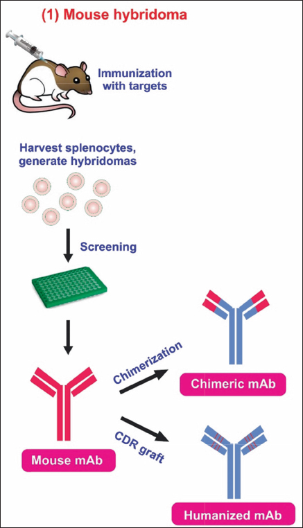
mAbs are versatile biomacromolecules that have high specificity for binding to various types of protein and non-protein substrates [5-7]. These mAbs can be produced using numerous approaches to enhance their functioning and utility [Figure 1] [8]. To date, there are currently over 130 therapeutically approved mAbs, with numerous others undergoing preclinical and clinical development [9]. Hybridoma technology is a widely employed technique for the production of mAbs. During this procedure, B lymphocytes that produce antibodies are separated from mice that have been immunized with a specific antigen. These B lymphocytes are then combined with immortal myeloma cell lines to create hybrid cells known as hybridoma cell lines. The hybridoma cells are cultivated in a controlled environment to generate mAbs that target a particular antigen.

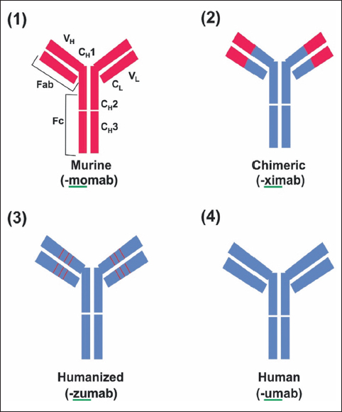
Figure 1: Structure of mAb and modification of murine mAb in therapy. (1) The murine monoclonal antibody. (2) Chimeric monoclonal antibody has variable regions of murine origin and the rest is of human origin. (3) Humanized monoclonal antibody has a hypervariable region of murine origin and the rest is of human origin. (4) Fully human monoclonal antibody.


[Click here to view]

mAbs in the market developed by hybridoma technology are given in Table 1. The human mAbs product developed by phage display, transgenic mice, and recombinant technology is given in Table 2. The top best-selling mAb drugs in the year 2022 are given in Table 3.

Table 1: US FDA-approved monoclonal antibody in the market developed by hybridoma technology.

S. No.AntibodyBrand nameYear of approvalTargetFormatIndicationCompanyReferences
1Muromonab-CD3Orthoclone OKT31986CD3Murine IgG2Kidney transplant rejectionCentocor Ortho Biotech Products L.P.[10]
2AbciximabReopro1994GPIIb/IIIaChimeric IgG1 FabPrevention of blood clots in angioplastyCentocor Inc.[10]
3RituximabMabThera, Rituxan1997CD20Chimeric IgG1Non-Hodgkin lymphomaBiogen Inc.[10]
4PalivizumabSynagis1998RSVHumanized IgG1Prevention of respiratory syncytial virus infectionMedImmune[10]
5InfliximabRemicade1998TNFαChimeric IgG1Crohn’s diseaseJanssen Biotech Inc.[10]
6TrastuzumabHerceptin1998HER2Humanized IgG1Breast cancerRoche, F. Hoffmann-La Roche, Ltd.[10]
7AlemtuzumabCampath, Lemtrada2001CD52Humanized IgG1Chronic myeloid leukemiaBerlex Inc.[10]
8Ibritumomab tiuxetanZevalin2002CD20Murine IgG1Non-Hodgkin lymphomaBiogen Inc.[10]
9OmalizumabXolair2003IgEHumanized IgG1AsthmaRoche, F. Hoffmann-La Roche, Ltd.[10]
10CetuximabErbitux2004EGFRChimeric IgG1Colorectal cancerBristol-Myers Squibb[10]
11BevacizumabAvastin2004VEGF-AHumanized IgG1Colorectal cancerRoche, F. Hoffmann-La Roche, Ltd.[10]
12NatalizumabTysabri2004ITGA4Humanized IgG4Multiple sclerosisBiogen Inc.[10]
13RanibizumabLucentis2006VEGF-AHumanized IgG1 FabMacular degenerationRoche, F. Hoffmann-La Roche Ltd.[10]
14EculizumabSoliris2007C5Humanized IgG2/4Paroxysmal nocturnal hemoglobinuriaAlexion Pharmaceuticals Inc.[10]
15Certolizumab pegolCimzia2008TNFαHumanized Fab, pegylatedCrohn’s diseaseCelltech, UCB.[10]
16TocilizumabRoActemra, Actemra2010IL-6RHumanized IgG1Rheumatoid arthritisChugai Pharmaceutical Co. Ltd.[10]
17Brentuximab vedotinAdcetris2011CD30Chimeric IgG1Hodgkin lymphomaSeattle Genetics Inc.[10]
18PertuzumabPerjeta2012HER2Humanized IgG1Breast CancerRoche, F. Hoffmann-La Roche, Ltd.[10]
19Trastuzumab emtansineKadcyla2012HER2Humanized IgG1Breast cancerRoche, F. Hoffmann-La Roche, Ltd.[10]
20ObinutuzumabGazyva, Gazyvaro2013CD20Humanized IgG1Chronic lymphocytic leukemiaBiogen Inc.[10]
21SiltuximabSylvant2014IL-6Chimeric IgG1Castleman diseaseCentocor Inc.[10]
22VedolizumabEntyvio2014α4β7 integrinHumanized IgG1Ulcerative colitis, Crohn diseaseGenentech Inc.[10]
23BlinatumomabBlincyto2014CD19, CD3Murine bispecific tandem scFvAcute lymphoblastic leukemiaAmgen[10]
24PembrolizumabKeytruda2014PD-1Humanized IgG4MelanomaMerck & Co. Inc.[10]
25IdarucizumabPraxbind2015DabigatranHumanized FabReversal of dabigatran-induced anticoagulationBIP[10]
26DinutuximabUnituxin2015GD2Chimeric IgG1NeuroblastomaUnited Therapeutics Corp.[10]
27MepolizumabNucala2015IL-5Humanized IgG1Severe eosinophilic asthmaCentocor Inc.[10]
28ElotuzumabEmpliciti2015SLAMF7Humanized IgG1Multiple myelomaBristol-Myers Squibb[10]
29IxekizumabTaltz2016IL-17αHumanized IgG4PsoriasisEli Lilly[10]
30ReslizumabCinqaero, Cinqair2016IL-5Humanized IgG4AsthmaCelltech, UCB[10]
31AtezolizumabTecentriq2016PD-L1Humanized IgG1Bladder cancerRoche, F. Hoffmann-La Roche, Ltd.[10]
32ObiltoxaximabAnthim2016Bacillus anthrasis PAChimeric IgG1Prevention of Inhalational anthraxElusys Therapeutics Inc.[10]
33Inotuzumab ozogamicinBesponsa2017CD22Humanized IgG4Acute lymphoblastic leukemiaWyeth Pharmaceuticals[10]
34OcrelizumabOcrevus2017CD20Humanized IgG1Multiple sclerosisBiogen Inc.[10]
35EmicizumabHemlibra2017Factor IXa, XHumanized IgG4, bispecificHemophilia AChugai Pharmaceutical Co., Ltd.[10]
36BenralizumabFasenra2017IL-5RαHumanized IgG1AsthmaMedImmune[10]
37Gemtuzumab ozogamicinMylotarg2017CD33Humanized IgG4; ADCAcute myeloid leukemiaPfizer[10]
38MogamulizumabPoteligeo2018CCR4Humanized IgG1Mycosis fungoides or Sézary syndromeKyowa Hakko Kirin[10]
39GalcanezumabEmgality2018CGRPHumanized IgG4Migraine preventionEli Lilly[10]
40TildrakizumabIlumya2018IL-23 p19Humanized IgG1Plaque psoriasisMerck & Co. Inc.[10]
41FremanezumabAjovy2018CGRPHumanized IgG2Migraine preventionTeva Pharmaceutical Industries, Ltd.[10]
42IbalizumabTrogarzo2018CD4Humanized IgG4HIV infectionTaimed Biologics Inc.[10]
43RavulizumabUltomiris2018C5humanized IgG2/4Paroxysmal nocturnal hemoglobinuriaAlexion Pharmaceuticals Inc.[10]
44CaplacizumabCablivi2019von Willebrand factorHumanized NanobodyAcquired thrombotic thrombocytopenic purpuraAblynx[10]
45RomosozumabEvenity2019SclerostinHumanized IgG2Osteoporosis inAmgen[10]
46RisankizumabSkyrizi2019IL-23 p19Humanized IgG1Plaque psoriasisBoehringer Ingelheim Pharmaceuticals[10]
47Polatuzumab vedotinPolivy2019CD79βHumanized IgG1 ADCDiffuse large B-cell lymphomaRoche, F. Hoffmann-La Roche, Ltd.[10]
48BrolucizumabBeovu2019VEGF-AHumanized scFvMacular degenerationNovartis Pharmaceuticals Corp.[10]
49CrizanlizumabAdakveo2019P-selectinHumanized IgG2Sickle cell diseaseNovartis Pharmaceuticals Corp.[10]
50Eptinezumab-jjmrVyepti2020CGRP blockHumanizedMigraineLundbeck[11]
51Isatuximab-irfcSarclisa2020Anti-CD38ChimericMultiple myelomaSANOFI AVENTIS[11]
52Sacituzumab govitecan-hziyTrodelvy2020Anti-Trop-2; SN-38;HumanizedmTNBCIMMUNOMEDICS[11]
53Daratumumab and hyaluronidase-fihjDarzalex Faspro2020Anti-CD38,HumanMultiple myelomaJANSSEN[11]
54Inebilizumab-cdonUplizna2020Anti-CD19HumanizedNMOSD (AQP4+)VIELA[11]
55Pertuzumab, trastuzumab, and hyaluronidase-zzxfPhesgo2020Anti-HER2;HumanizedBreast cancer (HER2+)GENENTECH[11]
56Belantamab mafodotin-blmBlenrep2020Anti-BCMAHumanizedMultiple MyelomaGLAXOSMITHKLINE[11]
57Satralizumab-mwgeEnspryng2020Anti-IL6 receptorHumanizedNMOSD (AQP4+)GENENTECH[11]
58ATOLTIVIMAB +2Inmazeb2020Zaire ebolavirus glycoproteinHumanized IgG1κZaire ebolavirus infection.REGENERON[11]
59NAXITAMAB-GQGKDanyelza2020Anti-glycolipid GD2Humanized IgG1NeuroblastomaY-MABS THERAPEUTICS[11]
60MARGETUXIMAB-CMKBMargenza2020Anti-HER2chimeric IgGBreast cancerMACROGENICS[11]
61RITUXIMAB-ARRXRiabni2020Anti-CD20chimeric IgGNon Hodgkin lymphomaAMGEN[11]
62Dostarlimab-gxlyJemperli2021PD-1HumanizedEndometrial cancerGSK[12]
63Loncastuximab tesirine-lpylZynlonta2021CD19Humanized IgG1B cell lymphomaADC[12]
64Dostarlimab-gxlyJemperli2021PD-1HumanizedSolid tumorGSK[12]
65Ranibizumab-nunaByooviz2021VEGFHumanized IgG1Macular degenerationSAMSUNG[12]
66RanibizumabSusvimo2021VEGFHumanized IgG1Macular degenerationGENEN TECH[12]
67Faricimab-svoaVabysmo2022VEGF; Ang-2Humanized IgG1nAMD; DMEGENENTECH[13]
68Sutimlimab-jomeEnjaymo2022CPHumanizedCold agglutinin diseaseBIOVERATIV[13]
69Bevacizumab-malyAlymsys2022VEGFHumanized IgG1mCRCAMNEAL[13]
70RisankizumabrzaaSkyrizi2022IL-23Humanized IgG1Plaque psoriasisABBVIE[13]
71Ranibizumab-eqrCimerli2022VEGFHumanized IgG1nAMD; RVO; DME;COHERUS[13]
72Spesolimab-sbzoSpevigo2022IL-36RHumanized IgG1Pustular psoriasisBOEHRINGER[13]
73Bevacizumab-adcVegzelma2022VEGFHumanized IgG1mCRC; NSCLC;CELLTRION[13]
74Mirvetuximab soravtansine-gynxElahere2022FRα-DM4Humanized IgG1Ovarian cancerIMMUNOGEN[13]
75Teplizumab-mzwvTzield2022CD3Humanized IgG1type 1 diabetesPROVENTION[13]
76Osunetuzumab-axgbLunsumio2022CD20; CD3Humanized IgG1Follicular lymphomaGENENTECH[13]
77Ublituximab-xiiyBriumv2022CD20Chimeric IgG1Multiple sclerosisTG[13]
78Teclistamab-cqyvTecvayli2022BCMA; CD3Humanized bispecific IgG4Multiple myelomaJANSSEN[13]
79ElranatamabElrexfio2023BCMA, CD3Humanized IgG2Multiple myelomaPfizer[10]
80RozanolixizumabRystiggo2023FcRn;Humanized IgG4Generalized myasthenia gravisUCB[10]
81TalquetamabTalvey2023G protein-coupled receptor 5D, CD3;Humanized IgG4 bispecificMultiple myelomaJanssen[10]
82EpcoritamabEpkinly2023CD20, CD3Bispecific humanized IgG1Diffuse large B cell lymphomaAbbvie[10]
83GlofitamabColumvi2023CD20, CD3eBispecificDiffuse large B-cell lymphomaGenentech[10]
84LecanemabLeqembi2023Amyloid beta protofibrils;Humanized IgG1Alzheimer’s diseaseBiogen[10]
85RetifanlimabZynyz2023PD-1Humanized IgG4Merkel cell carcinomaIncyte[10]

Table 2: US FDA-approved human monoclonal antibodies in the market.

S. No.AntibodyBrand nameYear of approvalTargetFormatTechnologyIndicationCompanyReferences
1AdalimumabHumira2002TNFαHuman IgG1Phage displayRheumatoid arthritisAbbVie Inc.[10]
2PanitumumabVectibix2006EGFRHuman IgG2Transgenic miceColorectal cancerAmgen[10]
3UstekinumabStelara2009IL-12/23Human IgG1Transgenic micePsoriasisCentocor Ortho Biotech Inc.[10]
4CanakinumabIlaris2009IL-1βHuman IgG1Transgenic miceMuckle-Wells syndromeNovartis Pharmaceuticals Corp.[10]
5GolimumabSimponi2009TNFαHuman IgG1Transgenic miceRheumatoid and psoriatic arthritis, ankylosing spondylitisCentocor Ortho Biotech Inc.[10]
6OfatumumabArzerra2009CD20Human IgG1Transgenic miceChronic lymphocytic leukemiaGenmab A/S[10]
7DenosumabXgeva, Prolia2010RANKLHuman IgG2Transgenic miceBone lossAmgen[10]
8BelimumabBenlysta2011BLySHuman IgG1Phage displaySystemic lupus erythematosusGlaxoSmithKline.[10]
9IpilimumabYervoy2011CTLA-4Human IgG1TransgenicMetastatic melanomaBristol-Myers Squibb[10]
10RaxibacumabAbthrax2012B. anthrasis
PA
Human IgG1Transgenic miceAnthrax infectionGlaxoSmithKline[10]
11RamucirumabCyramza2014VEGFR2Human IgG1Phage displayGastric cancerEli Lilly[10]
12NivolumabOpdivo2014PD-1Human IgG4Transgenic miceMelanoma, non-small cell lung cancerBristol-Myers Squibb[10]
13NecitumumabPortrazza2015EGFRHuman IgG1Phage displayNon-small cell lung cancerEli Lilly[10]
14SecukinumabCosentyx2015IL-17αHuman IgG1Transgenic micePsoriasisNovartis Pharmaceuticals Corp.[10]
15AlirocumabPraluent2015PCSK9Human IgG1Transgenic miceHigh cholesterolRegeneron Pharmaceuticals Inc.[10]
16EvolocumabRepatha2015PCSK9Human IgG2Transgenic miceHigh cholesterolAmgen[10]
17DaratumumabDarzalex2015CD38Human IgG1Transgenic miceMultiple myelomaGenmab A/S[10]
18OlaratumabLartruvo2016PDGFRαHuman IgG1Transgenic miceSoft tissue sarcomaEli Lilly[10]
19BezlotoxumabZinplava2016Clostridium difficile enterotoxin BHuman IgG1Transgenic micePrevention of Clostridium difficile infection recurrenceMerck & Co. Inc.[10]
20BrodalumabSiliq, Lumicef2017IL-17RHuman IgG2Transgenic micePlaque psoriasisMedImmune[10]
21GuselkumabTremfya2017IL-23 p19Human IgG1Phage displayPlaque psoriasisMorphoSys[10]
22DupilumabDupixent2017IL-4RαHuman IgG4Transgenic miceAtopic dermatitisRegeneron Pharmaceuticals Inc.[10]
23SarilumabKevzara2017IL-6RHuman IgG1Transgenic miceRheumatoid arthritisRegeneron Pharmaceuticals Inc.[10]
24AvelumabBavencio2017PD-L1Human IgG1Phage displayMerkel cell carcinomaMerck[10]
25DurvalumabImfinzi2017PD-L1Human IgG1Transgenic miceBladder cancerMedImmune[10]
26BurosumabCrysvita2018FGF23Human IgG1Transgenic miceX-linked hypophosphatemiaKyowa Hakko Kirin/Ultragenyx[10]
27LanadelumabTakhzyro2018Plasma kallikreinHuman IgG1Phage displayHereditary angioedema attacksDyax Corp.[10]
28ErenumabAimovig2018CGRPRHuman IgG2Transgenic miceMigraine preventionNovartis[10]
29CemiplimabLibtayo2018PD-1Human mAbTransgenic miceCutaneous squamous cell carcinomaRegeneron Pharmaceuticals Inc.[10]
30EmapalumabGamifant2018IFNγHuman IgG1Phage displayPrimary hemophagocytic lymphohistiocytosisNovImmmune[10]
31Moxetumomab pasudodoxLumoxiti2018CD22Murine IgG1 dsFvPhage displayHairy cell leukemiaMedImmune[10]
32Teprotumumab-trbTepezz2020IGF-1R blockHuman IgG1Transgenic miceThyroid eye diseaseHorizon therapeutics[11]
33Evinacu-mab-dgnbEvkeeza2021ANGPTL3HumanRecombinantHoFH LDL-CRegeneron[12]
34Amivantamab-vmjwRybrevan2021EGFR MET ReceptorHuman IgG1RecombinantNSCLCJanssen[12]
35Aducanumab-avwaAduhelm2021Amyloid betaHuman IgG1RecombinantAlzheimer’s diseaseBiogen[12]
36Anifro-lumab-fniaSaphnelo2021(I)IFNRHumanRecombinantSLEAstrazeneca[12]
37Tisotumab vedotin-tftvTivdak2021Tissue FactoHumanRecombinantCervicalSeagen[12]
38Adalimumab-aqvhYusimry2021TNFHuman IgG1RecombinantRACoherus[12]
39Tezepelumab-ekkoTezspire2021TSLPHumanRecombinantSevere asthmaAstra zeneca[12]
40Tralokinumab-ldrmadbry2021IL-13Human IgG4RecombinantEczemaLeo[12]
41Nivolumab; relatlimabrmbwOpdualag2022PD-1; LAG-3Human IgG4RecombinantmelanomaBms[13]
42TremelimumabactlImjudo bla 7612892022CTLA-4Human IgG2RecombinantLiver cancerAstra Zeneca[13]
43TremelimumabactlImjudo bla 7612702022CTLA-4Human IgG2RecombinantLiver cancerAstra Zeneca[13]
44Dalimumab-aacfIdacio2022TNFHuman IgG1RecombinantRA JIA PsAFresenius[13]
45PozelimabVeopoz2023Complement 5Human IgG4RecombinantCHAPLE diseaseRegeneron[10]
46NirsevimabBeyfortus2023RSVHuman IgG1RecombinantRSV infectionSanofi[10]

Table 3: Top five best-selling monoclonal antibody drugs in 2022.

S. No.AntibodyBrand nameCompanyTargetTechnology/
formats
IndicationRevenue (USD)References
1AdalimumabHumiraAbbVieTNFαPhage displayRheumatoid arthritis21.2 billion[14]
2PembrolizumabKeytrudaMerck & Co.PD-1Humanized IgG4Melanoma20.9 billion[14]
3UstekinumabStelaraJanssenIL-12/23Human IgG1Plaque psoriasis, psoriatic arthritis9.7 billion[14]
4DupilumabDupixentSanofi Genzyme, RegeneronIL-4RαHuman IgG4Atopic dermatitis, asthma, chronic rhinosinusitis with nasal polyps8.7 billion[14]
5NivolumabOpdivoBristol-Myers SquibbPD-1Transgenic miceMelanoma, non-small cell lung cancer8.2 billion[14]

mAb drugs have developed from clinical research to commercialization over the past few decades. In the past few years, the total number of antibody drugs approved for launch has proliferated, with 130 approved and available on the market [15]. mAb treatments were the fastest-growing category in the worldwide biopharmaceutical market in 2022, with four of the top ten best-selling biopharmaceutical products. Keytruda, which is a drug developed by Merck and introduced in 2014, garnered USD 20.9 billion in sales in 2022 [16]. In recent years, the global mAb market has grown, reaching USD 178.5 billion in 2021, which is an increase of 12% year over year. As of April 2022, there were 250 mAb therapies in Phase III clinical trials around the world [17]. Antibody medications are expected to be licensed and marketed in great numbers in the next few years, with a market value of USD 420–460 billion in 2028. The global mAb market has practically been split up by Roche, Johnson & Johnson, Merck, Novartis, AbbVie, and Amgen. The top five antibody drugs in the world by sales in 2022 were Humira, Keytruda, Stelara, Dupixent, and Opdivo, with a combined share of more than 50% of the global market [1]. Therefore, this review article discusses the rapid growth of therapeutic mAb products as well as the technological developments in producing these antibodies using various in vitro methods. Furthermore, the study offers a thorough synopsis of the approved mAbs currently accessible on the market, together with their distinct targets, structures, and permissible uses.


2. ANTIBODY STRUCTURE AND FUNCTION

Antibodies are large glycoproteins that belong to the immunoglobulin (Ig) superfamily that help the immune system to recognize foreign antigens, neutralize them, and trigger an immune response. Antibody molecules are made up of light chains (LCs) and heavy chains (HCs). Human immunoglobulins consist of two identical LCs and two identical HCs, forming a Y-shaped protein structure. In biological systems, the combination of one LC and one HC joins with another identical heterodimer creates the complete immunoglobulin. Moreover, antibodies are divided into five classes, namely, IgA, IgD, IgE, IgG, and IgM, are classified based on the type of HC they possess. Furthermore, in immunoglobulin G, disulfide bridges connect two HCs and two LCs to form a 150-kDa protein. One variable domain (VH or VL) and one to four constant domains (CH or CL) make up each chain. Each variable domain splits into three complementarity-determining regions (CDRs) with varying sequences and four framework portions with relatively constant sequences. Three constant domains are located in the HC of immunoglobulin G, while one is located in the LC. Y-shaped fundamental structure characterizes them. The binding location of the antibody, which can bind to the antigen, is determined by the CDRs of the variable chains. These CDRs are complementary to the epitope on the antigen. Variations in the amino acid sequences of the CDRs lend credence to antibodies. The Fc region, located at the base of the Y-shaped structure, enables interactions between antibodies and other components of the immune system [18].

The Fc region of antibodies is accountable for performing effector functions, including antibody-dependent cell-mediated cytotoxicity (ADCC) and complement-dependent cytotoxicity (CDC). Antibodies bind to Fc receptors (FcRs) on the surface of effector cells like natural killer (NK) cells and macrophages, causing them to phagocytose or lyse the target cells. The target cells are eliminated when antibodies in the CDC initiate the complement cascade on the cell surface. Human IgG1 exhibits superior efficacy in both CDC and ADCC, making it highly suitable for use in cell treatment to target infections or tumors. IgG1 and IgG3 possess the capacity to elicit ADCC and CDC. However, IgG2 and IgG4 do not possess this capability [19]. Moreover, a novel subset of recombinant antibodies, the single-chain Fv fragment, was first presented by Bird et al. [20]. In order to connect the VH and VL domains, a 15–20 amino acid flexible linker peptide was employed. Compared with intact antibodies, scFv is the smallest form of recombinant immunoglobulin that maintains full activity.

mAbs have significantly transformed biological studies and clinical diagnostics, and their therapeutic potential is currently being strategically positioned for application. The initial application of treatment involved mAbs derived from rodents, which resulted in notable endogenous antibody responses. Additionally, it often fails to consider the initiation of effector activities. These limitations have propelled the search for more compatible and effective alternatives.

In response to these challenges, humanization, which is a sophisticated form of genetic engineering, has emerged as a groundbreaking solution. By integrating only the rodent complementary determining regions with the human variable region framework and constant heavy and LC portions, humanized antibodies achieve 95% homology with genuine human antibodies. This innovative approach not only mitigates the immune response issues associated with rodent-derived antibodies but also enhances the efficacy of mAbs in therapeutic applications [21]. The implementation of antibody phage display enabled the direct selection of human-origin-specific antibodies, thus achieving a targeted antibody production approach. Antibody phage display has emerged as the primary method in antibody engineering due to its capacity to rapidly isolate single-chain Fv fragments derived from human offspring. This technology has been widely employed to choose antibodies targeting diverse compounds of interest. In recent years, new in vivo and in vitro approaches for the isolation of human antibodies have been discovered, expanding the spectrum of applications for antibody engineering technology. Transgenic mice having human IgG loci instead of natural Ig genes can be used to select human antibodies in vivo [22,23]. Bacterial surface display, yeast surface display, and cell-free ribosome display are all in vitro possibilities for picking single-chain Fv fragments, and they are all discussed elsewhere.


3. VARIOUS METHODOLOGIES EMPLOYED IN THE DEVELOPMENT OF ANTIBODIES THAT EXHIBIT REACTIVITY TOWARD THE INTENDED TARGET

3.1. Immunize an Animal

The process of creating mAbs is both intricate and fascinating. Initially, an animal (usually a mouse or rat) can be vaccinated with the target antigen. This phase is essential because it primes the animal’s system to generate the required antibodies. During the subsequent stage, B cells derived from the animal’s immune system are meticulously combined with myeloma cells, leading to the creation of a hybridoma. This hybridoma is a biological combination that merges the specificity of the B cell with the lifespan of the myeloma cell, resulting in immortality. Subsequently, by selecting clones that produce mAb with the required specificity for the target antigen, hybridomas can produce an unlimited number of mAb [Figure 2]. Muromonab-CD3 is one of the mAbs created by this method (Orthoclone OKT3).

Figure 2: Development of mouse monoclonal antibody by hybridoma technology. Chimeric antibodies are developed by joining a sequence of murine variable domains with the human constant domain region. The humanized antibodies are developed by transplanting the murine CDR sequence to the human framework sequence. This technique is termed CDR grafting.


[Click here to view]

A significant limitation of this technology is the immune response triggered by mouse antibodies in certain individuals, leading to the development of human-anti-mouse antibodies (HAMA). The primary drawback of this method is the activation of the immune system by mouse antibodies in specific individuals, resulting in the formation of HAMA. This immune response not only reduces the effectiveness of the original mouse mAbs but also prevents the use of any mAbs that have similar sequences to mice because of the potential for allergic reactions and decreased availability in the body. As a result, the use of mouse mAbs in medical conditions is significantly limited.

Furthermore, the process of genetic engineering was employed to produce chimeric antibodies by merging the variable regions of mice with the constant segments of humans. This measure was implemented in order to reduce the probability of mouse antibodies eliciting an immunological response in humans [Figure 2] [24]. Despite the fact that chimeric antibodies are less immunogenic than murine MAbs, human anti-chimeric antibody reactions have been found [25]. Approaches to engineer alterations to the immunoglobulin molecule, such as humanizing the antibody or producing a chimeric antibody, have been established and used in the majority of mAbs selected in animals. In order to minimize the presence of mouse elements, the process of humanizing non-human antibodies involves grafting the CDRs from non-human antibodies onto human frames. For the development of these humanized antibodies, human frameworks that have the closest similarity to the framework regions of non-human antibodies as recipients for CDR grafting were selected [26,27]. However, CDR grafting by this method results in a loss of affinity for their specific targets [28]. Therefore, key framework residues that support the conformations of CDR loops in the murine antibody are also grafted onto the human template in order to restore the affinity of the parental murine antibody. Based on these progressions in antibody engineering, the development of recombinant mAbs signifies another significant achievement in the pursuit of more effective and ethically manufactured therapeutic agents.

3.2. Recombinant mAbs Development

Recombinant antibodies refer to mAbs that are artificially created in laboratory conditions through the use of synthetic genes. In contrast to mAbs, recombinant antibodies are produced using advanced technology that does not employ hybridomas or animals. Recombinant mAbs offer effective therapies for cancer, autoimmune disorders, and various other disorders. Additionally, these antibodies can be employed in biomedical and toxicological studies. mAbs are extensively used in biomedical science and medicine because they have the capacity to attach to, neutralize, or eliminate antigens that are particular to certain cells [29]. However, the ascites method of manufacturing causes considerable agony and misery to the animals involved. Basically, recombinant antibodies are mAbs generated in vitro using synthetic genes [Figure 3]. The technology involves recovering antibody genes from source cells, amplifying and cloning the genes into an appropriate vector, introducing the vector into a host (bacteria, yeast, or mammalian cell lines), and achieving the expression of adequate amounts of functional antibody [30,31].

Figure 3: Schematic overview of production and development OF Recombinant antibodies. Recombinant antibodies are a type of monoclonal antibodies that are generated in vitro from a synthetic gene without immunizing any animals or cultivating any hybridomas.


[Click here to view]

3.3. Phage Display

3.3.1. Phage-display vectors

Phages are viruses that infect bacterial cells, and many of the vectors used in recombinant DNA studies are phages that infect Escherichia coli, which is the most common recombinant DNA host. Recombinant DNA vectors, including phages, have the capacity to include segments of “foreign” DNA, which can be derived from human DNA or chemically synthesized. The foreign “insert” is copied alongside the vector DNA in its E. coli host as a guest. The distribution of phages seems to be entirely stochastic. Phage capsid fusions are used to provide extensive collections of encoded peptides or proteins in combinatorial libraries. The library can be used to identify phages that carry the appropriate peptides or proteins, which can then be decoded via phage DNA sequencing [32,33]. By employing panning and selection techniques, it is possible to isolate specific phage clones that produce desired recombinant antibodies from vast populations of phages, which may consist of tens of millions of different clones [Figure 4]. The utilization of genetically modified antibody fragments in combination with the phage display technique highlights the adaptability and effectiveness of this procedure in the selection of targeted antibodies.

Figure 4: The phage display cycle. (1) A library of DNA variant sequences encoding peptides or proteins is created and (2) thus cloned into phage genomes as fusions to a coat protein gene. (3) The phage library exhibiting variant peptides or proteins is revealed to target molecules and phages with appropriate specificity are captured. (4) Non-binding phages are washed off. (5) Bound phage is eluted by disrupting the interaction between the displayed peptide or protein and the target. (6) Eluted phage is infected into host bacterial cells and thereby amplified. (7) This amplified phage population results in a secondary library that is greatly enriched in phage-displaying peptides or proteins that bind to the target. On repeating the bio-panning steps (3–6), the phage population becomes less and less diverse as the population becomes more and more enriched in the limited number of variants with binding capacity. (8) After several (usually three to five) rounds of bio-panning, monoclonal phage populations may be selected and analyzed individually.


[Click here to view]

Figure 5: Development of Human monoclonal antibody by phage display and transgenic mouse technology. The characteristic of phages is that they house human DNA, this insert replicates along with vector DNA in E. coli host, and then DNA sequences are analyzed to construct and express human IgGs. The transgenic mouse is genetically modified by replacing endogenous Ig with human Ig genes to produce human IgGs.

Additional methods for presenting information, including displaying it on the surface of E. coli or Saccharomyces cerevisiae, as well as employing ribosome display, were created based on the phage display principle. The efficacy of in vitro display methods in finding binders specific to antigens is undisputed, and in the future, these methods may be combined to mutually enhance each other. Despite the advantages of antibody phage display, such as the capacity to avoid animal vaccination, isolate antibodies against hazardous or non-immunogenic antigens, and manufacture conformation-specific antibodies, vaccinated mice approaches provide the vast majority of licensed therapeutic antibodies. This is due to the immune system’s filtration mechanism, which allows human antibodies to possess superior biophysical characteristics compared with phage display antibodies [33].

3.4. Transgenic Mouse

Transgenic mouse strains with human immunoglobulin repertoires provide an alternative approach to select therapeutic mAbs that have a lower likelihood of causing an immune response. Transgenic technology is used to genetically modify mice strains in order to produce human sequence antibodies, unlike antibody engineering techniques that primarily involve modifying and optimizing individual protein components. These antibodies can be directly used in drug discovery and can be moved into clinical use without the need for additional optimization. The immunoglobulin transgenic mice have been validated as drug discovery platforms with the regulatory approval of their first product, panitumumab, 12 years after its initial publication in the scientific literature [34-36].

In 1994, the HuMabMouse [37] and the Xeno-Mouse [22] were the first transgenic mouse lines released using this technology. Genetic modifications of these lines were done in a manner, where human immunoglobulin (Ig) genes were inserted into the genome, replacing the indigenous Ig genes and allowing the animals to manufacture fully human antibodies [Figure 5] [35,37]. Following a comparable approach, the generation of neutralizing human antibodies from human B cells has also exhibited encouraging results for the treatment of infectious diseases. If feasible, it is preferable to obtain antibodies directly from humans rather than humanizing them from other animals for the treatment of infectious diseases. Human B cells are currently widely employed as a starting material for isolating human mAbs using techniques such as in vitro display, B cell immortalization, and single B cell expression cloning [38]. Human antibodies that may neutralize infectious pathogens may exhibit enhanced efficacy when derived directly from human B cells. This can be caused by the fact that the coupling of heavy and LCs has already been selected in vivo through rearrangement and has been well-tolerated in individuals, either following infection or immunization.

The progress of bispecific antibodies offers new possibilities for the advancement of novel protein treatments. Bispecific antibodies, through virtue of their ability to bind to two separate targets, have long held the potential of broadening the possibilities of mAb treatments. Bispecific act through a range of different mechanisms, including forming immunological synapses—or interfaces—between immune effector cells (macrophages, or T or NK cells) and tumor cells, staging a double blockade of disease-related pathways, cross-linking receptors, or bridging a gap in the coagulation cascade. The future use of transgenic animals that produce antibodies in their milk remains uncertain, although they have already been successfully engineered.


4. RECOMBINANT EXPRESSION AND PURIFICATION STRATEGIES OF ANTIBODIES

Currently, extensive bioreactors are employed to sustain mammalian cell culture for the production of clinically applicable antibodies. Consequently, the cost of a refined antibody for therapeutic use may exceed up to USD 1000 per gram, in contrast to only USD 5 per gram for standard small molecules produced through chemical syntheses [5].

The efficacy of a therapeutic antibody is not solely determined by its specificity and affinity. Regarding mAb lead candidates, additional quality attributes, including solubility, viscosity, expression yield, heat, and long-term stability, are crucial [39,40]. The amino acid sequence has a significant impact on the physicochemical properties of antibodies [41]. Some mAbs may exhibit unfavorable developability traits such as increased immunogenicity, physical instability, self-aggregation, higher viscosity, non-specific binding, short half-life, and low expression levels [42,43]. mAb potency, bioavailability, and immunogenicity can be affected by low solubility during biomanufacturing [44-48]. Structural and functional integrity as well as intrinsic qualities depend on thermal stability [48,49]. In addition, immunogenicity problems have made aggregation a major obstacle in the progress of therapeutic mAbs [51-54].

Antibodies and antibody fragments can be produced by other expression systems, including bacteria, yeasts, and plants. E. coli bacteria are best at producing small, nonglycosylated, affinity-purifiable fragments of Fab and scFv [55,56]. If there is a need to avoid using affinity tags, alternative purification methods that involve size exclusion chromatography, ion-exchange chromatography, and ammonium sulfate precipitation can be employed to further refine the purification of full-length antibodies and scFv fragments. These methods can be used after the initial purification using the Protein A- and Protein L-binding matrices [57]. The lack of post-translational modifications in bacteria could pose a problem for antibody fragments or fusion proteins that require glycosylation.


5. CONCLUSION

mAbs are treasured for their specificity, selectivity, and binding affinity. All these properties, combined with their ease of production in recombinant mammalian systems, make them successful in a wide array of applications. Antibodies are the preferred choice in therapy due to their minimal toxicity and high specificity, making them advantageous for treating a diverse array of human diseases including asthma, rheumatoid arthritis, Crohn’s disease, multiple sclerosis, infectious diseases, and some types of cancers. In addition to this, mAbs, which are a type of drug, can be used to treat a number of other diseases. Considering the higher production costs compared to small molecules synthesized chemically, antibody-based therapeutics provide various advantages, such as their specificity and the ease of selecting antigen-specific binders. Therapeutic mAbs have gained significant recognition as a type of biological substance that is extensively used in research, development, and commercialization. The significance of mAbs in both the biopharmaceutical sector and the market is undeniable. As mAbs can be tailored to specific targets and pathways, they have transformed the approach to treating human diseases. New methods and techniques are being created to develop novel therapeutic mAbs. mAbs in advanced stages of development for new therapeutic applications are expected to significantly alter previously unexplored targets [58,59]. Besides new mAbs, biosimilars have become increasingly popular due to the impending expiration of patents for numerous highly successful mAbs. For instance, the patent on rituximab, which is an extensively used drug in the fields of cancer, hematology, rheumatology, nephrology, and other related areas, expired in Europe in 2013 and in the United States in 2018 [60,61]. Following the expiration of patents on original products, the creation of biosimilars is intended to make biotherapeutics cheaper. The production of a biosimilar entails a series of progressive comparability exercises, beginning with a comparison of the quality features of biosimilars and reference. Regulatory agencies approve biosimilars based on their demonstrated similarity to a relevant reference in terms of quality, nonclinical, and clinical criteria.


6. AUTHOR CONTRIBUTIONS

All authors made substantial contributions to conception and design, acquisition of data, or analysis and interpretation of data; took part in drafting the article or revising it critically for important intellectual content; agreed to submit to the current journal; gave final approval of the version to be published; and agree to be accountable for all aspects of the work. All the authors are eligible to be an author as per the international committee of medical journal editors (ICMJE) requirements/guidelines.


7. FINANCIAL SUPPORT AND SPONSORSHIP

There is no funding to report.


8. CONFLICTS OF INTEREST

The authors report no financial or any other conflicts of interest in this work.


9. ETHICAL APPROVALS

This study does not involve experiments on animals or human subjects.


10. DATA AVAILABILITY

All the data is available with the authors and shall be provided upon request.


11. USE OF ARTIFICIAL INTELLIGENCE (AI)-ASSISTED TECHNOLOGY

The authors confirm that there was no use of artificial intelligence (AI)-assisted technology for assisting in the writing or editing of the manuscript and no images were manipulated using AI.


12. PUBLISHER’S NOTE

All claims expressed in this article are solely those of the authors and do not necessarily represent those of the publisher, the editors and the reviewers. This journal remains neutral with regard to jurisdictional claims in published institutional affiliation.


REFERENCES

1.  Schwaber J, Cohen EP. Human mouse somatic cell hybrid clone secreting immunoglobulins of both parental types. Nature 1973;244:444–7.

2.  Köhler G, Milstein C. Continuous cultures of fused cells secreting antibody of predefined specificity. Nature 1975;256:495–7.

3.  Shin SU, Morrison SL. Production and properties of chimeric antibody molecules. Methods Enzymol 1989;178:459–76.

4.  Weiner GJ. Building better monoclonal antibody-based therapeutics. Nat Rev Cancer 2015;15:361–70. [CrossRef]

5.  Schoonbroodt S, Steukers M, Viswanathan M, Frans N, Timmermans M, Wehnert A, et al. Engineering antibody heavy chain CDR3 to create a phage display Fab library rich in antibodies that bind charged carbohydrates. J Immunol 2008;181:6213–21. [CrossRef]

6.  Zhang P, Sun F, Liu S, Jiang S. Anti-PEG antibodies in the clinic: Current issues and beyond PEGylation. J Control Release 2016;244:184–93. [CrossRef]

7.  Watanabe H, Nakanishi T, Umetsu M, Kumagai I. Human anti-gold antibodies: Biofunctionalization of gold nanoparticles and surfaces with anti-gold antibodies. J Biol Chem 2008;283:36031–8. [CrossRef]

8.  Saeed AF, Wang R, Ling S, Wang S. Antibody engineering for pursuing a healthier future. Front Microbiol 2017;8:495. [CrossRef]

9.  Kaplon H, Muralidharan M, Schneider Z, Reichert JM. Antibodies to watch in 2020. MAbs 2020;12:1703531. [CrossRef]

10.  The Antibody Society. Therapeutic monoclonal antibodies approved or in review in the EU or US. (date accessed); Available from: www.antibodysociety.org/resources/approved-antibodies

11.  Cai HH, Pandit A. Therapeutic monoclonal antibodies approved by FDA in 2020. Clin Res Immunol 2021;4:1–2.

12.  Cai HH, Pandit A. Therapeutic monoclonal antibodies approved by FDA in 2021. Acta Sci Microbiol 2022;5(11): 81–3.

13.  Cai HH, Pandit A. Therapeutic monoclonal antibodies approved by FDA in 2022. J Clin Exp Immunol 2022;8(1);533–5.

14.  Buntz B. Drug Discovery and Development Report: Best selling pharmaceuticals of 2022. Available from: https://www.drugdiscoverytrends.com/

15.  Urquhart L. Top companies and drugs by sales in 2019. Nat Rev Drug Discov 2020;19:228. [CrossRef]

16.  Walsh G. Biopharmaceutical benchmarks 2018. Nat Biotechnol 2018;36:1136–45. [CrossRef]

17.  Grilo AL, Mantalaris A. The increasingly human and profitable monoclonal antibody market. Trends Biotechnol 2019;37:9–16. [CrossRef]

18.  Murphy, K. Immunobiology, 9th ed. New York, NY: Garland Science; 2017.

19.  Weiner LM, Surana R, Wang S. Monoclonal antibodies: Versatile platforms for cancer immunotherapy. Nat Rev Immunol 2010;10:317–27.

20.  Bird RE, Hardman KD, Jacobson JW, Johnson S, Kaufman BM, Lee SM, et al. Single-chain antigen-binding proteins. Science 1988;242 (4877):423–6. [CrossRef]

21.  Queen C, Schneider WP, Selick HE, Payne PW, Landolfi NF, Duncan JF, et al. A humanized antibody that binds to the interleukin 2 receptor. Proc Natl Acad Sci U S A 1989;86:10029–33.

22.  Mendez MJ, Green LL, Corvalan JR, Jia XC, Maynard-Currie CE, Yang XD, et al. Functional transplant of megabase human immunoglobulin loci recapitulates human antibody response in mice [published erratum appears in Nat Genet 1997 Aug;16(4):410]. Nat Genet 1997;15:146–156.

23.  Ishida I, Tomizuka K, Yoshida H, Tahara T, Takahashi N, Ohguma A, et al. Production of human monoclonal and polyclonal antibodies in TransChromo animals. Cloning Stem Cells 2002;4:91–102; [CrossRef]; PMID:12006160.

24.  Morrison SL, Johnson MJ, Herzenberg LA, Oi VT. Chimeric human antibody molecules: Mouse antigen-binding domains with human constant region domains. Proc Natl Acad Sci U S A Nov 1984;81(21):6851–5. [CrossRef]

25.  Bell SJ, Kamm MA. Review article: The clinical role of anti-TNFalpha antibody treatment in Crohn’s disease. Aliment Pharmacol Ther 2000;14(5):501–14. [CrossRef]; PMID: 10792111.

26.  Pelat T, Bedouelle H, Rees AR, Crennell SJ, Lefranc MP, Thullier, P. Germline humanization of a non-human primate antibody that neutralizes the anthrax toxin, by in vitro and in silico engineering. J Mol Biol 2008;384:1400–07.

27.  Robert R, Streltsov VA, Newman J, Pearce LA, Wark KL, Dolezal O. Germline humanization of a murine Abeta antibody and crystal structure of the humanized recombinant Fab fragment. Protein Sci 2010;19:299–308.

28.  Pavlinkova G, Colcher D, Booth BJ, Goel A, Wittel UA, Batra SK. Effects of humanization and gene shuffling on immunogenicity and antigen binding of anti-TAG-72 single-chain Fvs. Int J Cancer 2001;94:717–26.

29.  Kettleborough CA, Saldanha J, Heath VJ, Morrison CJ, Bendig MM. Humanization of a mouse monoclonal antibody by CDR-grafting: The importance of framework residues on loop conformation. Protein Eng 1991;4(7):773–83. [CrossRef]

30.  Dhankar R, Rathee P, Jain AK, Arora S, Kumar MS, Rath G, et al. HER-2-targeted immunonanoparticles for breast cancer chemotherapy. J Appl Pharm Sci 2011;132–9.

31.  Nabiel A, Yosua Y, Sriwidodo S, Maksum IP. Overview of refolding methods on misfolded recombinant proteins from Escherichia coli inclusion bodies. J Appl Biol Biotech 2023;11(3):47–52. [CrossRef]

32.  Smith GP. Filamentous fusion phage: Novel expression vectors that display cloned antigens on the virion surface. Science 1985;228(4705):1315–7. [CrossRef]

33.  Hoess RH. Protein design and phage display. Chem Rev 2001;101(10):3205–18.

34.  Lonberg N, Taylor LD, Harding FA, Trounstine M, Higgins KM, Schramm SR, et al. Antigen-specific human antibodies from mice comprising four distinct genetic modifications. Nature 1994;368(6474):856–9. [CrossRef]; PMID: 8159246.

35.  Green LL, Hardy MC, Maynard-Currie CE, Tsuda H, Louie DM, Mendez MJ, et al. Antigen-specific human monoclonal antibodies from mice engineered with human Ig heavy and light chain YACs. Nat Genet 1994;7(1):13–21.

36.  Gibson TB, Ranganathana A, Grothey A. Randomized phase III trial results of panitumumab, a fully human anti-epidermal growth factor receptor monoclonal antibody, in metastatic colorectal cancer. Clin Colorectal Cancer 2006;6(1):29–31. [CrossRef]

37.  Lonberg N, Taylor LD, Harding FA, Trounstine M, Higgins KM, Schramm SR, et al. Antigen-specific human antibodies from mice comprising four distinct genetic modifications. Nature 1994;368:856–9.

38.  Wilson PC, Andrews SF. Tools to therapeutically harness the human antibody response. Nat Rev Immunol 2012;12(10):709–19.

39.  Chennamsetty N, Voynov V, Kayser V, Helk B, Trout BL. Prediction of aggregation prone regions of therapeutic proteins. J Phys Chem B 2010;114:6614–24. [CrossRef]

40.  Jain T, Sun T, Durand S, Hall A, Houston NR, Nett JH, et al. Biophysical properties of the clinical-stage antibody landscape. Proc Natl Acad Sci U S A 2017;114:944–9. [CrossRef]

41.  Lecerf M, Kanyavuz A, Lacroix-Desmazes S, Dimitrov JD. Sequence features of variable region determining physicochemical properties and polyreactivity of therapeutic antibodies. Mol Immunol 2019;112:338–46. [CrossRef]

42.  Lavoisier A, Schlaeppi J-M. Early developability screen of therapeutic antibody candidates using Taylor dispersion analysis and UV area imaging detection. MAbs 2015;7:77–83. [CrossRef]

43.  Dobson CL, Devine PWA, Phillips JJ, Higazi DR, Lloyd C, Popovic B, et al. Engineering the surface properties of a human monoclonal antibody prevents self-association and rapid clearance in vivo. Sci Rep 2016;6:38644. [CrossRef]

44.  Bye JW, Platts L, Falconer RJ. Biopharmaceutical liquid formulation: A review of the science of protein stability and solubility in aqueous environments. Biotechnol Lett. 2014;36:869–75. [CrossRef]

45.  Garidel P, Kuhn AB, Schäfer LV, Karow-Zwick AR, Blech M. High-concentration protein formulations: How high is high? Eur J Pharm Biopharm 2017;119:353–60. [CrossRef]

46.  Trevino SR, Scholtz JM, Pace CN. Measuring and increasing protein solubility. J Pharm Sci 2008;97:4155–66. [CrossRef]

47.  Gibson TJ, Mccarty K, Mcfadyen IJ, Cash E, Dalmonte P, Hinds KD, et al. Application of a high-throughput screening procedure with PEG-induced precipitation to compare relative protein solubility during formulation development with IgG1 monoclonal antibodies. J Pharm Sci 2011;100:1009–21. [CrossRef]

48.  Sormanni P, Amery L, Ekizoglou S, Vendruscolo M, Popovic B. Rapid and accurate in silico solubility screening of a monoclonal antibody library. Sci Rep 2017;7:8200. [CrossRef]

49.  Seeliger D, Schulz P, Litzenburger T, Spitz J, Hoerer S, Blech M, et al. Boosting antibody developability through rational sequence optimization. MAbs 2015;7:505–15. [CrossRef]

50.  Chennamsetty N, Voynov V, Kayser V, Helk B, Trout BL. Design of therapeutic proteins with enhanced stability. Proc Natl Acad Sci U S A 2009;106:11937–42. [CrossRef]

51.  Lowe D, Dudgeon K, Rouet R, Schofield P, Jermutus L, Christ D. Aggregation, stability, and formulation of human antibody therapeutics. Adv Protein Chem Struct Biol 2011;84:41–61. [CrossRef]

52.  Perchiacca JM, Tessier PM. Engineering aggregation-resistant antibodies. Annu Rev Chem Biomol Eng 2012; 3:263–86. [CrossRef]

53.  Rosenberg AS. Effects of protein aggregates: An immunologic perspective. AAPS J 2006;8:E501–7. [CrossRef]

54.  Li W, Prabakaran P, Chen W, Zhu Z, Feng Y, Dimitrov DS. Antibody aggregation: Insights from sequence and structure. Antibodies (Basel) 2016;5:19. [CrossRef]

55.  Skerra A, Pfitzinger I, Plückthun A. The functional expression of antibody fv fragments in Ischhuchia coli: Improved vectors and a generally applicable purification technique. Biotechnology (N Y) 1991;9(3):273–8.

56.  Terpe K. Overview of tag protein fusions: From molecular and biochemical fundamentals to commercial systems. Appl Microbiol Biotechnol 2003;60:523–33.

57.  Bouvet JP. Immunoglobulin Fab fragment-binding proteins. Int J Immunopharmacol 1994;16(5-6):419–24.

58.  Verma V. Leveraging monoclonal antibodies as therapeutics to address antimicrobial resistance in bacteria. J App Biol Biotech 2023;11(3):53–60. [CrossRef]

59.  Adnyana IMO, Tedyanto EH, Widyadharma IPE. Efficacy and safety of eptinezumab as migraine preventive therapy: A systematic review and meta-analysis. J Appl Pharm Sci 2023;13(02):011–20. [CrossRef]

60.  DUBLIN--(BUSINESS WIRE)--May 2, 2019--The “Global and China Monoclonal Antibody Industry Report, 2019–2025”. Available from: https://www.businesswire.com

61.  First World Pharma, Truxima, The First Biosimilar mAb in Oncology, Granted EU Marketing Authorisation, 2017.

Reference

1. Schwaber J, Cohen EP. Human mouse somatic cell hybrid clone secreting immunoglobulins of both parental types. Nature 1973;244:444-7. https://doi.org/10.1038/244444a0

2. Köhler G, Milstein C. Continuous cultures of fused cells secreting antibody of predefined specificity. Nature 1975;256:495-7. https://doi.org/10.1038/256495a0

3. Shin SU, Morrison SL. Production and properties of chimeric antibody molecules. Methods Enzymol 1989;178:459-76. https://doi.org/10.1016/0076-6879(89)78034-4

4. Weiner GJ. Building better monoclonal antibody-based therapeutics. Nat Rev Cancer 2015;15:361-70. https://doi.org/10.1038/nrc3930

5. Schoonbroodt S, Steukers M, Viswanathan M, Frans N, Timmermans M, Wehnert A, et al. Engineering antibody heavy chain CDR3 to create a phage display Fab library rich in antibodies that bind charged carbohydrates. J Immunol 2008;181:6213-21. https://doi.org/10.4049/jimmunol.181.9.6213

6. Zhang P, Sun F, Liu S, Jiang S. Anti-PEG antibodies in the clinic: Current issues and beyond PEGylation. J Control Release 2016;244:184-93. https://doi.org/10.1016/j.jconrel.2016.06.040

7. Watanabe H, Nakanishi T, Umetsu M, Kumagai I. Human anti-gold antibodies: Biofunctionalization of gold nanoparticles and surfaces with anti-gold antibodies. J Biol Chem 2008;283:36031-8. https://doi.org/10.1074/jbc.M805547200

8. Saeed AF, Wang R, Ling S, Wang S. Antibody engineering for pursuing a healthier future. Front Microbiol 2017;8:495. https://doi.org/10.3389/fmicb.2017.00495

9. Kaplon H, Muralidharan M, Schneider Z, Reichert JM. Antibodies to watch in 2020. MAbs 2020;12:1703531. https://doi.org/10.1080/19420862.2019.1703531

10. The Antibody Society. Therapeutic monoclonal antibodies approved or in review in the EU or US. (date accessed); Available from: www.antibodysociety.org/resources/approved-antibodies

11. Cai HH, Pandit A. Therapeutic monoclonal antibodies approved by FDA in 2020. Clin Res Immunol 2021;4:1-2.

12. Cai HH, Pandit A. Therapeutic monoclonal antibodies approved by FDA in 2021. Acta Sci Microbiol 2022;5(11): 81-3. https://doi.org/10.31080/ASMI.2022.05.1166

13. Cai HH, Pandit A. Therapeutic monoclonal antibodies approved by FDA in 2022. J Clin Exp Immunol 2022;8(1);533-5. https://doi.org/10.33140/JCEI.08.01.03

14. Buntz B. Drug Discovery and Development Report: Best selling pharmaceuticals of 2022. Available from: https://www.drugdiscoverytrends.com/.

15. Urquhart L. Top companies and drugs by sales in 2019. Nat Rev Drug Discov 2020;19:228. https://doi.org/10.1038/d41573-019-00049-0

16. Walsh G. Biopharmaceutical benchmarks 2018. Nat Biotechnol 2018;36:1136-45. https://doi.org/10.1038/nbt.4305

17. Grilo AL, Mantalaris A. The increasingly human and profitable monoclonal antibody market. Trends Biotechnol 2019;37:9-16. https://doi.org/10.1016/j.tibtech.2018.05.014

18. Murphy, K. Immunobiology, 9th ed. New York, NY: Garland Science; 2017.

19. Weiner LM, Surana R, Wang S. Monoclonal antibodies: Versatile platforms for cancer immunotherapy. Nat Rev Immunol 2010;10:317-27. https://doi.org/10.1038/nri2744

20. Bird RE, Hardman KD, Jacobson JW, Johnson S, Kaufman BM, Lee SM, et al. Single-chain antigen-binding proteins. Science 1988;242 (4877):423-6. https://doi.org/10.1126/science.3140379

21. Queen C, Schneider WP, Selick HE, Payne PW, Landolfi NF, Duncan JF, et al. A humanized antibody that binds to the interleukin 2 receptor. Proc Natl Acad Sci U S A 1989;86:10029-33. https://doi.org/10.1073/pnas.86.24.10029

22. Mendez MJ, Green LL, Corvalan JR, Jia XC, Maynard-Currie CE, Yang XD, et al. Functional transplant of megabase human immunoglobulin loci recapitulates human antibody response in mice [published erratum appears in Nat Genet 1997 Aug;16(4):410]. Nat Genet 1997;15:146-156. https://doi.org/10.1038/ng0297-146

23. Ishida I, Tomizuka K, Yoshida H, Tahara T, Takahashi N, Ohguma A, et al. Production of human monoclonal and polyclonal antibodies in TransChromo animals. Cloning Stem Cells 2002;4:91-102; https://doi.org/10.1089/153623002753632084

24. Morrison SL, Johnson MJ, Herzenberg LA, Oi VT. Chimeric human antibody molecules: Mouse antigen-binding domains with human constant region domains. Proc Natl Acad Sci U S A Nov 1984;81(21):6851-5. https://doi.org/10.1073/pnas.81.21.6851

25. Bell SJ, Kamm MA. Review article: The clinical role of anti- TNFalpha antibody treatment in Crohn's disease. Aliment Pharmacol Ther 2000;14(5):501-14. https://doi.org/10.1046/j.1365-2036.2000.00777.x

26. Pelat T, Bedouelle H, Rees AR, Crennell SJ, Lefranc MP, Thullier, P. Germline humanization of a non-human primate antibody that neutralizes the anthrax toxin, by in vitro and in silico engineering. J Mol Biol 2008;384:1400-07. https://doi.org/10.1016/j.jmb.2008.10.033

27. Robert R, Streltsov VA, Newman J, Pearce LA, Wark KL, Dolezal O. Germline humanization of a murine Abeta antibody and crystal structure of the humanized recombinant Fab fragment. Protein Sci 2010;19:299-308. https://doi.org/10.1002/pro.312

28. Pavlinkova G, Colcher D, Booth BJ, Goel A, Wittel UA, Batra SK. Effects of humanization and gene shuffling on immunogenicity and antigen binding of anti-TAG-72 single-chain Fvs. Int J Cancer 2001;94:717-26. https://doi.org/10.1002/ijc.1523

29. Kettleborough CA, Saldanha J, Heath VJ, Morrison CJ, Bendig MM. Humanization of a mouse monoclonal antibody by CDR-grafting: The importance of framework residues on loop conformation. Protein Eng 1991;4(7):773-83. https://doi.org/10.1093/protein/4.7.773

30. Dhankar R, Rathee P, Jain AK, Arora S, Kumar MS, Rath G, et al. HER-2-targeted immunonanoparticles for breast cancer chemotherapy. J Appl Pharm Sci 2011;132-9.

31. Nabiel A, Yosua Y, Sriwidodo S, Maksum IP. Overview of refolding methods on misfolded recombinant proteins from Escherichia coli inclusion bodies. J Appl Biol Biotech 2023;11(3):47-52. https://doi.org/10.7324/JABB.2023.112204

32. Smith GP. Filamentous fusion phage: Novel expression vectors that display cloned antigens on the virion surface. Science 1985;228(4705):1315-7. https://doi.org/10.1126/science.4001944

33. Hoess RH. Protein design and phage display. Chem Rev 2001;101(10):3205-18. https://doi.org/10.1021/cr000056b

34. Lonberg N, Taylor LD, Harding FA, Trounstine M, Higgins KM, Schramm SR, et al. Antigen-specific human antibodies from mice comprising four distinct genetic modifications. Nature 1994;368(6474):856-9. https://doi.org/10.1038/368856a0

35. Green LL, Hardy MC, Maynard-Currie CE, Tsuda H, Louie DM, Mendez MJ, et al. Antigen-specific human monoclonal antibodies from mice engineered with human Ig heavy and light chain YACs. Nat Genet 1994;7(1):13-21. https://doi.org/10.1038/ng0594-13

36. Gibson TB, Ranganathana A, Grothey A. Randomized phase III trial results of panitumumab, a fully human anti-epidermal growth factor receptor monoclonal antibody, in metastatic colorectal cancer. Clin Colorectal Cancer 2006;6(1):29-31. https://doi.org/10.3816/CCC.2006.n.01

37. Lonberg N, Taylor LD, Harding FA, Trounstine M, Higgins KM, Schramm SR, et al. Antigen-specific human antibodies from mice comprising four distinct genetic modifications. Nature 1994;368:856-9. https://doi.org/10.1038/368856a0

38. Wilson PC, Andrews SF. Tools to therapeutically harness the human antibody response. Nat Rev Immunol 2012;12(10):709-19. https://doi.org/10.1038/nri3285

39. Chennamsetty N, Voynov V, Kayser V, Helk B, Trout BL. Prediction of aggregation prone regions of therapeutic proteins. J Phys Chem B 2010;114:6614-24. https://doi.org/10.1021/jp911706q

40. Jain T, Sun T, Durand S, Hall A, Houston NR, Nett JH, et al. Biophysical properties of the clinical-stage antibody landscape. Proc Natl Acad Sci U S A 2017;114:944-9. https://doi.org/10.1073/pnas.1616408114

41. Lecerf M, Kanyavuz A, Lacroix-Desmazes S, Dimitrov JD. Sequence features of variable region determining physicochemical properties and polyreactivity of therapeutic antibodies. Mol Immunol 2019;112:338-46. https://doi.org/10.1016/j.molimm.2019.06.012

42. Lavoisier A, Schlaeppi J-M. Early developability screen of therapeutic antibody candidates using Taylor dispersion analysis and UV area imaging detection. MAbs 2015;7:77-83. https://doi.org/10.4161/19420862.2014.985544

43. Dobson CL, Devine PWA, Phillips JJ, Higazi DR, Lloyd C, Popovic B, et al. Engineering the surface properties of a human monoclonal antibody prevents self-association and rapid clearance in vivo. Sci Rep 2016;6:38644. https://doi.org/10.1038/srep38644

44. Bye JW, Platts L, Falconer RJ. Biopharmaceutical liquid formulation: A review of the science of protein stability and solubility in aqueous environments. Biotechnol Lett. 2014;36:869-75. https://doi.org/10.1007/s10529-013-1445-6

45. Garidel P, Kuhn AB, Schäfer LV, Karow-Zwick AR, Blech M. High-concentration protein formulations: How high is high? Eur J Pharm Biopharm 2017;119:353-60. https://doi.org/10.1016/j.ejpb.2017.06.029

46. Trevino SR, Scholtz JM, Pace CN. Measuring and increasing protein solubility. J Pharm Sci 2008;97:4155-66. https://doi.org/10.1002/jps.21327

47. Gibson TJ, Mccarty K, Mcfadyen IJ, Cash E, Dalmonte P, Hinds KD, et al. Application of a high-throughput screening procedure with PEG-induced precipitation to compare relative protein solubility during formulation development with IgG1 monoclonal antibodies. J Pharm Sci 2011;100:1009-21. https://doi.org/10.1002/jps.22350

48. Sormanni P, Amery L, Ekizoglou S, Vendruscolo M, Popovic B. Rapid and accurate in silico solubility screening of a monoclonal antibody library. Sci Rep 2017;7:8200. https://doi.org/10.1038/s41598-017-07800-w

49. Seeliger D, Schulz P, Litzenburger T, Spitz J, Hoerer S, Blech M, et al. Boosting antibody developability through rational sequence optimization. MAbs 2015;7:505-15. https://doi.org/10.1080/19420862.2015.1017695

50. Chennamsetty N, Voynov V, Kayser V, Helk B, Trout BL. Design of therapeutic proteins with enhanced stability. Proc Natl Acad Sci U S A 2009;106:11937-42. https://doi.org/10.1073/pnas.0904191106

51. Lowe D, Dudgeon K, Rouet R, Schofield P, Jermutus L, Christ D. Aggregation, stability, and formulation of human antibody therapeutics. Adv Protein Chem Struct Biol 2011;84:41-61. https://doi.org/10.1016/B978-0-12-386483-3.00004-5

52. Perchiacca JM, Tessier PM. Engineering aggregation-resistant antibodies. Annu Rev Chem Biomol Eng 2012; 3:263-86. https://doi.org/10.1146/annurev-chembioeng-062011-081052

53. Rosenberg AS. Effects of protein aggregates: An immunologic perspective. AAPS J 2006;8:E501-7. https://doi.org/10.1208/aapsj080359

54. Li W, Prabakaran P, Chen W, Zhu Z, Feng Y, Dimitrov DS. Antibody aggregation: Insights from sequence and structure. Antibodies (Basel) 2016;5:19. https://doi.org/10.3390/antib5030019

55. Skerra A, Pfitzinger I, Plückthun A. The functional expression of antibody fv fragments in Ischhuchia coli: Improved vectors and a generally applicable purification technique. Biotechnology (N Y) 1991;9(3):273-8. https://doi.org/10.1038/nbt0391-273

56. Terpe K. Overview of tag protein fusions: From molecular and biochemical fundamentals to commercial systems. Appl Microbiol Biotechnol 2003;60:523-33. https://doi.org/10.1007/s00253-002-1158-6

57. Bouvet JP. Immunoglobulin Fab fragment-binding proteins. Int J Immunopharmacol 1994;16(5-6):419-24. https://doi.org/10.1016/0192-0561(94)90031-0

58. Verma V. Leveraging monoclonal antibodies as therapeutics to address antimicrobial resistance in bacteria. J App Biol Biotech 2023;11(3):53-60. https://doi.org/10.7324/JABB.2023.90087

59. Adnyana IMO, Tedyanto EH, Widyadharma IPE. Efficacy and safety of eptinezumab as migraine preventive therapy: A systematic review and meta-analysis. J Appl Pharm Sci 2023;13(02):011-20. https://doi.org/10.7324/JAPS.2023.130202

60. DUBLIN--(BUSINESS WIRE)--May 2, 2019--The "Global and China Monoclonal Antibody Industry Report, 2019-2025". https://doi.org/10.1016/j.focat.2019.08.007

61. First World Pharma, Truxima, The First Biosimilar mAb in Oncology, Granted EU Marketing Authorisation, 2017.

Article Metrics
195 Views 118 Downloads 313 Total

Year

Month

Related Search

By author names