
1. INTRODUCTION
Tissue engineering is a rapidly emerging field in regenerative medicines. It is an upcoming field that includes tissue regeneration and is also used in organ development. The main focus in tissue regeneration is on the scaffold preparation and the tissue repair mechanism. Scaffolds are used as a matrix for the cell’s growth and give support to cell proliferation and cell-to-cell communication. Synthetic polymeric scaffolds are used for bone and tissue grafting [1]. As synthetic scaffolds do not have biocompatibility, researchers are concerned about the development of natural scaffolds. Natural scaffolds such as cellulose, chitin/chitosan, and collagen are widely used in the tissue repair process. Cellulose is a natural biomaterial available abundantly in plants and wood. This cellulose cannot be used as a scaffold because of the presence of hemicellulose and lignin [2]. Many purification steps and alkali treatment are required, and it is difficult to be used as a scaffold material in tissue engineering [2]. Our research aims at the production of scaffolds using bacterial sources.
The cellulose obtained from the microbial source is considered the purest form of cellulose and can be used in various medical applications. The bacterial cellulose (BC) produced is the extracellular polysaccharides. Azotobacter, Acetobacter, and Rhizobium are some of the important genera of microorganisms that produce microbial cellulose. The isolation and purification of the BC are easy when compared to that of the plant cellulose [3]. The cellulose produced by the bacteria is by the metabolism of glucose, sugar, glycerol, and other organic substrates [4]. Acetobacter xylinum is a Gram-negative aerobic bacteria that assimilate various sugars such as glucose and yield a high amount of cellulose as microfibrils in the liquid medium [4]. It produces cellulose in both synthetic and natural mediums by oxidative fermentation [2]. The microbial cellulose produced from Acetobacter sp. consists of a straight-chain β (1-4) linear polysaccharide chain with the molecular formula (C6H10O5)n [5]. These bacteria produce glucose chains and are eliminated through the tiny pores present in the bacterial cell envelope. These eliminated glucose chains will gather together and form needle-like structures. These structures are represented as microfibrils, which again cluster to form cellulose ribbons with pores on their surface. These ribbon-like structures form a web-shaped network on the surface of the liquid medium called BC [6]. Various forms of cellulose could be produced by various types of fermentation methods. Three dimensional interconnected reticular cellular pellicles can be produced from the bacteria cultured in the static culture method and irregular spheres like cellulose pellicles are produced from stirred and agitated culture methods [7]. Under Static conditions, A. xylinum produces interspaced BC with high porosity, high polymerization, high water holding capacity, and high mechanical strength [7].
The nature and concentration of the carbon source play an important role in cellulose production [8]. BC forms the white leather-like pellicle at the top layer of the medium. Its structure is identical to vegetal cellulose. In comparison to vegetal cellulose BC has high purity, higher polymerization, high crystallinity index, tensile strength, and high water absorption and water-retaining capacity. It can be used for the immobilization of enzymes, such as horseradish peroxidase, glucose oxidase, and laccase for biosensors, bioanalysis, and enzymatic biofuel cell [9]. BC is composed mainly of 99% of water when compared to vegetable cellulose. BC is free from lignin, hemicellulose, which affects the degradation of the polymer, and is also 100 times thinner than plant cellulose [10]. BC is highly porous and the absence of impurities accelerates its degradation than the vegetal cellulose [11]. Because of these properties, it has a broad range of applications. BC can be used in wound healing for burnt skin, artificial blood vessels, and polymeric scaffolds [12]. It also has a wide range of applications in the food industry as a packing film [13]. The present study aims to focus on the enhanced yield of microbial cellulose using A. xylinum sp. through media optimization. A cytotoxicity study was carried out to check the toxic nature of the BC and to confirm its use in the field of tissue engineering.
2. MATERIALS AND METHODS
Our present study is focused on the production of BC based scaffolds for tissue engineering purposes.
2.1. Microorganism
A. xylinum The National Collection of Industrial Microorganisms (NCIM) 2526 culture was obtained from NCIM, Pune, India. The strain was revived using the selective medium Acetobacter suboxydans and sub-cultured in nutrient broth.
2.2. Culturing of Microorganism
A. xylinum was cultured in the nutrient broth and incubated for 2 days at 30°C [14].
2.3. Media Optimization for the BC Production
A. xylinum was cultured in the standard Hestrin Schramm (HS) medium which is composed of glucose 2% (w/v), peptone 0.5% (w/v), yeast extract 0.5% (w/v), Na2HPO4 0.27% (w/v), and citric acid 1.15 (g/L) [15]. The growth and efficacy of A. xylinum and media optimization for cellulose production were performed using different carbon sources. HS medium was used only for the production of cellulose and NB medium was used for culture maintenance and to check the growth rate of the organism at 600 nm wavelength. For purification, the cellulose pellicle from the HS medium was harvested under sterile conditions, washed with water to remove the cellular debris. Pellicles were immersed in NaOH for 6 h and rinsed with double distilled water and dried at 80°C [16]. The dried cellulose was used for further studies. Optimization for the production of cellulose in HS medium was done by Design-expert software, where each component of the medium components was optimized and cellulose production was estimated [16].
2.4. Confirmation Test for Cellulose
Confirmation of cellulose was done by Schulze’s reagent [17]. The obtained cellulose was dried in the hot air oven at 80°C for 3 h until all the water evaporated and dried cellulose was obtained. It is dissolved in Dimethyl Sulfoxide (DMSO). Few drops of Schulze’s reagent were added to the sample. The presence of cellulose was confirmed by the formation of purple color in the sample.
2.5. Estimation of BC
Anthrone reagent was prepared by dissolving 200 mg in 100 mL concentrated H2SO4 and stored at 4°C. The stock solution was prepared using cellulose at a concentration of 100 mg/mL. 3mL of Acetic acid was added and the mixture was placed in the water bath at 100°C for 30 min. Samples were cooled and centrifuged for 20 min. The supernatant was discarded and pellets were washed with distilled water. 100 mL of 67% sulfuric acid was added to the pellet and incubated for 1 h. The working standard was prepared by diluting the cellulose stock solution with 100 mL of water. For the 1mL of working standard 10 mL of anthrone reagent were added and the samples were boiled at the water bath for 10 min and absorbance was measured at 630 nm [18].
2.6. Characterization and Nanofibers Formation
Characterization was done by Scanning Electron Microscope (SEM) and Fourier Transform Infrared Spectroscopy (FTIR). For cellulose nanofiber formation, electrospinning method was used. 5 g of dried cellulose was dissolved in 5 mL (DMSO) and injected into the capillary tube of the electrospinning machine. A high voltage electric field was applied and nanofiber was produced at the metal plate and ethanol was used for coagulation [17]. This nanofiber was characterized by the SEM. Purified cellulose pellicle was dried at 80°C and dried cellulose was given for FTIR and SEM analysis.
2.7. Cytotoxicity Assay
MTT assay is a sensitive, quantitative and solid colorimetric assay that estimates the cell viability, proliferation, and activation of cells. The assay depends on the mitochondrial dehydrogenase enzyme in living cells to convert the soluble substrate 3-(4, 5 dimethylthiazol-2-yl) 2, 5-diphenyl tetrazolium bromide into a dark blue formazan substrate which is insoluble in water. The total number of formazan produced is directly proportional to the cell number in cell lines [19,20].
Vero cell lines were procured from the National Centre for Cell Science, Pune, India. These cell lines were subcultured in Dulbecco Modified Eagle medium (DMEM).
In 96 well cell culture plates, cells were seeded with 10, 00,000 cells/well and incubated at a 5% CO2 incubator for 24 h. After the incubation 20 μL of cellulose solution (1 mg/mL) was added and incubated for 24 h. 20 μL of MTT (1 mg/mL) is added and incubated for 4 h then add 100 μL of DMSO, once the purple formazan crystals are visible under the microscope, measure the absorbance at 570 nm in ELISA reader.
The percentage of viable cells is calculated, from the data using the formula

The absorbance of the samples was measured at 570 nm.
2.8. Culturing of Fibroblast cells in Cellulose
The eggshell was cut at the top, the embryo was decapitated and the internal organs were removed and the tissues were chopped into small pieces and washed with phosphate buffer solution and 0.25% of trypsin was added and trypsinization was done. 1% Fetal Bovine serum was added. Cells were centrifuged at 1000 rpm for 10 min and the supernatant was filtered using a muslin cloth. The cells were again suspended in DMEM growth medium [21]. These fibroblast cells were allowed to grow in the electrospinning cellulose nanofiber.
3. RESULTS AND DISCUSSION
BC is an exopolysaccharide produced by some bacteria and it has a unique structure and mechanical properties and is highly pure and crystalline. The production of cellulose was carried over by the microorganism A. xylinum.
3.1. Culturing of Microorganism
A. xylinum is a Gram-negative bacteria; the culture was revived in the medium called A. suboxydans and the production of cellulose was done in HS medium [22].
3.2. Cellulose Production before Media Optimization
A. xylinum is grown statically in HS broth at 30°C for 7 days and the supernatant was estimated for the presence of cellulose. As there was no positive result, it was further incubated for 14 days for the growth of cellulose. The incubation time affects the production of cellulose. From the results obtained it was interesting to note that the production of cellulose was observed maximum only on the 14th day of incubation of A. xylinum. [Figure 1] shows the isolated cellulose after 14 days of incubation. When the culture was grown in rotary shaking, it resulted in the production of spherical cellulose rather than the flat. A sheet-like pellicle was formed on the top of the medium in static cultivation. The doubling time for A. xylinum held in static culture is between 8 and 10 h, while in aerated culture (by shaking) the organism doubles every 4–6 h. Cellulose pellicles (4. 86 g/L) were formed at 30°C in the incubator with the static condition for 14 days. After that, the BC was taken out and purified by immersing it in running water or 2% w/v NaOH. In the previous studies, they got 0.47g/30 mL, this may be due to the presence of Vitamin C (Ascorbic acid) which has an antioxidant behavior [4]. In our studies before optimization, we got 4.86 g/L, this may be due to the lower concentration of carbon sources. So to improve the cellulose production, optimization of media components was done by Response Surface Methodology using Design-Expert software.
 | Figure 1: Isolated cellulose from the culture after 14 days of incubation [Click here to view] |
3.3. Cellulose Production after Media Optimization
Media optimization was performed to identify the best carbon source and the optimum composition of the medium components. The medium components include Glucose as a carbon source, yeast extract, and peptone as the nitrogen source. Appropriate nutrients such as citric acid and disodium hydrogen phosphate were added to the medium.
The effect of various media components on the production of cellulose was assessed by Plackett Burman statistical design using Design-Expert software. 12 trials were carried out and the effect of selected media components on cellulose production was ranked. In [Figure 2] Pareto chart was used to find the positive and negative effects of the medium components, if the components having t-value more than 2.44691, it shows significant effects on cellulose production. Components having negative effects are represented in blue color and positive effects are in orange color. Components such as Yeast extract, Glucose, and Peptone are more significant for cellulose production as their t-value is more than 2.44691 but yeast extract shows negative effect on cellulose production. Out of 5 compounds, 1 compound (Yeast extract) was found to have a negative effect.
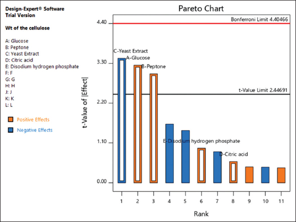 | Figure 2: Pareto chart for Plackett Burman method [Click here to view] |
In [Table 1] media components such as Glucose, Peptone, Yeast extract, Citric acid, and Disodium hydrogen phosphate are taken in different compositions and the trials were performed as per the Plackett Burman Design and the components were ranked based on the response [23,24], using Design-Expert Software. All components are in the concentration of g/100 mL [Table 1].
Table 1: Media optimization by Plackett Burman method.
| Run | A: Glucose(g/100 ml) | B: Peptone(g/100 ml) | C: Yeast extract(g/100 ml) | D: Citric acid(g/100 ml) | E: Disodium hydrogen phosphate(g/100 ml) | Weight of cellulose(g/100 ml) |
|---|---|---|---|---|---|---|
| 1 | 5 | 1 | 0.25 | 0.3 | 0.5 | 0.486 |
| 2 | 5 | 1 | 0.25 | 0.05 | 0.1 | 0.4272 |
| 3 | 5 | 0.25 | 0.25 | 0.05 | 0.5 | 0.2516 |
| 4 | 1 | 1 | 0.25 | 0.3 | 0.5 | 0.3824 |
| 5 | 1 | 0.25 | 0.25 | 0.3 | 0.1 | 0.0864 |
| 6 | 1 | 1 | 1 | 0.05 | 0.5 | 0.102 |
| 7 | 5 | 0.25 | 1 | 0.3 | 0.5 | 0.228 |
| 8 | 5 | 0.25 | 1 | 0.3 | 0.1 | 0.18 |
| 9 | 5 | 1 | 1 | 0.05 | 0.1 | 0.26 |
| 10 | 1 | 1 | 1 | 0.3 | 0.1 | 0.146 |
From the result [Table 1 and Figure 2], the yeast extract harms the production of cellulose. Thus, the concentration of yeast extract is reduced to optimize the production of cellulose in the subsequent steps. Optimization of media components was carried by Box Behnken design using Design-Expert software [25]. The parameters selected for media optimization were yeast extract, Glucose, and Peptone. The trials are given in [Table 2].
Table 2: RSM by Box-Benkn method.
| Run | A: Yeast extract(g/100 ml) | B: Glucose(g/100 ml) | C: Peptone(g/100 ml) | Weight of cellulose.(g/100 ml) |
|---|---|---|---|---|
| 1 | 0.375 | 7.5 | 1 | 0.4566 |
| 2 | 0.375 | 5 | 0.75 | 0.4184 |
| 3 | 0.375 | 7.5 | 1 | 0.5507 |
| 4 | 0.375 | 5 | 1.25 | 0.6838 |
| 5 | 0.375 | 7.5 | 1 | 0.4903 |
| 6 | 0.25 | 7.5 | 0.75 | 0.4457 |
| 7 | 0.5 | 5 | 0.75 | 0.6593 |
| 8 | 0.5 | 10 | 1 | 0.582 |
| 9 | 0.25 | 10 | 1 | 0.5738 |
| 10 | 0.5 | 5 | 1 | 0.5355 |
| 11 | 0.375 | 5 | 0.75 | 0.6927 |
| 12 | 0.5 | 7.5 | 1.25 | 0.5563 |
| 13 | 0.25 | 5 | 1 | 0.447 |
| 14 | 0.375 | 5 | 1.25 | 0.5737 |
| 15 | 0.25 | 5 | 1.25 | 0.6581 |
| 16 | 0.375 | 7.5 | 1 | 0.4572 |
| 17 | 0.375 | 7.5 | 1 | 0.4426 |
Optimized media concentration:
Glucose – 5 g
Yeast Extract – 0.25 g
Peptone – 1.24 g
Citric acid – 0.3 g
Disodium hydrogen phosphate – 0.5 g
Distilled water – 100 mL
In [Table 3] the Model F-value of 7.07 means the model is significant. Here, P < 0.0500; this indicates the model terms are significant. In this case, B, AC, and C² are significant model terms. When the values are >0.1000, then the model terms are not significant. When the lack of fit value is not significant then the model is good.
Table 3: ANOVA for quadratic model.
| Source | Sum of squares | Df | Mean square | F-value | P-value |
|---|---|---|---|---|---|
| Model | 0.1213 | 9 | 0.0135 | 7.07 | 0.0086 |
| A-Yeast extract | 0.0054 | 1 | 0.0054 | 2.85 | 0.1351 |
| B-Glucose | 0.0389 | 1 | 0.0389 | 20.40 | 0.0027 |
| C-Peptone | 0.0082 | 1 | 0.0082 | 4.29 | 0.0770 |
| AB | 0.0016 | 1 | 0.0016 | 0.8459 | 0.3883 |
| AC | 0.0249 | 1 | 0.0249 | 13.05 | 0.0086 |
| BC | 0.0067 | 1 | 0.0067 | 3.54 | 0.1021 |
| A² | 0.0019 | 1 | 0.0019 | 1.01 | 0.3480 |
| B² | 0.0048 | 1 | 0.0048 | 2.51 | 0.1572 |
| C² | 0.0263 | 1 | 0.0263 | 13.78 | 0.0075 |
| Residual | 0.0133 | 7 | 0.0019 | ||
| Lack of Fit | 0.0058 | 3 | 0.0019 | 1.02 | 0.4734 not significant |
| Pure Error | 0.0076 | 4 | 0.0019 | ||
| Cor Total | 0.1346 | 16 |
The predicted versus observed cellulose yields are plotted in [Figure 3] and a good linear correlation is observed. Residual analysis is performed to determine model adequacy. The residual is the difference between the observed and the predicted responses to determine model adequacy. [Figure 4] shows the normal probability plot of the residuals and indicates that the errors were distributed normally in a straight line. The cellulose production was high when yeast extract was used at 0.25 g and glucose at 5 g and it showed the 3D image of the cellulose produced and inferred the required concentration of glucose, yeast extract, and peptone for the production of cellulose [Figure 5]. It was interesting to observe that the cellulose production was recorded at 8 g/L. This result was approximately double when compared to the production of cellulose before optimization (4.8 g/L). 5.56 g/L of BC was produced from Gluconacetobacter persimmonis when fructose is used as a carbon source and when combinations of carbon sources such as galactose + lactose, galactose + maltose, galactose + fructose gave the cellulose yield of 6.89 g/L, 6.28 g/L, and 5.82 g/L, respectively. When galactose + sucrose is used as a carbon source, the yield was 7.67 g/L [26]. BC produced by Gluconacetobacter xylinium from HS medium with distiller’s grain enzymatic hydrolysate yielded 7.42 g/L [27]. But in our study, while using an optimized medium for cellulose production, we obtained the yield of 8 g/L which was greater than the reported research of Hungund. et al and He. et al. The carbon sources used for BC production affect the physical properties of BC such as water retention capacity, porosity, polymerization, and crystallinity index [28]. The yield of BC is greater when carbon sources such as fructose, glycerol, ethanol, lactic acid, and malic acid are used. However, it will alter the physical and chemical properties. This results in decreased crystallinity index and porosity. During BC production, bi-products like gluconic acids are formed, this decreases the yield of production. To overcome this, we formulated the medium, which increases the crystallinity index, and porosity [28]. Although our BC production is less, our BC can be more stable with good mechanical properties such as high crystallinity index, high porosity, and water retention capacity. The production was higher in our study this may be due to increasing the peptone, glucose concentration, and decreasing the concentration of yeast extract which harms the production of cellulose. Most of the commercial cellulose is prepared from plant lignocellulose. Vegetal cellulose contains lignin and hemicellulose apart from cellulose. To remove this, lignocellulose is subjected to chemical treatment. These chemical residues present in the cellulose are toxic to the embryonic cells. Because of this reason commercial cellulose was not used. Since microbial cellulose does not need any chemical treatment its cell toxicity is low and it is preferred for the growth of embryonic cells.
 | Figure 3: Cellulose production predicted vs. Actual cellulose production [Click here to view] |
 | Figure 4: Cellulose production Normal vs. Externally Studentized Residuals [Click here to view] |
 | Figure 5: Effect of Glucose vs. Yeast extract in the cellulose production [Click here to view] |
3.4. Estimation of Cellulose
Estimation of cellulose is done by the anthrone method. It is the colorimetric way of determining the cellulose content in the sample [18]. Cellulose undergoes acetolysis to form the glucose molecule on treatment with 67% sulfuric acid. The glucose molecule present in the cellulose gets dehydrated, which leads to the formation of the green-colored product and the absorbance were measured at 630 nm.
3.5. Confirmation Test for Cellulose
The saturated aqueous solution of potassium chlorate and nitric acid forms a complex with the glucose and produces an intense purple color. If the sample contains cellulose, it produces purple color otherwise it remains yellow [17]. From the observed results, the presence of purple color confirmed the presence of cellulose.
3.6. SEM
The surface pellicle formed by A. xylinum in the HS medium was observed by SEM. Fibril structure was observed directly rising from the cell surface. The microfibrils are uniformly observed. SEM analysis was carried out using a JEOL JSM-6390 microscope to investigate the diameter of cellulose. The sample is deposited on carbon tape before mounting on a sample holder for SEM analysis. The analysis shows the fiber morphology of the network. While, the plant cellulose has a microfibril bundle [5]. SEM image of BC was shown in [Figure 6]. White-colored crystal-like structure, porous, thin cellulose fibrils, and thin longer cellulose fibrils are shown in [Figure 6]. BC produced from acetic acid bacteria grown in glucose medium will have a fiber-like structure with high water retention capacity [29]. In our present study, we observed porous, thin fiber like cellulose fibrils, this structure will act as an environment for water retention. SEM image of electrospinned scaffold was shown in [Figure 7], here individual 3D cellulose thin nanofibrils with even pores act as the external microenvironment for cells to adhere and grow. In tissue engineering, the extracellular matrix (ECM) allows cell attachment and provides biochemical and biophysical cues to the nascent cells and tissues. In this study, the BC was taken as the ECM for the growth of embryonic cells. Cellulose, which is a biopolymer with tunable properties, could be a promising platform for biomaterial development and tissue engineering. In order for cells to sense and respond to their physical environment, they must first establish a physical connection. The hydrophilic hydroxyl moieties of the cellulose and specialized cellulose-binding domains allow cells to attach to cellulose. The surface area of the BC has been increased by converting it into a nanofiber. It enhanced the adhesion of embryonic cells in the ECM – BC [30]. This character makes BC a unique biopolymer.
 | Figure 6: SEM image of bacterial cellulose. [Click here to view] |
 | Figure 7: SEM image of electrospinned cellulose scaffold [Click here to view] |
3.7. FTIR
FTIR was used to find the different functional groups present in the cellulose sample and compare them with the property of already existing cellulose [10]. FTIR results are given in [Figure 8] which was confirmed with the [Figure 9] standard FTIR graph with functional group values [31].
 | Figure 8: FTIR results of bacterial cellulose [Click here to view] |
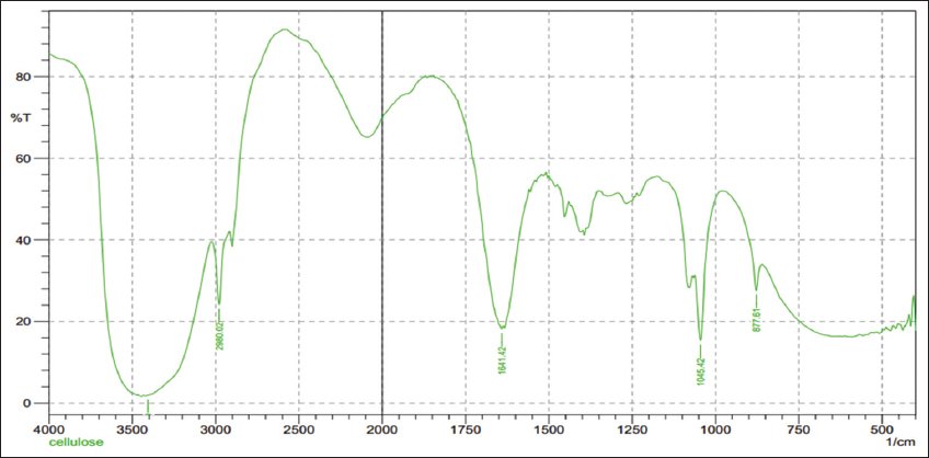 | Figure 9: FTIR results of standard cellulose. [Click here to view] |
For pure cellulose O-H stretch lies between 3100 and 3400 cm−1, C=O stretch lies between 1640 and 1800 cm−1, adsorbed water lies between 1620 and 1650 cm−1 [32]. In our present study, FTIR results of BC were shown as n(OH) of the hydroxyl groups around 3153.61cm−1 the amount of absorbed water bond around 1641.42 cm−1, and n(OH) at 1112.93 cm−1. Obtained results were similar to the standard spectroscopy. This confirmed the purity of the BC produced after the media optimization. The FTIR result of the BC has the same O-H stretching bond and absorbed water in it. Hence, it is confirmed that the compound obtained is cellulose.
3.8. Cytotoxicity Assay
The cytotoxicity assay was performed to identify the toxicity of the cellulose obtained from the A. xylinum for the production of scaffolds. It was found that cellulose produced from A. xylinum is less toxic and it is suitable for the production of scaffolds. MTT results are shown in [Figure 10]. At the lower concentration of cellulose 20 mg and 40 mg, the percentage of the viable cells is 97.6% and 96.78%, when the concentration of the BC increased to 100 mg, the percentage of viable cells is 73.56%. When L929 (mouse fibroblast cell line) cells were incubated with BC for 24 h the percentage of cell viability is noted as 70% and according to the ISO 10993 cell viability above 70% will be considered as non-cytotoxic [33]. In our present study, BC produced from A. xylinum showed 73.56% of cell viability at the highest concentration of 100 μg. Based on the ISO 10993 produced BC is non-cytotoxic. Hence, the cellulose produced from A. xylinum is less toxic so it can be used in tissue engineering to produce scaffolds.
 | Figure 10: MTT assay graph showing the percentage of viable cells. [Click here to view] |
3.9. Culturing of Fibroblast Cells on the BC
The embryonic fibroblast culture was done on the BC. The cell was seeded at the concentration of 1 × 10⁶ cells/mL. After incubating, the confluency of cells was observed and the multiplication of cells was found to be increased after 24 h. In [Figure 11] the growth of fibroblast cells on the BC was observed. Cellulose produced by A. xylinum in buffered HS medium and RSV-doped BC scaffold was prepared and Human adipose stem cells were incubated in the BC scaffold for 7 days, then the growth of human adipose stem cells was seen only in 7th day [34]. In our present study, we could see the growth of fibroblast cells on the BC scaffolds from 24 h, it was noted that multiplication of cells increased rapidly after 24 h. The cellulose produced by the A. xylinum cultured in the optimized media possessed a thin nanofiber-like structure with pores, this acts as the microenvironment for cells to adhere and multiply rapidly, this could be the reason for the rapid growth of fibroblast cells over the BC scaffolds. Thus, cellulose produced by the A. xylinum was confirmed to be biocompatible which can be further used for medical purposes.
 | Figure 11: Culturing of Fibroblast cells on the electrospinned cellulose [Click here to view] |
4. CONCLUSION
From the study, it was understood that media optimization played an important role in the production of cellulose from the bacteria A. xylinum. By optimizing the medium the production of cellulose reached 8 g/L. The characterization using SEM and FT-IR proved that the product obtained is cellulose. The cytotoxic study confirmed that cellulose is less toxic to the cells. Hence, we conclude that cellulose can be used as a biopolymer in the field of tissue engineering.
5. ACKNOWLEDGMENTS
We would like to thank the Department of Biotechnology, Karunya Institute of Technology and Sciences, Coimbatore for carrying out the work.
6. AUTHORS’ CONTRIBUTIONS
All authors listed have made a substantial, direct, and intellectual contribution to the work, and approved it for publication.
7. FUNDING
There is no funding to report.
8. CONFLICT OF INTEREST
The author declares that they have no conflicts of interest.
9. ETHICAL APPROVALS
This article does not contain any studies with human participants or animals performed by any of the authors.
10. DATA AVAILABILITY
All datasets generated or analyzed during this study are included in the manuscript.
11. PUBLISHER’S NOTE
This journal remains neutral with regard to jurisdictional claims in published institutional affiliation.
REFERENCES
1. Courtenay JC, Sharma RI, Scott JL. Recent advances in modified cellulose for tissue culture applications. Molecules 2018;23:654. [CrossRef]
2. Esa F, Tasirin SM, Rahman NA. Overview of bacterial cellulose production and application. Agric Agric Sci Procedia 2014;2:113-9. [CrossRef]
3. Rangaswamy BE, Vanitha KP, Hungund BS. Microbial cellulose production from Bacteria isolated from rotten fruit. Int J Polym Sci 2015;2015:1-8. [CrossRef]
4. Keshk SM. Bacterial cellulose production and its industrial applications. J Bioprocess Biotech 2014;4:1-10. [CrossRef]
5. Lestari P, Elfrida N, Suryani A, Suryadi Y. Study on the production of bacterial cellulose from Acetobacter xylinum using agro-waste. Jordan J Biol Sci 2014;7:75-80. [CrossRef]
6. Shah N, Ul-Islam M, Khattak WK, Park JK. Overview of bacterial cellulose composites?:A multipurpose advanced material. Carbohydr Polym 2013;98:1585-98. [CrossRef]
7. Castro C, Zuluaga R, Putaux JL, Caro G, Mondragon I, Gañán P. Structural characterization of bacterial cellulose produced by Gluconacetobacter swingsii sp from colombian agroindustrial wastes. Carbohydr Polym 2011;84:96-102. [CrossRef]
8. Tanskul S, Amornthatree K, Jaturonlak N. A new cellulose-producing bacterium, Rhodococcus sp. MI 2?:Screening and optimization of culture conditions. Carbohydr Polym 2013;92:421-8. [CrossRef]
9. Torgbo S, Sukyai P. Bacterial cellulose-based scaffold materials for bone tissue engineering. Appl Mater Today 2018;11:34-49. [CrossRef]
10. Gayathry G, Gopalaswamy G. Production and characterization of microbial cellulosic fiber from Acetobacter xylinum. Indian J Fibre Text Res 2014;39:93-6.
11. Schröpfer SB, Bottene MK, Bianchin L, Robinson LC, De Lima V, Jahno VD, et al. Biodegradation evaluation of bacterial cellulose, vegetable cellulose, and poly (3-hydroxybutyrate) in soil. Polimeros 2015;25:154-60. [CrossRef]
12. Petersen N, Gatenholm P. Bacterial cellulose-based materials and medical devices:Current state and perspectives. Appl Microbiol Biotechnol 2011;91:1277-86. [CrossRef]
13. Tang XZ, Kumar P, Alavi S, Sandeep KP. Recent advances in biopolymers and biopolymer-based nanocomposites for food packaging materials. Crit Rev Food Sci Nutr 2012;52:426-42. [CrossRef]
14. Brown RM, Willison JH, Richardson CL. Cellulose biosynthesis in Acetobacter xylinum:Visualization of the site of synthesis and direct measurement of the? in vivo process. Proc Natl Acad Sci U S A 1976;73:4565-9. [CrossRef]
15. Schramm M, Hestrin S. Factors affecting the production of cellulose at the air/liquid interface of a culture of Acetobacter xylinum. J Gen Microbiol 1954;11:123-9. [CrossRef]
16. Rani MU, Rastogi NK, Anu Appaiah KA. Statistical optimization of medium composition for bacterial cellulose production by Gluconacetobacter hansenii UAC09 using coffee cherry husk extract-an agro-industry waste. J Microbiol Biotechnol 2011;21:739-45. [CrossRef]
17. Paloheimo L, Herkola E, Kero ML. A method for cellulose determination. Agric Food Sci 1962;34:57-65. [CrossRef]
18. Updegraff DM. Semi micro determination of cellulose in biological nano paper synthesized materials. Anal Biochem 1969;32:420-4. [CrossRef]
19. Kim H, Yoon SC, Lee TY, Jeong D. Discriminative cytotoxicity assessment based on various cellular damages. Toxicol Lett 2009;184:13-7. [CrossRef]
20. Chen YM, Xi T, Zheng Y, Guo T, Hou J, Wan Y, et al. In vitro cytotoxicity of bacterial cellulose scaffolds used for tissue-engineered bone. J Bioact Compat Polym 2009;24:137-45. [CrossRef]
21. Lixin D, Jing A. The Investigation of cell culture conditions to maintain chicken embryonic stem cells as totipotent cells. Asian-Aust J Anim Sci 2003;16:1102-7. [CrossRef]
22. Pourramezan GZ, Roayaei AM, Qezelbash QR. Optimization of culture conditions for bacterial cellulose production by Acetobacter sp. 4B-2. Biotechnology 2009;8:150-4. [CrossRef]
23. Mandal S, Bhunia B, Kumar A, Dasgupta D, Mandal T, Datta S, Bhattacharya P. A statistical approach for optimization of media components for phenol degradation by Alcaligenes faecalis using plackett-burman and response surface methodology. Desalin Water Treat 2013;51:6058-69. [CrossRef]
24. Bhadri G, Hegde S, Narsapur K, Koppal S, Oswal P, Turmuri N, et al. Statistical optimization of medium components by response surface methodology for enhanced production of bacterial cellulose by Gluconacetobacter persimmonis. J Bioprocess Biotech 2013;04;1. [CrossRef]
25. Bae S, Shoda M. Statistical optimization of culture conditions for bacterial cellulose production using box-behnken design. Biotechnol Bioeng 2005;90:20-8. [CrossRef]
26. Hungund B. Production of bacterial cellulose from Gluconacetobacter persimmonis GH-2 using dual and cheaper carbon sources. J Microb Biochem Technol 2013;5:31-3. [CrossRef]
27. He F, Yang H, Zeng L, Hu H, Hu C. Production and characterization of bacterial cellulose obtained by Gluconacetobacter xylinus utilizing the by-products from Baijiu production. Bioprocess Biosyst Eng. 2020;43:927-36. [CrossRef]
28. Molina Ramirez C, Enciso C, Torres Taboeda M, Zuluaga R, Ganan P, Rojas OJ, et al. Effects of alternative energy sources on bacterial cellulose characteristics produced by Komagataeibacter medellinensis. Int J Biol Macromol 2018;117:735-41. [CrossRef]
29. Rozenberga L, Skute M, Belkova L, Sable I, Vikele L, Semjonovs P, et al. Characterisation of films and nanopaper obtained from cellulose synthesised by acetic acid Bacteria. Carbohydr Polym 2016;144:33-40. [CrossRef]
30. Hickey RJ, Pelling AE. Cellulose biomaterials for tissue engineering. Front Bioeng Biotechnol 2019;7:1-15. [CrossRef]
31. Moutta R, Silva RC, Reis Corrales RC, Cerullo MA, Ferreira-Leitao VS, Da Silva Bon EP. Comparative response and structural characterization of sugarcane bagasse, straw and bagasse-straw 1?:1 Mixture subjected to hydrothermal pretreatment and enzymatic conversion. J Microb Biochem 2013;5:S12-005.
32. Jia Y, Wang X, Huo M, Zhai X, Li F, Zhong C. Preparation and characterization of a novel bacterial cellulose/chitosan bio-hydrogel. Nanomater Nanotechnol 2017;7:1-8. [CrossRef]
33. Leal S, Cristelo C, Silvestre S, Fortunato E, Sousa A, Alves A, et al. Hydrophobic modification of bacterial cellulose using oxygen plasma treatment and chemical vapor deposition. Cellulose 2020;27:10733-46. [CrossRef]
34. Meng E, Chen CL, Liu CC, Liu CC, Chang SJ, Cherng JH, et al. Bioapplications of bacterial cellulose polymers conjugated with resveratrol for epithelial defect regeneration. Polymers (Basel) 2019;11:1048. [CrossRef]