
1. INTRODUCTION
Cordyceps militaris is a heterothallic entomopathogenic fungus that has attracted considerable attention due to its diverse medicinal properties, including antioxidant, anticancer, immunomodulatory, and neuroprotective activities. These biological effects are mainly attributed to a variety of bioactive compounds, such as cordycepin, adenosine, carotenoids, and polysaccharides [1–4]. In response to increasing market demand, commercial cultivation of C. militaris has expanded rapidly. However, large-scale production remains challenged by strain degeneration resulting from repeated subculturing, which leads to reduced yield and compromised quality of fruiting bodies [5–7].
Degeneration in C. militaris is commonly manifested by decreased mycelial biomass, delayed or failed fruiting body formation, and alterations in pigmentation and enzymatic activity [8,9]. Although environmental stresses can exacerbate degenerative processes, accumulating evidence indicates that genetic factors play a central role in degeneration, with alterations in the mating-type (MAT) locus being recognized as a major contributing genetic factor [5,10].
Cordyceps militaris exhibits a bipolar heterothallic mating system, in which successful sexual reproduction requires the coexistence of two distinct MAT idiomorphs: MAT1-1 and MAT1-2. The MAT1-1 locus contains the MAT1-1-1 and MAT1-1-2 genes, whereas the MAT1-2 locus harbors the MAT1-2-1 gene [11]. The presence of both MAT loci is essential for fruiting body initiation and perithecial development, and strains carrying only a single MAT idiomorph are generally incapable of forming fruiting bodies [12–14]. Consequently, disruptions or imbalances in MAT loci are considered a critical factor underlying reproductive failure and degeneration during prolonged cultivation of C. militaris.
Several studies have provided molecular evidence linking MAT locus alterations to degeneration. Yin et al. [15] reported that degenerated strains exhibited loss of the MAT1-2-1 region and mutations in MAT1-1-1 and MAT1-1-2, highlighting the involvement of MAT genes in degeneration-associated reproductive defects. More recently, the effects of the MAT locus imbalance on degeneration have been investigated. Vu et al. [14] demonstrated that when the proportion of MAT1-2 spores exceeded that of MAT1-1 spores by more than 15-fold, fruiting body formation was markedly suppressed, whereas an increased proportion of MAT1-1 spores resulted in only a slight decline in reproductive capacity. Similarly, Wang et al. [10] reported that a MAT1-1:MAT1-2 ratio of 9:1 led to a severe reduction in fruiting body dry weight. These findings suggest that not only the presence but also the relative band-intensity profile of MAT loci is critical for normal fruiting body development. Nevertheless, the precise mechanisms by which MAT locus imbalance contributes to degeneration remain unclear, and the relative contributions of individual MAT loci to reproductive competence have yet to be fully elucidated.
Importantly, previous studies have primarily relied on artificial spore mixing at defined ratios to investigate the effects of MAT locus imbalance [10,14]. While this approach provides valuable insights, it does not fully reflect the dynamic genetic changes that may occur during repeated subculturing. To date, no studies have systematically monitored changes in MAT locus abundance across successive cultivation generations to elucidate how such alterations accumulate and contribute to degeneration in C. militaris.
In the present study, we investigated the relationship between MAT locus imbalance and degeneration in C. militaris across seven consecutive subculture generations. Phenotypic traits related to mycelial growth and fruiting body development were evaluated, and the relative PCR band intensity of MAT1-1 and MAT1-2 loci was analyzed using multiplex PCR. This study aims to investigate the relationship between progressive changes in MAT loci and degeneration during prolonged cultivation.
2. MATERIALS AND METHODS
2.1. Strain and Culture Conditions
A C. militaris strain designated L40 was obtained from the Cell Biology Laboratory, University of Science Education–University of Danang, Vietnam. The strain was maintained on potato dextrose agar (PDA) and propagated in potato dextrose (PD) liquid medium containing 200 g/l potato extract, 20 g/l glucose, 5 g/l peptone, 1 g/l K2HPO4, and 1 g/l MgSO4, as previously described [16]. Liquid cultures were incubated in the dark at 23°C ± 2°C, with a relative humidity of 62%–65%, under constant agitation at 180–200 rpm.
2.2. Successive Subculturing and Fruiting Body Induction
Seven consecutive subculture generations (G1–G7) were established by transferring actively growing bulk mycelium from liquid cultures into fresh PD medium at each subculturing cycle. Subculturing was performed using homogenized mycelial inoculum to maintain the original population structure of the strain. For each generation, three independent subculture lines were maintained and processed in parallel to ensure biological replication.
Fruiting body induction was performed using a modified cultivation method based on previously reported protocols [16]. Briefly, polypropylene culture boxes were filled with 50 g of brown rice supplemented with 50 ml of nutrient solution containing 60 g/l silkworm pupae powder, 30 g/l soybean powder, 10 g/l peptone, 10 g/l glucose, 1 g/l K2HPO4, and 1 g/l MgSO4. For each generation, three cultivation jars per subculture line (total n = 9) were prepared, and each jar was treated as an independent biological replicate in the fruiting body experiment.
Following complete substrate colonization under dark conditions at 25°C, cultures were exposed to light for primordium induction at an intensity of 1,800 lux. Fruiting body development and maturation were subsequently conducted under a reduced light intensity of 800 lux, following established cultivation conditions [17].
All subcultures were maintained under identical environmental and nutritional conditions throughout the experiment to minimize external variation. As no single-spore isolation or genetic stabilization step was applied, the observed generational changes primarily reflect the combined effects of physiological aging and potential genetic drift during serial subculturing.
2.3. Phenotypic Characterization
Mycelial growth and morphological characteristics were evaluated in both liquid culture and PDA plates across all subculture generations. Phenotypic parameters included mycelial pellet size, fresh and dry mycelial biomass, radial mycelial growth rate on PDA, time to primordium emergence, fruiting body height and diameter, and total fruiting body yield (g per jar) [18]. All measurements were performed using three independent biological replicates unless otherwise stated.
2.4. DNA Extraction and Multiplex PCR Amplification
Genomic DNA was extracted from mycelia harvested from liquid cultures using a modified cetyltrimethylammonium bromide method [19]. DNA concentration and purity were assessed using a NanoDrop spectrophotometer, and all samples were adjusted to a final concentration of 100 ng/µl.
Multiplex PCR was conducted to simultaneously amplify the MAT genes MAT1-1-1, MAT1-1-2, and MAT1-2-1 using primer sets previously reported by Nguyen et al. [20]. Each 20 µl PCR reaction mixture contained 1× Biosharp Master Mix, 1 pmol of each forward and reverse primer, and 100 ng of genomic DNA. Thermal cycling conditions were as follows: initial denaturation at 95°C for 60 seconds; 30 cycles of denaturation at 95°C for 30 seconds, annealing at 58°C for 30 seconds, and extension at 72°C for 30 seconds, followed by a final extension at 72°C for 5 minutes.
2.5. Agarose Gel Electrophoresis and Quantification of PCR Products
PCR products were separated by electrophoresis on 1% agarose gels prepared in 1× TAE buffer at 100 V for 30 minutes. DNA bands were stained with RedSafe and visualized under ultraviolet illumination. Band intensities were quantified using GelAnalyzer software version 23.1.1 (http://www.gelanalyzer.com) [21]. The relative PCR band intensity of each MAT locus across generations was normalized to the corresponding MAT locus band intensity in the first generation (G1).
2.6. DNA Sequencing and Sequence Alignment
Representative PCR amplicons of the MAT1-1-1, MAT1-1-2, and MAT1-2-1 loci from generations G1 and G4 were excised from agarose gels and purified using a commercial PCR purification kit according to the manufacturer’s instructions. For generation G7, only the MAT1-2-1 amplicon was detected, excised, and purified for sequencing. Purified products were submitted for Sanger sequencing (Macrogen Inc., Seoul, South Korea). Sequencing reactions were performed using the corresponding forward primers employed in the multiplex PCR amplification.
Raw chromatogram files were visually inspected to ensure sequencing quality and trimmed to remove low-quality regions at both ends. The resulting sequences were then compared with reference MAT locus sequences available in the NCBI GenBank database using the BLASTn algorithm. Multiple sequence alignment was performed using MEGA version 11 with the ClustalW algorithm to detect potential nucleotide substitutions, insertions, or deletions among generations [22].
The resulting sequence alignment files have been deposited in the Figshare repository (https://figshare.com/).
2.7. Statistical Analysis
All experiments were performed using a completely randomized design with three independent biological replicates. Differences between the two groups were analyzed using an unpaired two-tailed Student’s t-test. For comparisons among multiple groups, one-way ANOVA followed by Tukey’s multiple comparisons test was used. Statistical significance was defined as p < 0.05.
Statistical analyses were conducted using GraphPad Prism version 6 (GraphPad Software, USA). Data are presented as mean ± standard error (SE) in tables and mean ± SD in figures. For experiments including technical replicates, values represent the mean of three independent experiments with three technical replicates each (n = 9).
3. RESULTS AND DISCUSSION
3.1. Degeneration of Mycelial Growth and Biomass During Successive Subculturing of C. militaris
Successive subculturing of C. militaris over seven generations resulted in progressive alterations in mycelial growth characteristics and biomass production (Fig. 1, Table 1). Radial mycelial growth on PDA plates remained statistically unchanged among the first three generations (G1–G3), indicating stable vegetative growth during early subcultures (Fig. 1a). In contrast, a marked reduction in growth rate was observed from G4 onward, and G4–G7 exhibited approximately a two-fold decrease compared with G1–G3.
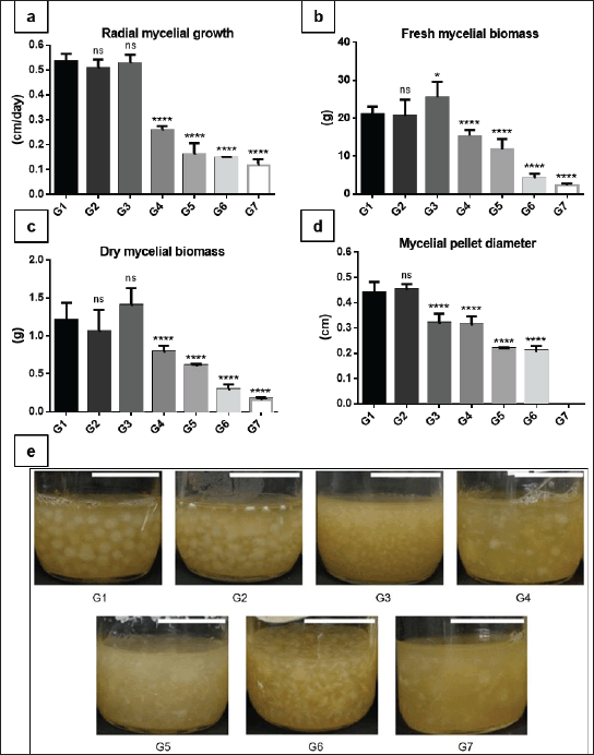 | Figure 1. Radial mycelial growth on agar (a), fresh mycelial biomass (b), dry mycelial biomass (c), mycelial pellet size in liquid culture (d), and morphological traits and biomass yield (e) were evaluated across seven subculture generations derived from a single parental strain. Scale bar = 2 cm. Data are presented as mean ± SD. Statistical significance was indicated as ns (no significant), *p < 0.05; **p < 0.01; ***p < 0.001; ****p < 0.0001. [Click here to view] |
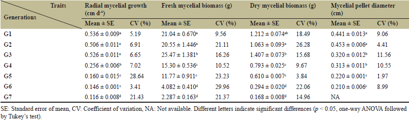 | Table 1. Growth characteristics of C. militaris mycelia across successive subculture generations. [Click here to view] |
A similar generational pattern was observed for mycelial biomass accumulation in liquid culture. Both fresh and dry biomass showed no significant differences among G1–G3 but declined progressively from G4 to G7 (Fig. 1b and c). These trends were summarized in Table 1 and collectively indicated a loss of vegetative vigor after repeated subculturing.
Changes in mycelial pellet morphology were detected earlier than biomass-related parameters. Pellet diameter did not differ significantly between G1 and G2, but a gradual decrease became evident from G3 onward (Fig. 1d). Representative images revealed that pellets from later generations lost compactness and spherical integrity (Fig. 1e). In the final generation (G7), severe degeneration resulted in highly dispersed and irregular mycelial aggregates, preventing reliable measurement of pellet diameter.
Macroscopic observations of colony morphology and biomass yield further supported the quantitative data (Fig. 1e). Collectively, these results indicated that while early subcultures of C. militaris maintained relatively stable growth characteristics, repeated subculturing progressively reduced vegetative growth performance, with clear degeneration becoming apparent after the fourth generation.
3.2. Progressive Reproductive Degeneration During Serial Subculturing
Degenerative effects observed during vegetative growth also extended to the reproductive stage of C. militaris, affecting developmental timing, fruiting body morphology, and yield across successive subculture generations (Fig. 2; Table 2).
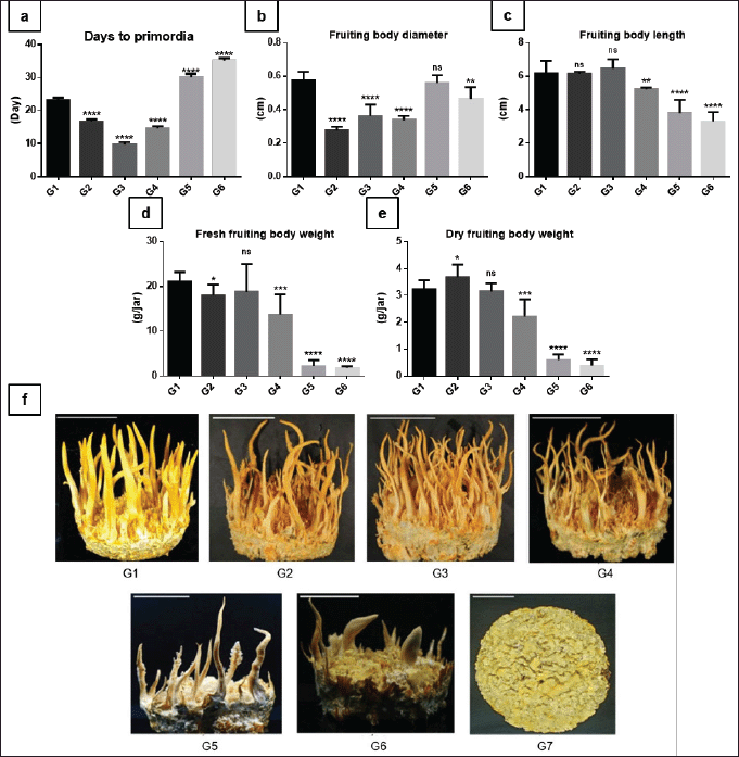 | Figure 2. Time to primordia formation (a), fruiting body diameter (b), fruiting body length (c), fresh fruiting body weight (d), and dry fruiting body weight (e) were evaluated across six consecutive subculture generations. (f) Representative fruiting bodies of C. militaris across seven subculture generations. Data are presented as mean ± SD. Statistical significance was indicated as ns (no significant), *p < 0.05; **p < 0.01; ***p < 0.001; ****p < 0.0001. [Click here to view] |
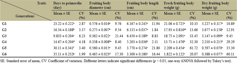 | Table 2. Developmental and yield-related traits of C. militaris fruiting bodies across generations. [Click here to view] |
The number of days required for primordium formation exhibited a biphasic pattern across generations (Fig. 2a). From G1 to G3, the time required for primordium emergence decreased markedly, indicating accelerated reproductive initiation during early generations. However, from G4 onward, primordium formation became progressively delayed, and later generations required substantially longer cultivation periods to initiate reproductive development.
Fruiting body size parameters displayed generation-dependent but non-uniform trends. Fruiting body diameter decreased from G2 to G4 but showed no significant difference between G5–G6 and G1 (Fig. 2b). In contrast, fruiting body length declined consistently from G4 to G6 (Fig. 2c), indicating impaired stromatal elongation in later generations.
Fruiting body productivity was more strongly affected than morphological size parameters. Fresh and dry fruiting body weights remained relatively stable during early generations but decreased sharply in G5 and G6 (Fig. 2d and e), indicating a substantial reduction in reproductive yield. Macroscopic observations further supported these quantitative findings (Fig. 2f). Fruiting bodies from G1 to G3 appeared well-formed, upright, and pigmented, whereas morphological abnormalities, including shortened, curved, or poorly differentiated stromata, became increasingly evident from G4 onward. In the final generations, fruiting bodies were sparse or failed to develop entirely. The high coefficient of variation (CV) for both fresh and dry fruiting body weights in later generations resulted from severe fruiting body degradation. This stochastic degradation led to substantial discrepancies among the recorded data in subsequent replicates.
In G7, severe degeneration resulted in highly dispersed and irregular mycelial aggregates, indicating a substantial loss of mycelial integrity. Consequently, no fruiting body development was observed, and reproductive traits could not be evaluated for this generation (Fig. 2f). These observations further support the notion that advanced degeneration not only affects vegetative growth but ultimately disrupts the developmental competence necessary for sexual reproduction in C. militaris.
3.3. Progressive Alteration of MAT Loci During Serial Subculturing of C. militaris
Multiplex PCR analysis revealed clear alterations in the distribution of MAT loci across seven successive subculture generations (G1–G7) of C. militaris (Fig. 3a). The MAT1-2-1 locus was consistently detected in all generations. In contrast, MAT1-1-1 and MAT1-1-2 were strongly amplified only in early generations.
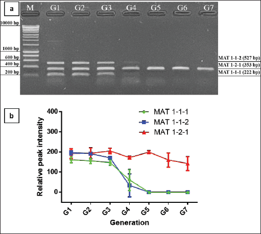 | Figure 3. Alterations in MAT loci of C. militaris during serial subculturing. Representative multiplex PCR profiles (a) and densitometric quantification of MAT1-1-1, MAT1-1-2, and MAT1-2-1 across generations G1–G7 (b). [Click here to view] |
Distinct bands corresponding to all three loci were observed from G1 to G3, indicating the presence of both MAT idiomorphs during early subcultures. In G4, amplification signals for MAT1-1-1 and MAT1-1-2 became markedly weaker, whereas MAT1-2-1 remained clearly detectable. From G5 to G7, the MAT1-1-1 and MAT1-1-2 bands were no longer detected. Densitometric analysis of band intensities supported these observations (Fig. 3b). Relative signal intensities showed a progressive decline of MAT1-1-1 and MAT1-1-2 from G1 to G4 and became PCR-undetectable from G5 onward. In contrast, MAT1-2-1 bands remained consistently detectable across generations. It should be noted that densitometric analysis was performed using image-based band intensity measurement and was used only to support the electrophoretic observations; therefore, the results should be interpreted as indicative of relative band presence rather than absolute quantification of MAT locus abundance.
Sequencing of partial MAT amplicons from G1, G4, and G7 revealed no nucleotide substitutions or structural variations in MAT1-1-1, MAT1-1-2, or MAT1-2-1. The sequences obtained from the analyzed samples were identical, with no detectable variation across the examined MAT loci. The corresponding multiple sequence alignment files have been deposited in Figshare and are publicly available at https://doi.org/10.6084/m9.figshare.31742305.
4. DISCUSSION
Sexual reproduction in C. militaris was controlled by MAT loci that regulated meiotic development and sexual differentiation [23]. As a heterothallic fungus, normal sexual reproduction typically requires the interaction between two compatible MATs: MAT1-1 and MAT1-2 [13]. These MAT loci have been considered functionally analogous to sex chromosomes in C. militaris [24]. Each ascospore contained a single haploid nucleus carrying either the MAT1-1 idiomorph (MAT1-1-1 and MAT1-1-2) or the MAT1-2 idiomorph (MAT1-2-1) [25]. Therefore, the balanced coexistence of both MATs within the mycelial population was generally required to complete the sexual cycle and produce normal fruiting bodies with perithecia.
Previous studies demonstrated that strains containing both MAT1-1 and MAT1-2 loci were capable of forming fertile fruiting bodies with perithecia, whereas strains carrying only a single MAT locus often failed to develop normal reproductive structures [12]. However, the precise roles of individual MAT loci in fruiting body development have remained somewhat controversial. Cordyceps militaris was the first ascomycete species in which a strain containing only MAT1-1 could still produce fruiting bodies, although these structures lacked perithecia. Similarly, single-ascospore isolates carrying either MAT1-1 or MAT1-2 were able to form fruiting bodies individually but failed to produce perithecia. When spores with opposite MATs were mixed, the highest frequency of perithecium formation occurred at a MAT1-1:MAT1-2 ratio of 1:9, whereas the opposite ratio (9:1) resulted in much lower reproductive success [26]. Because these results were not entirely consistent, the specific functional contributions of individual MAT loci to fruiting body formation remained unresolved.
Subsequent studies suggested that both MAT1-1 and MAT1-2 loci contributed to fruiting body development [27]. More recent molecular analyses further clarified that MAT1-1-1 and MAT1-2-1 were essential for fruiting body formation, whereas MAT1-1-2 played an important role in ascospore production. Together, MAT1-1-1, MAT1-1-2, and MAT1-2-1 were required for the completion of a full sexual cycle during outcrossing [24]. These findings highlighted the importance of maintaining a balanced MAT locus composition within the mycelial population to sustain reproductive competence.
Interestingly, several studies indicated that MAT1-1 often appeared to be dominant or more prevalent in natural populations of C. militaris. For example, analysis of ascospore progeny from a hybrid strain revealed that 28 out of 30 randomly isolated spores carried the MAT1-1 locus, whereas only two spores contained MAT1-2 [26]. Similarly, a recent survey of Vietnamese C. militaris strains showed that seven out of eight isolates contained only MAT1-1, while only one strain possessed both MAT1-1 and MAT1-2 loci [28]. These observations suggested that MAT1-1 might play a particularly important role in fruiting body development and may be preferentially maintained during strain propagation.
In addition to the presence of MAT loci, their relative proportions also appeared to influence reproductive performance. Several studies reported that a near-balanced MAT1-1:MAT1-2 ratio was favorable for fruiting body formation. An approximately 1:1 ratio of MAT1-1 to MAT1-2 was optimal for C. militaris cultivation [29]. Similar ratios were observed in other studies analyzing ascospore populations [30]. More recently, the proportion of MAT1-1 to MAT1-2 asexual spores in normal fruiting strains was reported to be approximately 1:2. Importantly, deviations from this balance could significantly affect reproductive capacity [31]. Another study demonstrated that when MAT1-2 spores exceeded MAT1-1 spores by more than fifteen-fold, fruiting body formation was strongly suppressed. In contrast, increases in MAT1-1 spores caused only a modest reduction in fruiting efficiency [14]. These findings suggested that excessive dominance of MAT1-2 could be particularly detrimental to fruiting body formation.
The results of our study were consistent with these previous observations. During successive subculturing, the PCR-based band-intensity distribution of MAT1-1 loci gradually declined and eventually could not be detected in later generations (Fig. 3). This imbalance coincided with the onset of severe degenerative phenotypes, including reduced mycelial growth, delayed primordium formation, abnormal stromatal morphology, and decreased fruiting body yield (Figs. 1 and 2). These findings suggested MAT1-1 loci became undetectable under the conventional multiplex PCR conditions used, contributing to degeneration in C. militaris during prolonged subculturing.
Several mechanisms may explain the observed alterations in the MAT locus composition. One possible explanation involves spontaneous genetic mutations or genomic rearrangements arising during repeated vegetative propagation. Fungal mycelia possess relatively short cell cycles and considerable genetic plasticity, which may lead to elevated mutation rates during prolonged cultivation [23]. Consistent with this hypothesis, deletion mutations affecting MAT loci have been reported in degenerated C. militaris strains [15]. However, no mutations were detected in the MAT sequences in the present study, suggesting that the observed changes are unlikely to result from sequence-level alterations of MAT genes.
Another potential mechanism involves changes in nuclear composition during heterokaryotic growth. Degeneration may occur when heterokaryotic mycelia containing nuclei of both MATs gradually shift toward homokaryotic populations carrying only a single MAT nucleus. Such transitions may arise through clonal selection, nuclear segregation, or altered nuclear competition during vegetative propagation [27]. Because fungal mycelia exhibit dynamic nuclear organization and high genetic plasticity, prolonged subculturing may facilitate shifts in the relative abundance of MAT1-1 and MAT1-2 nuclei. The dominance of homokaryotic nuclei could ultimately disrupt the balanced interaction between MATs required for normal sexual development and fruiting body formation.
In addition, instability of MAT signaling pathways may contribute to these changes. Mutations affecting pheromone signaling or mating recognition systems may allow hyphae carrying identical MAT loci to fuse and undergo same-sex mating, a phenomenon known as homothallic or unisexual reproduction [32,33]. Similar transitions between heterothallism and homothallism have been proposed in several fungal lineages, where gene loss or MAT switching may occur under selective pressures [34]. Such processes could contribute to the gradual destabilization of MAT composition during prolonged artificial cultivation.
Degeneration of C. militaris has previously been associated with multiple physiological changes, including reduced mycelial growth, decreased pigmentation, diminished fruiting body formation, abnormal stromatal morphology, reduced spore production, and altered secondary metabolite production [5]. Changes in pellet morphology may provide additional insight into the early stages of degeneration during serial subculturing. In filamentous fungi, pellet formation in submerged culture is largely determined by hyphal branching frequency, extension rate, and inter-hyphal adhesion [35]. A reduction in pellet diameter may therefore reflect alterations in hyphal architecture or aggregation behavior that occur prior to measurable declines in biomass production. Pellet cohesion is also strongly influenced by extracellular polysaccharides that mediate hyphal adhesion and structural integrity of fungal aggregates. Changes in extracellular matrix production could therefore contribute to the reduced pellet size observed in later generations.
Furthermore, morphological alterations in fungal pellets have been reported to accompany metabolic or physiological shifts during prolonged cultivation. Such structural changes may precede detectable reductions in growth or productivity, suggesting that pellet morphology could serve as an early indicator of physiological stress or degeneration [36]. In the present study, the decrease in pellet diameter from generation G3 onward occurred before significant reductions in biomass and growth rate, supporting the hypothesis that morphological changes in submerged culture may represent an early manifestation of degeneration in C. militaris during serial subculturing.
The accelerated primordia formation observed in the early generations may not solely reflect a mild stress response. It may also indicate an initial physiological adaptation of the mycelium to the cultivation conditions, resulting in temporarily enhanced developmental activity. In filamentous fungi, early subcultures can favor the selection of fast-growing or highly responsive mycelial sectors, which may transiently accelerate developmental processes. However, this initial vigor is frequently followed by progressive degeneration during prolonged vegetative propagation. Similar patterns of early stimulation followed by functional decline have been reported in several fungal cultivation systems, suggesting that the early acceleration of primordia formation may represent a transient adaptive phase preceding degeneration [5,23,35].
Taken together, these results suggest that degeneration in C. militaris is associated with complex interactions among MAT locus instability, physiological stress, and metabolic reprogramming during repeated subculturing. The progressive imbalance of MAT loci, particularly the depletion of MAT1-1 nuclei, may disrupt the heterokaryotic equilibrium required for normal sexual development and thereby contribute to the loss of reproductive capacity observed in later generations. However, the heterokaryotic status of the strain was inferred from the simultaneous detection of both MAT loci in early generations rather than directly demonstrated at the nuclear level. Further studies using single-spore isolation or nuclear-level analyses will be required to confirm the heterokaryotic composition of the strain.
These findings may also have practical implications for the commercial cultivation of C. militaris. Monitoring early morphological indicators, such as pellet size and developmental timing, together with periodic assessment of MAT locus distribution, may provide a useful strategy for the early detection of degeneration during serial subculturing. Such monitoring could facilitate timely strain rejuvenation or replacement, thereby helping to maintain stable productivity in large-scale cultivation systems.
5. CONCLUSION
Serial subculturing of C. militaris was accompanied by progressive degeneration, manifested by delayed primordia formation, reduced fruiting body development, and decreased biomass. These phenotypic changes were associated with alterations in MAT locus composition. Molecular analyses revealed a gradual decline of the MAT1-1 idiomorph after the fourth generation, while MAT1-2-1 remained detectable throughout the cultivation period, and no sequence variations were detected in the analyzed MAT loci. These observations suggest that degeneration during prolonged subculturing may be associated with a ratio imbalance of MAT loci rather than detectable mutations in these regions. Fruiting body formation was mainly observed in early generations when both MAT loci remained detectable, highlighting the potential importance of MAT equilibrium for sustaining reproductive capacity. Collectively, these findings suggest that the MAT locus imbalance may contribute to degeneration during prolonged cultivation. Monitoring MAT locus distribution may therefore provide a useful approach for the early detection and management of strain deterioration in C. militaris cultivation systems.
6. AUTHORS’ CONTRIBUTIONS
All authors made substantial contributions to conception and design, acquisition of data, or analysis and interpretation of data; took part in drafting the article or revising it critically for important intellectual content; agreed to submit to the current journal; gave final approval of the version to be published; and agree to be accountable for all aspects of the work. All the authors are eligible to be authors as per the International Committee of Medical Journal Editors' requirements/guidelines.
7. FUNDING
This work did not receive any external funding.
8. CONFLICTS OF INTEREST
The authors report no financial or any other conflicts of interest in this work.
9. ETHICAL APPROVALS
This study does not involve experiments on animals or human subjects.
10. DATA AVAILABILITY
All the data are available and shall be provided upon request.
11. USE OF AI TOOLS
AI tools were used solely for language editing purposes, including improving grammar, readability, and clarity of the manuscript. No AI-generated data or unverified AI-generated content was included in this study.
12. PUBLISHER’S NOTE
All claims expressed in this article are solely those of the authors and do not necessarily represent those of the publisher, the editors, and the reviewers. This journal remains neutral with regard to jurisdictional claims in published institutional affiliation.