
1. INTRODUCTION
The plant-insect interaction is one of the most fundamental and extensively studied biotic interactions in terrestrial ecosystems, which significantly influences the patterns of biodiversity, community structure, and ecosystem stability [1]. Interestingly, plants are the primary producers, supplying everything from food to shelter and even oviposition sites for most of the arthropods. Insects are key drivers of plant reproduction through pollination regimes, in turn regulating the population and evolutionary changes [2]. These mutual interactions are the result of long-term coevolutionary processes that have produced remarkable diversity in insect feeding strategies and highly specialized plant defense systems [3]. Both plants and insects have undergone active evolution in adaptive associations over evolutionary time [4]. Insect infestation can also significantly change plant physiology, leading to changes in metabolic pathways, affecting lipid metabolism, and adding another dimension to primary metabolic reprogramming. Certain fatty acids and lipid-derived metabolites extend their functions beyond signaling by directly impacting insect feeding and growth, diminishing tissue palatability or nutritional quality [5,6]. These primary metabolic alterations frequently trigger and influence the activation of secondary defense mechanisms. After insect infestation, plants undergo extensive changes in primary metabolism, in which fatty acids become key regulators of both defense and signaling [7]. Insect infestation induces rapid remodeling of membrane lipids and fatty acid pools to maintain cellular integrity, allocate energy, and adapt to stress [8–11]. Polyunsaturated fatty acids (PUFA), primarily linoleic (18:2Δ9,12) and α-linolenic acid (18:3Δ9,12,15), serve as vital precursors for oxylipin biosynthesis and are catalytically converted in the lipoxygenase pathway [12]. α-Linolenic acid is the direct precursor for jasmonic acid biosynthesis; linoleic acid may contribute indirectly after desaturation [13,14]. Previous studies have reported significant changes in plant fatty acid composition in response to whitefly and leafminer infestation [15]. This study was conducted to assess how infestation by the Indian lac insect (Kerria lacca) alters the fatty acid profile of its primary host plant, Flemingia semialata. It is a perennial, shrub-like legume belonging to the Fabaceae family (subfamily Faboideae) and is found throughout tropical and subtropical areas of India and Southeast Asia, and is also one of the most significant host plants for the lac insect [16]. The lac insect, K. lacca, is an economically important resin-producing scale insect from the order Hemiptera, superfamily Coccoidea, and family Kerriidae. It feeds on plant phloem sap and infests more than 400 plant species [17]. Interestingly, lac resin is primarily composed of polyesterified hydroxy fatty acids and sesquiterpenic acids. Thus, the fatty acid fraction can play a structural and functional role in resin formation, as the insect, being sedentary/immobile, survives on the plant phloem as its sole source of nutrition, and can also play a major role in defense mechanisms. As lac resin is rich in polyesterified hydroxy fatty acids and the insect relies exclusively on host phloem sap [18], host fatty acid remodeling may simultaneously reflect defense activation and supply essential precursors for resin biosynthesis, thereby shaping the plant–insect interaction.
2. MATERIAL AND METHODS
2.1. Sample Collection
The Kusumi strain of K. lacca (Kerr) was used to inoculate the lac host plant F. semialata Roxb. ex W.T. Aiton for the present investigation. In natural field conditions, approximately 80 g of broodlac was inoculated on each plant to ensure uniform infestation. All host plants selected for the study were of uniform age and maintained under identical field conditions, receiving the same nutritional and agronomic management to minimize environmental variability. A non-infested F. semialata plant maintained under the same conditions served as the control. Following infestation, bark samples were collected at four time intervals: 1 (control- non-infested), 7, 14, and 21 days post-infestation with three biological replicates at each sampling point. Bark tissues were excised from young, green, and tender shoots, immediately frozen in liquid nitrogen, and stored for subsequent biochemical and Gas Chromatography-Mass Spectrometry (GC-MS) analysis.
2.2. Sample Preparation
The stored samples were lyophilized to enhance stability for subsequent analytical procedures. Lyophilized powdered materials (200 mg) were thoroughly mixed with 600 µl of cold methanol (GC-MS grade) using a vortex. The mixture was chilled on ice for 10 minutes, then vortexed again. The material was then centrifuged at 15,000 g for 10 minutes. The supernatant was transferred to a new Micro Centrifuge Tube, and the extraction process was repeated. The same tube was used to combine the obtained supernatant.
2.3. Derivatization Process
To the dried oil extract, 300 µl of acidified methanol was added. The mixture was stirred for 2 minutes using a vortex and heated at 70°C for 1 hour. After that, the sample was cooled down to room temperature for 10 minutes. Then, 300 µl of NaCl solution was added, followed by 300 µl of hexane. The mixture was agitated in a vortex for 1 minute to ensure complete mixing. Finally, the material was centrifuged at 3,000 g for three minutes.
2.4. GC-MS Analysis
Following centrifugation, 100 µl of the hexane layer was carefully transferred into a GC-MS vial. After that, the vial was sealed and kept cold until it was injected into the GC-MS apparatus. The prepared plant sample was analyzed using GC-MS to identify the compounds. This procedure followed the methodology described in [19], with minor adjustments. The GC-MS analysis was conducted using a Shimadzu GC-MS-QP2010 system (Shimadzu, Kyoto, Japan) with an auto-injector (AOC-20i), a headspace sampler (AOC-20s), and a silica capillary column (Rtx-5). The oven temperature was originally set at 50°C and gradually increased to 280°C. 1 microliter of the plant extract at 1 mg/ml was loaded onto the column in crude form. The carrier gas was helium (99.99% purity), circulating at 1.2 ml/min. The mass spectrometer detected unique chromatographic peaks, and compound identification was accomplished using retention time (RT) and mass spectra collected under standardized GC-MS conditions. Compounds were quantified using peak area. The peak spectral data were compared with mass spectral libraries, specifically NIST14 and WILEY8, to identify chemicals. The relative fraction of each ingredient was calculated using peak area normalization.
2.5 Statistical Analysis
For the current study, the experiment was carried out with biological triplicates (N = 3). The final value was determined as the average and SE derived from these triplicates. The IBM SPSS version 21.0 software was used for one-way ANOVA with Duncan post hoc analysis at a 95% confidence level (p < 0.05) to compare temporal intervals. The data were graphically presented in Microsoft Word.
3. RESULT AND DISCUSSION
The present study revealed that infestation by the lac insect, K. lacca, resulted in significant temporal changes in the fatty acid profile of F. semialata. These changes in fatty acids align with typical herbivore-induced lipid signalling responses in plants and may be associated with the activation of the classical herbivore-mediated lipid pathway [20]. The GC-MS analysis of the infested and non-infested young bark of the tender shoots of F. semialata identified thirteen fatty acids, comprising saturated (lauric 12:0, myristic 14:0, palmitic 16:0, stearic 18:0, nonadecanoic 19:0, heneicosanoic 21:0, docosanoic 22:0, tricosanoic 23:0, pentacosanoic 25:0, hexacosanoic 26:0), monounsaturated (oleic 18:1Δ9), and PUFAs (linoleic 18:2Δ9,12 and α-linolenic 18:3Δ9,12,15) (Table 1; Supplementary Fig. 1). A distinct temporal shift in fatty acid profiles was evident when comparing non-infested plant samples (day 1) with lac-infested plants after 7th, 14th, and 21st day of the infestation. Most saturated fatty acids (SFAs) showed declining or fluctuating patterns during infestation. Palmitic acid (16:0), the dominant SFA, decreased steadily from 22.39% in non-infested bark to 21.25%, 20.95%, and 19.65% on Day 7, Day 14, and Day 21, respectively. Stearic acid (18:0) followed a similar pattern, declining from 10.71% to 10.37%, 9.60%, and 8.21% across the same period (Fig. 1A and Table 1). Lauric acid (12:0) showed minor fluctuations (0.16–0.24%), while nonadecanoic acid (19:0), heneicosanoic acid (21:0), docosanoic acid (22:0), tricosanoic acid (23:0), and pentacosanoic acid (25:0) exhibited slight reductions or remained relatively stable. Hexacosanoic acid (26:0) decreased sharply at Day 7 (0.02% compared to 0.32% in controls) but subsequently recovered to 0.35% and 0.46% by Days 14 and 21. In contrast to these general declines, myristic acid (14:0) showed a pronounced increase from 0.61% in non-infested bark to 2.73%, 3.17%, and 2.72% during infestation (Fig. 1A and Table 1). The decrease of these major SFAs, such as palmitic acid (16:0) and stearic acid (18:0), may reflect redistribution of membrane lipid carbon toward unsaturated fatty acid pools involved in defense signalling [21]. On the other side, the significant increase of myristic acid (14:0) may suggest that de novo fatty-acid synthesis is more pronounced in the case of cell wall reinforcement and wound healing, as was reported with herbivores during wound response [22,23].
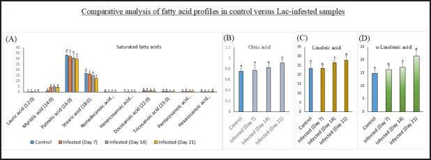 | Figure 1. Comparative analysis of fatty acid profiles in Flemingia semialata bark under control (non-infested) and K. lacca infested conditions. (A) Changes in saturated fatty acids (C12:0–C26:0) in control and infested samples at 7, 14, and 21 days post-infestation. (B–D) Temporal variation in major unsaturated fatty acids: (B) oleic acid, (C) linoleic acid, and (D) α-linolenic acid in control and infested bark. Data are presented as mean ± SE of three independent biological replicates. Lowercase letters indicate significant differences between days (p < 0.05, one-way ANOVA, Duncan’s post hoc test). [Click here to view] |
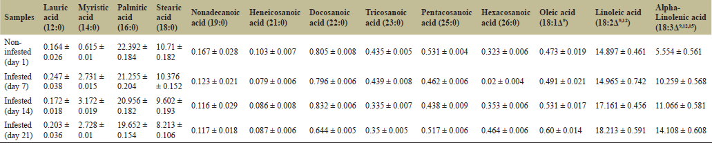 | Table 1. The changes in fatty acid composition of Flemingia semialata during K. lacca infestation at different sampling days (Day 1, 7, 14, and 21). Values are presented in mean percentage ± SE (n = 3). [Click here to view] |
Oleic acid (18:1Δ9), the only MUFA detected, showed a moderate but consistent increase from 0.47% in non-infested bark to 0.49%, 0.53%, and 0.60% on Days 7, 14, and 21. This increase in oleic acid (18:1Δ9) concentrations towards compensatory stability and repair of the membrane during infestation, with MUFAs frequently serving as a buffer to oxidative damage, may be associated with promoting bilayer maintenance in stress conditions [21]. The PUFAs showed pronounced, progressively increasing trends during infestation (Fig. 1C, D). Linoleic acid (18:2Δ9,12) increased from 14.89% in non-infested bark to 14.96%, 17.16%, and 18.21% on Day 7, 14 and 21, respectively (Fig. 1C). Alpha-linolenic acid (18:3Δ9,12,15) showed a sharper rise, increasing from 5.55% in the non-infested sample to 10.25%, 11.06%, and 14.10% over the same period (Fig. 1D and Table 1). By Day 21, the total PUFA fraction had increased by approximately 58% relative to non-infested bark (Fig. 1C, D and Table 1). Several studies have documented the elevation of free and esterified PUFAs following herbivory infestation as an initial response to the activation of the octadecanoid pathway [10]. These may also be associated with the enhancement of green leaf volatiles (GLVs) and oxylipins, which can modulate both direct and indirect defenses [13,24]. The observed steady rise in PUFAs in F. semialata might improve the preparation of distal tissues for a more rapid and systemic defensive response away from the local feeding site. However, the lac insect can still successfully colonise the host, despite these biochemical defense responses.
Additionally, the total SFAs decreased by approximately 9% from Day 1 to Day 21, while unsaturated fatty acids (MUFA + PUFA) increased substantially. Consequently, the SFA:UFA ratio declined from 1.73 in non-infested bark to 1.00 at Day 21, which may suggest a substantial shift toward higher unsaturation. The PUFA: MUFA ratio increased throughout infestation, driven by stronger induction of linoleic (18:2Δ9,12) and α-linolenic (18:3Δ9,12,15) acids relative to oleic acid (18:3Δ9). Furthermore, the 18:2/18:3 ratio declined from 2.68 on Day 1 to 1.29 on Day 21, reflecting proportionally greater accumulation of α-linolenic acid over linoleic acid, which may be associated with membrane remodelling resulting from biotic stress and has previously been associated with increased desaturase activity and enhanced membrane fluidity, which have been linked to signal transduction in previous studies. In addition to insect infestation, other factors such as plant developmental stages, wound responses, changes in carbon allocation during phloem sap feeding, and insect nutritional needs are also known to impact fatty acid composition [11,21,25]. Thus, changes in fatty acid composition should be viewed as multifactorial responses rather than the outcome of a single influencing factor.
These findings may be associated with mechanisms observed in other plant-insect systems, in which herbivores are recognised as affecting plant defensive responses. [26,27]. Interestingly, insects with a piercing-sucking behaviour, such as scale insects, also cause comparatively minimal tissue damage, compared to chewing herbivores, as reported in previous studies [28]. Furthermore, various hemipterans have been reported to detoxify or neutralise lipid-derived defense molecules through gut peroxidases or symbiotic microbes, further compromising the plant’s defense mechanisms [29,30]. The lac insect infestation caused the following major changes in the fatty acid profiles of host plant: (i) progressive decreases in major SFAs (16:0 and 18:0), (ii) a marked increase in PUFAs, especially α-linolenic acid (18:3Δ9,12,15), (iii) moderate increases in oleic acid (18:1Δ9), and (iv) significant reductions in the saturated to unsaturated fatty acid (SFA: UFA) and the 18:2Δ9,12/18:3Δ9,12,15 ratio. These findings demonstrate substantial remodelling of fatty acid composition throughout the 21-day infestation period.
4. CONCLUSION
These findings suggest that K. lacca infestation may be related to lipid profile changes associated with defensive responses in F. semialata, such as the accumulation of PUFA, oxylipin precursor activation, membrane remodeling, and alteration in certain fatty acid ratios. Nevertheless, the lac insect’s adaptations to stylet feeding, salivary inhibition and biochemical detoxification may help the insect adapt to host plant responses and maintain successful colonization. This interaction highlights a complex relationship in which temporal changes in lipid profiles occur during infestation, while the insect continues to colonize the host successfully. Such metabolic adaptability in the plant, combined with effective counter-defense strategies in the lac insect, likely underlies their long-term compatibility. Understanding this balance may provide novel insights into host compatibility and the sustainable management of the host plant–lac insect system.
5. ACKNOWLEDGMENTS
All authors acknowledge the financial support from the Department of Science and Technology (DST), New Delhi, to Indira Gandhi National Tribal University (IGNTU), Amarkantak, through the DST-SERB (CRG/2021/004163) project. The authors also acknowledge the National Institute of Plant Genome Research (NIPGR), New Delhi, for providing the GC-MS facility.
6. AUTHOR CONTRIBUTIONS
All authors made substantial contributions to conception and design, acquisition of data, or analysis and interpretation of data; took part in drafting the article or revising it critically for important intellectual content; agreed to submit to the current journal; gave final approval of the version to be published; and agree to be accountable for all aspects of the work. All the authors are eligible to be author as per the International Committee of Medical Journal Editors (ICMJE) requirements/guidelines.
7. CONFLICT OF INTEREST
The authors report no financial or any other conflicts of interest in this work.
8. ETHICAL APPROVALS
This study does not involve experiments on animals or human subjects.
9. DATA AVAILABILITY
All the data is available with the authors and shall be provided upon request.
10. PUBLISHER’S NOTE
All claims expressed in this article are solely those of the authors and do not necessarily represent those of the publisher, the editors and the reviewers. This journal remains neutral with regard to jurisdictional claims in published institutional affiliation.
11. USE OF ARTIFICIAL INTELLIGENCE (AI)-ASSISTED TECHNOLOGY
The authors declare that they have not used artificial intelligence (AI)-tools for writing and editing of the manuscript, and no images were manipulated using AI.
12. SUPPLEMENTARY MATERIAL
The supplementary material can be accessed at the journal’s website: https://jabonline.in/admin/php/uploadss/1491_pdf.pdf
REFERENCES
1. Schowalter TD. Insect ecology: an ecosystem approach. London: Academic Press; 2022 Feb 24. CrossRef
2. Lee Y, Choi BY. The relationship between plants and insects. Social lives of plants: how plants connect—as friends or foes—with other plants, microorganisms, animals, and humans. Singapore, Springer Nature Singapore, pp 81–108, 2025. CrossRef
3. Beran F, Petschenka G. Sequestration of plant defense compounds by insects: from mechanisms to insect–plant coevolution. Annu Rev Entomol 2022;67(1):163–80. CrossRef
4. Bruce TJA. Interplay between insects and plants: dynamic and complex interactions that have coevolved over millions of years but act in milliseconds. J Exp Botany 2015;66(2):455–65. CrossRef
5. Howe GA, Jander G. Plant immunity to insect herbivores. Annu Rev Plant Biol 2008;59:41–66. CrossRef
6. Bertoni G. Oxylipins and plant palatability. Plant Cell 2012;24(4):1305. CrossRef
7. Mahanta DK, Komal J, Samal I, Bhoi TK, Kumar PVD, Mohapatra S, et al. Plant Defense Responses to Insect Herbivores Through Molecular Signaling, Secondary Metabolites, and Associated Epigenetic Regulation. Plant-Environment Interact 2025;6(1):70035. CrossRef
8. Upchurch RG. Fatty acid unsaturation, mobilization, and regulation in the response of plants to stress. Biotechnol Lett 2008;30(6):967–77. CrossRef
9. Walley JW, Kliebenstein DJ, Bostock RM, Dehesh K. Fatty acids and early detection of pathogens. Curr Opinion Plant Biol 2013;16(4):520–6. CrossRef
10. He M, Ding NZ. Plant unsaturated fatty acids: multiple roles in stress response. Front Plant Sci 2020;11:562785. CrossRef
11. Cook R, Lupette J, Benning C. The role of chloroplast membrane lipid metabolism in plant environmental responses. Cells 2021;10(3):706. CrossRef
12. Wasternack C, Song S. Jasmonates: biosynthesis, metabolism, and signaling by proteins activating and repressing transcription. J Exp Bot 2017; 68(6):1303–1321. CrossRef
13. He Y, Borrego EJ, Gorman Z, Huang PC, Kolomiets MV. Relative contribution of LOX10, green leaf volatiles and JA to wound-induced local and systemic oxylipin and hormone signature in Zea mays (maize). Phytochemistry 2020;174:112334. CrossRef
14. León J, Sánchez-Serrano JJ. Molecular biology of jasmonic acid biosynthesis in plants. Plant Physiol Biochem 1999;37(5):373–380. CrossRef
15. Kumaraswamy S, Yogendra K, Sotelo-Cardona P, Shivanna A, Hemalatha S, Mohan M, et al. Non-targeted metabolomics reveals fatty acid and associated pathways driving resistance to whitefly and tomato leaf miner in wild tomato accessions. Scientific Rep 2025;15(1):3754. CrossRef
16. Press BR. Mabberley's plant-book: a portable dictionary of plants, their classification and uses. J Botanical Res Inst Texas 2018;12(2):578. CrossRef
17. Sharma KK. Lac insects. In: Omkar (ed.). Commercial insects, Boca Raton, FL, CRC Press, pp 1–16, 2023. CrossRef
18. Sarkar PC, Shrivastava AK. FT-IR spectroscopic studies on degradation of lac resin–part I: thermal degradation. Pigment & Resin Technol 2000;29(1):23–8. CrossRef
19. Semwal P, Painuli S. Antioxidant, antimicrobial, and GC-MS profiling of Saussurea obvallata (Brahma Kamal) from Uttarakhand Himalaya. Clin Phytoscience 2019;5(1):12. CrossRef
20. Shyam P, Mansoori A, Raikwar A, Kaushik S, Shweta S, Kumar A, et al. Assessing the infestation-induced response on the plant host by the Indian lac insect. Plant Sci Today 2024;11(2):363–72. CrossRef
21. Yu L, Zhou C, Fan J, Shanklin J, Xu C. Mechanisms and functions of membrane lipid remodeling in plants. Plant J 2021;107(1):37–53. CrossRef
22. Kloth KJ, Dicke M. Rapid systemic responses to herbivory. Curr Opinion Plant Biol 2022;68:102242. CrossRef
23. Mostafa S, Wang Y, Zeng W, Jin B. Plant responses to herbivory, wounding, and infection. Int J Mol Sci 2022;23(13):7031. CrossRef
24. Matsui K, Engelberth J. Green leaf volatiles—the forefront of plant responses against biotic attack. Plant Cell Physiol 2022;63(10):1378–90. CrossRef
25. Laureano G, Cavaco AR, Matos AR, Figueiredo A. Fatty acid desaturases: uncovering their involvement in grapevine defense against downy mildew. Int J Mol Sci 2021;22(11):5473. CrossRef
26. Du H, Xu HX, Wang F, Qian LX, Liu SS, Wang XW. Armet from whitefly saliva acts as an effector to suppress plant defenses by targeting tobacco cystatin. New Phytologist 2022;234(5):1848–62. CrossRef
27. Wu X, Ye J. Manipulation of jasmonate signaling by plant viruses and their insect vectors. Viruses 2020;12(2):148. CrossRef
28. Ali J, Ton?a A, Islam T, Mir S, Mukarram M, Konôpková AS, et al. Defense strategies and associated phytohormonal regulation in Brassica plants in response to chewing and sap-sucking insects. Front Plant Sci 2024;15:1376917. CrossRef
29. Holt JR, Cavichiolli De Oliveira N, Medina RF, Malacrinò A, Lindsey ARI. Insect–microbe interactions and their influence on organisms and ecosystems. Ecol Evol 2024;14(7):e11699. CrossRef
30. Kline O, Joshi NK. Microbial symbiont-based detoxification of different phytotoxins and synthetic toxic chemicals in insect pests and pollinators. J Xenobiotics 2024;14(2):753–1. CrossRef