Research Article | Volume 14, Issue 3, May, 2026

Isolation, screening, and optimization of CMCase production from Bacillus pumilus A5 isolated from the fecal sample of Bos indicus

Hitesha Panchal Mohammedfaizan Shaikh Nishu Yadav Depali Tadvi Sanskriti Tiwari Choudhary Krishan Kumar   

Open Access   

Published:  Apr 05, 2026

DOI: 10.7324/JABB.2026.289138
Abstract

Bacterial cellulase plays a vital role in industries, such as bioethanol, textiles, paper and pulp, etc. Although numerous cellulolytic bacteria have already been isolated from diverse ecological niches, for the exploration of more potential bacteria, a fecal sample of Bos indicus was collected from Godhra, Gujarat, India. The serially diluted fecal sample was cultured on Bushnell Haas carboxymethyl cellulase (CMC) agar using the spreading technique, resulting in the isolation of five morphologically different bacterial colonies. Among these, isolate A5 was selected for further characterization, as it showed the best result in both qualitative and quantitative tests. The comprehensive investigation, which included colony morphology, biochemical profiling, and 16S Ribosomal RNA gene sequencing, identified isolate A5 as Bacillus pumilus. Optimal cellulase synthesis occurred at neutral pH (7.0) and 40°C after 72 h of incubation in a 2% (w/v) CMC-enriched production medium. Crude extract of this strain possessed activity against multiple substrates, including CMC, pectin, starch, 4-nitrophenyl β-D-glucopyranoside, and xylan, which highlights its potential for many industries, including bioethanol, textile, paper and pulp, etc.


Keyword:     Cellulolytic bacteria Cow feces CMCase activity β-glucosidase Lignocellulosic biomass


Citation:

Panchal H, Shaikh MS, Yadav N, Tadvi D, Kumar K. Isolation, screening, and optimization of CMCase production from Bacillus pumilus A5 isolated from the fecal sample of Bos indicus. J Appl Biol Biotech 2026;14(3):165-172. http://doi.org/10.7324/JABB.2026.289138

Copyright: Author(s). This is an Open Access article distributed under the terms of the Creative Commons Attribution-NonCommercial-ShareAlike license.

HTML Full Text

1. INTRODUCTION

Lignocellulosic biomass (LCB) is the most ubiquitous polymer on the earth, and its plentiful source makes it very convenient for usage [1]. Agricultural residues are one of the primary sources of LCB. Burning of lignocellulosic crop residues causes remarkable pollution in the atmosphere and huge nutritional losses in the soil. To overcome these problems, these crop residues can be used to produce second-generation biofuels [2]. LCB consists solely of three structural polymers: Cellulose, hemicellulose, and lignin, organized within a highly resistant matrix. Cellulose, the predominant carbohydrate component, is a linear homopolysaccharide composed of repeated D-glucose monomers linked by β-1,4-glycosidic bonds. Enzymatic and chemical hydrolysis methods are used for the breaking down of cellulose into fermentable sugar monomers, which are essential substrates for further bioethanol synthesis. This conversion mostly relies on the class of enzyme known as cellulases, which is a group of synergistic enzymes, namely, endoglucanases (E.C. 3.2.1.4), cellobiohydrolase (E.C. 3.2.1.91), and β-glucosidase (E.C. 3.2.1.21) [3-5]. Endoglucanases break cellulose chains at random internal sites, producing shorter fragments known as cello-oligosaccharides. Cellobiohydrolases then act on the ends of these fragments and release cellobiose units. Finally, β-glucosidase hydrolyses cellobiose into glucose.

Till now, a lot of research has been done on the enzymatic depolymerization of LCB by various organisms, including bacteria and fungi. Cellulase enzymes produced by fungi are widely used in commercial sectors due to their high enzymatic activity, but recently, researchers have been focusing on bacterial cellulase enzymes due to their rapid growth, multi-enzyme complex expression, stability in extreme environments, and resistance to various environmental stresses, which sets bacteria apart from their fungal counterparts [6,7]. Depolymerization of LCB has been reported for various cellulolytic bacterial species isolated from a variety of sources, such as Bacillus cereus A49 [8], Klebsiella oxytoca M21WG [7], and Klebsiella spp. Z6WG [7], Klebsiella pneumoniae [9], and Enterococcus faecium BS5 [10]. Some other recently isolated cellulolytic bacterial strains with high CMCase activity include Stutzerimonas stutzeri CS7, isolated from the Solanum melongena soil [10]; Bacillus pumilus XM, which is isolated from Père David’s deer feces [11]; and Enterobacter xiangfangensis isolated from the gut of the grasshopper Eyprepocnemis alacris alacris [9].

Many cellulolytic bacterial strains are isolated from diverse sources, such as municipal waste, industrial waste, agricultural leftovers, animal excreta, crop-processing trash, logging slash, and sawdust from timber extraction [12]. However, there is still research ongoing where potential strains of bacteria possessing multiple activities (CMCase, glucosidases, etc.) in the mesophilic range are studied, as there is a growing demand of mesophilic bacteria in simultaneous saccharification and fermentation, which helps to reduce the cost of bioethanol production [13]. Bacterial cellulases play a major role in several industries, especially in food and beverage production, paper and pulp processing, and textile treatments, including biostoning and biopolishing. These enzymes are extensively commercialized through submerged fermentation by many biotechnology companies.

Therefore, in the present research, a cellulase producing bacterial strain was isolated from cow (Bos indicus) fecal matter collected in Godhra, Gujarat, India. Cow dung is undigested food residue that is excreted by herbivorous bovine animals after the consumption of food. Cow dung is a mixture of feces and urine in the ratio of 3:1, and it mainly contains cellulose, hemicellulose, and lignin [14]. Cow dung is the source for various microorganisms, which contain different species of bacteria (Bacillus spp., Corynebacterium spp., etc.), protozoa, and yeast [15,16].

The potential cellulase-producing bacteria were screened out based on the hydrolytic zone on Bushnell Haas (BH) agar plates having carboxymethyl cellulase (CMC) and initial CMCase activity of crude enzyme. B. pumilus A5 was determined to be the bacterial isolate that was chosen after it was subjected to morphological analysis, biochemical profiling, and molecular analyses. Cellulase production of this strain was optimized using the one factor at a time (OFAT) method, after which the activity of the resulting crude enzyme extract was further evaluated and optimized.


2. MATERIALS AND METHODS

2.1. Substrates and Chemicals used in Experiments

CMC; medium viscosity and BH broth were obtained from Sigma-Aldrich, USA. Media and reagents, including Luria-Bertani (LB) broth, yeast extract, agar powder, peptone, tryptone broth, glucose phosphate broth (GPB), Congo red, Lugol’s iodine, and apple pectin, along with Simmon’s citrate agar and triple sugar iron (TSI) agar, were obtained from HiMedia Pvt. Ltd., India. 4-nitrophenyl β-D-glucopyranoside (pNPG) was purchased from Sisco Research Laboratories Pvt. Ltd (SRL), India. All chemicals and reagents used in this study were of analytical grade.

2.2. Sample Collection and Screening of Bacterial Strain

A fecal sample from B. indicus (Malvi breed) was collected from Godhra, Panchmahal, Gujarat, India (22.7788° N, 73.6143° E) using sterile forceps and was kept at 4°C until further use. 1 g of fecal sample was mixed with 10 mL of sterile 0.85 % (w/v) saline solution, and was placed on an orbital shaker (120 rpm) for 1 h until the mixture was homogenized. The homogenized mixture was serially diluted up to 10−8 using saline solution and from each dilution, 100 μL of sample was spread onto CMC-BH agar plates composed of g/L: (CMC (10.0); K2HPO4 (1.0); MgSO4.7H2O (0.2); KH2PO4 (1.0); FeCl3.6H2O (0.05); NH4NO3 (1.0); CaCl2 (0.02); and agar powder (15.0) and incubated at 37°C for 96 h [5]. Morphologically distinct bacterial colonies were selected and further subcultured by streaking on BH-CMC agar plates, followed by incubation at 37°C for 2 days. Each purified colony was grown into fresh LB broth and incubated at 37°C for 16 h. For the qualitative analysis, 3 μL of each overnight-grown culture was placed on CMC-BH agar plates and incubated at 37°C for 48 h. After incubation, agar plates were flooded with 0.3% Congo-red solution for 20 min and counterstained with 1M NaCl for 10 min [17]. For quantitative analysis, the selected isolates were cultured in 50 mL of cellulase production medium (pH 7.0) composed of g/L: (CMC (10.0); K2HPO4 (1.0); MgSO4.7H2O (0.2); KH2PO4 (1.0); FeCl3.6H2O (0.05); NH4NO3 (1.0); CaCl2 (0.02) and yeast extract (5.0) at 37°C, 180 rpm for 72 h. The freshly grown culture was subjected to centrifugation at 10,000 × g for 20 min at 4°C, and the resultant cell-free crude supernatant was utilized to measure CMCase activity. CMCase activity of each crude enzyme was determined using the 3,5-dinitrosalicylic acid (DNS) method [18].

2.3. Identification of Potent Cellulolytic Bacteria

Selected bacterial isolates were subjected to morpho-biochemical characterization and compared with the Bergey’s manual of systematic bacteriology [19]. The Gram stain method was performed to differentiate between Gram-negative and Gram-positive bacteria [20]. The starch hydrolysis property of the selected strain was done by streaking on a starch agar plate, followed by incubation at 37°C for 24 h. Starch agar plates were soaked with Lugol’s iodine solution and examined for a distinct, colorless zone [21]. An isolated bacterial colony was placed onto a sterile, dry glass slide for the catalase test using a sterile nichrome wire loop. After adding a few drops of 3% hydrogen peroxide (H2O2) to the colony, the reaction was examined for the immediate release of gas bubbles (effervescence), which indicated a positive catalase activity [22]. The oxidase test was performed by impregnating a filter paper disc with a 1% (w/v) tetramethyl-p-phenylenediamine dihydrochloride solution, a synthetic electron donor referred to as Wurster’s reagent, and then air drying it. A small portion of the bacterial colony was subsequently put onto the treated disc, and color development was detected within 10 s [23]. Bacterial culture was grown into GPB with incubation of 24–48 h at 37°C. In the methyl red test, a few drops of methyl red were added into GPB and observed for the appearance of a red color. For the Voges-Proskauer (VP) test, 0.6 mL of α-naphthol followed by 0.4 mL of KOH solution were added and observed for the red color development [22]. The bacterial culture was inoculated into peptone nitrate broth and incubated at 37°C for 24 h, for the nitrate reduction test. Following incubation, 0.5 mL of sulfanilic acid and α-naphthylamine were added, and development of a red color was observed, which signifies the nitrate reduction [21]. The indole production was evaluated by inoculating a loopful of the test culture into tryptone broth and incubated at 37°C for 24 h. After incubation, 1 mL of Kovac’s reagent was added, and the appearance of a pink ring at the surface was examined [22]. The TSI test involved streaking a loopful of test culture onto the TSI agar and incubated at 37°C for 24 h. Acid production, gas formation, and H2S production were observed in both the butt and the slant [22]. A loopful of test culture was streaked onto Simmon’s citrate agar slants and incubated at 37°C for 24 h, and the color change from green to blue was observed [23].

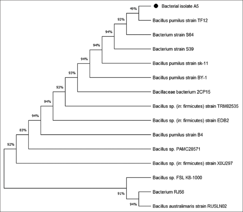
2.4. Analysis of 16s Ribosomal RNA (rRNA) of Cellulolytic Bacteria

16S rRNA amplification, sequencing, and molecular identification of the bacterial isolate A5 was conducted at Yaazh Xenomics, Coimbatore, Tamil Nadu, India. The Qiagen DNA extraction kit was used to isolate DNA from bacterial samples. The bacterial strain was identified using polymerase chain reaction using universal primers 27F (5’-CCAGAGTTTGATCMTGGCTCAG-3’) and 1492R (5’-TACGGYTACCTTGTTACGACTT-3’) [24]. The 16S gene sequence was then subjected to basic local alignment search tool (BLAST) analysis at the National Center for Biotechnology Information (NCBI) GenBank database, followed by multiple sequence alignment using Thompson et al. [25]. A phylogenetic tree was constructed using the maximum likelihood method, utilizing molecular evolutionary genetics analysis (MEGA) 11.0 software [26].

2.5. Crude Enzyme Production and Endo-β-1, 4-Glucanase Enzyme Assay

Bacterial isolate (A5) with the highest initial CMCase activity was selected and cultured into LB broth and incubated at 37°C, 180 rpm for 16 h. 1 mL of freshly grown bacterial culture was inoculated into 100 mL of enzyme production media (g/L: CMC (10.0); BH (3.27); yeast extract (5.0) at pH 7.0) and incubated at 37°C at 180 rpm for 72 h. The enzyme production medium was subject to centrifugation at 10,000 × g for 10 min after the incubation and the cell-free extract was collected for further analysis [27,28]. CMCase activity was determined by adding 300 μL of crude enzyme along with 600 μL of substrate (1% CMC in 50 mM phosphate buffer, pH 7.0) and incubated for 20 min at 37°C. After incubation, 900 μL of DNS solution was added into the reaction mixture, followed by 10 min incubation in a boiling water bath. The reaction mixture was cooled down after incubation, and 900 μL of distilled water was added before measuring the absorbance using a spectrophotometer at 540 nm and compared to a blank containing all the reagents except the crude enzyme [29]. One unit of CMCase activity was defined as 1 mL of enzyme solution that catalyzes CMC hydrolysis and produces 1 μmoL of glucose/min under given assay conditions [30].

2.6. CMCase Production Optimization

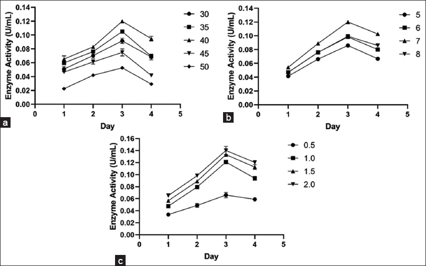
The influence of temperature, incubation time, pH, and CMC concentration was determined by using OFAT. The effect of incubation time on the CMCase production was analyzed by incubating freshly grown bacterial culture in enzyme production media at 37°C, pH 7.0 for 96 h. Production medium was withdrawn at regular intervals and centrifuged at 10,000 × g for 10 min to obtain crude extract. CMCase activity of crude extract was determined through the DNS method as described in section 2.5 [11]. The effect of incubation temperature on the production of CMCase was analyzed by incubating 1 mL of freshly grown bacterial culture into enzyme production media (100 mL), followed by incubation at temperatures ranging from 35 to 50°C at 180 rpm for 96 h. The impact of varying pH levels on CMCase production was assessed by inoculating 1 mL of freshly grown culture into enzyme production medium of different pH levels (5.0, 6.0, 7.0, and 8.0), followed by incubation at 180 rpm for 96 h at the optimal temperature. The enzyme production medium (100 mL) containing different volumes of CMC concentration (0.5, 1.0, 1.5, and 2.0%) was incubated at the optimal incubation temperature and pH to examine the influence of substrate concentration on CMCase production [31].

2.7. Crude Enzyme Activity Characterization

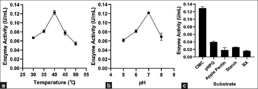
The optimal temperature for endo-β-1,4-glucanase activity from the bacterial isolate was determined by incubating 300 μL of the crude enzyme with 600 μL of CMC (1% w/v in 50 mM phosphate buffer, pH 7.0) at temperatures ranging from 30°C to 50°C for 20 min. The enzymatic process was halted by the addition of 900 μL of DNSA reagent, and the mixture was then incubated in a boiling water bath for 10 min to develop the color. The pH optimum was determined by incubating 300 μL of the crude enzyme with 600 μL of CMC (1% w/v in buffer of pH from 5.0 to 8.0 using 50 mM citrate-phosphate or Tris-HCl buffer) at 40°C for 20 min, followed by the addition of DNSA and a subsequent boiling period of 10 min. Upon cooling, each reaction mixture was diluted by adding 900 μL of distilled water, and absorbance was recorded at 540 nm to evaluate enzyme activity across varying temperature and pH parameters [32].

2.8. Substrate Specificity

The crude B. pumilus A5 extract was tested against substrates: CMC, BX, pNPG, starch, and apple pectin. 900 μL reaction mixture containing 600 μL substrate (1% in 50 mM phosphate buffer, pH 7.0) and 300 μL crude enzyme was incubated for 20 min at 40°C. After incubation, 900 μL of DNS reagent was added to the reaction mixture and further incubated in a boiling water bath for 10 min, followed by the addition of 900 μL of distilled water, and the absorbance was measured at 540 nm using a spectrophotometer [33,34]. All assays were carried out in triplicate. One unit of CMCase, xylanase, amylase, and pectinase were defined as the amount of enzyme used to produce 1 μmoL of glucose, xylose, maltose, and galacturonic acid, respectively, per minute under defined assay conditions [35]. The β-glucosidase activity was assessed by adding crude enzyme (500 μL) with 1,000 μL of 5 mM pNPG, prepared in 50 mM phosphate buffer (pH 7.0), and incubated for 20 min at its optimum temperature. After incubation, 1,500 μL of Na2CO3 (1 M) was added to the reaction mixture to terminate the process. Absorbance was measured at 405 nm using a spectrophotometer using para-nitrophenol (pNP) as a standard [30,36]. One unit of β-glucosidase activity is defined as the amount of enzyme used to produce 1 μmol of pNP/min under specific assay conditions [35].

2.9. Data Analysis

All graphs were generated using GraphPad Prism version 8.0. Cellulase production under varying conditions was evaluated by two-way analysis of variance at p < 0.0001. A phylogenetic tree was created using MEGA 11.0 software.


3. RESULTS AND DISCUSSION

3.1. Isolation and Screening of Cellulolytic Bacteria

Five bacterial isolates were selected on the basis of morphological differences after spreading the B. indicus feces sample on CMC-BH agar plates. Out of these five isolates, only isolate A5 demonstrated a visible hydrolytic zone after staining with 0.3% Congo red solution [Figure 1a]. All isolates were also investigated quantitatively, and isolate A5 demonstrated notable initial CMCase activity (0.064 ± 0.0045 U/mL) after 72 h of incubation at 37°C [Figure 1b]. The initial CMCase activity of isolate A5 was comparable with some other known cellulolytic producing strains isolated from various environments, which include Cellulomonas spp. (0.033 U/mL) [36], Micrococcus spp. (0.015 U/mL) [36], Brevibacillus spp. JXL (0.02 U/mL) [37], Geobacillus spp. (0.012 U/mL) [38], Pseudomonas spp. NE (0.061 U/mL) [32] and Bacillus subtilis AS3 (0.07 U/mL) [39].

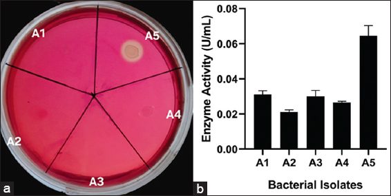
Figure 1: Primary screening of cellulase producing bacterial isolates on Bushnell Haas Carboxymethyl cellulose agar plates (a) Plates showing clear hydrolytic zone surrounding bacterial isolates after staining with 0.3% Congo red solution (b) Initial CMCase activity of bacterial isolates isolated from the fecal sample of cow.


[Click here to view]

3.2. Identification of Potent Cellulolytic Bacterial Strain

Selected bacterial strain A5 was subjected to cultural characterization, such as size, shape, and pigmentation. Microscopic examination under an oil immersion lens (×100) revealed that bacterial isolate A5 was Gram-positive rod-shaped in nature [Figure 2]. According to biochemical characterization, isolate A5 indicated negative results for the indole, starch hydrolysis, and nitrate reduction tests, while yielding positive results for the VP, methyl red, oxidase, catalase, and citrate utilization tests. In the case of the TSI test, the color of the butt and slant remained orange with no H2S production [Table 1]. Similar biochemical profiles were reported by Parvathi et al. [40] and Kapilan and Arasaratnam [41] for B. pumilus. These morpho-biochemical characteristics, together with a comparison with the Bergey’s manual of systematic bacteriology, imply that isolate A5 is most likely B. pumilus.

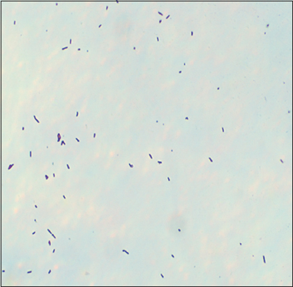
Figure 2: Microscopic observation of bacterial cells of B. pumilus A5 after Gram staining method


[Click here to view]

Table 1: Morphological and biochemical characterization of B. pumilus A5 isolated from the feces of a cow.

CharacterizationReference reactionsA5
ShapeShort rod
Gram staining+
PigmentationOff white
Colony textureOpaque
Colony marginIrregular
Methyl RedPositive (pink color)+
Voges ProskauerPositive (pink color)+
Indole productionNegative (no color change)-
CatalasePositive (effervesces observed)+
OxidasePositive (purple color)+
Nitrate reductionNegative (no color observed after A and B, but after the addition of zinc)-
Starch hydrolysisNegative (no clear zone)-
Citrate utilizationPositive (blue color)+
TSI (H2S)Negative-
Gas productionNo gas production-
TSI (Butt color)Orange
TSI (Slant color)Orange

TSI: Triple sugar iron, +: Positive, −: Negative.

3.3. Molecular Identification of the Cellulolytic Bacteria

Bacterial isolate A5 was subjected to 16S gene sequence analysis for molecular characterization. The 16S rRNA gene sequence of bacterial isolate A5 was compared with nucleotide sequences of closely related bacteria through the BLAST function in the NCBI GenBank database. The gene sequence alignment revealed 99.39% similarity to B. pumilus, indicating that isolate A5 is genetically very close to this species. Similarly, the phylogenetic tree demonstrated that the 16S rRNA sequence of isolate A5 clustered closely with B. pumilus strains [Figure 3]. Furthermore, these results are in line with preliminary identification (morpho-biochemical characterization), indicating that bacterial isolate A5 belongs to the genus B. pumilus. The 16S rRNA nucleotide sequence of bacterial isolate A5 was submitted to NCBI’s GenBank database with the accession number PX517296.

Figure 3: A phylogenetic tree of isolate A5 indicating bacterial species to be Bacillus pumilus.


[Click here to view]

3.4. CMCase Production Optimization using OFAT

The OFAT method was employed to optimize enzyme production parameters, such as incubation time, temperature, medium pH, and substrate concentration. Optimization of the incubation time is critical to achieve the maximal CMCase production. B. pumilus A5 produced maximum enzyme at 72 h [Figure 4]. Enzyme production significantly decreased beyond the optimum incubation period, most likely as a result of nutritional exhaustion, byproduct formation in the culture medium, and death of bacterial cells. Growth rates and catalytic activity profiles of each isolate vary based on the duration of incubation, which emphasizes the necessity of maximizing incubation times to get the anticipated results. A similar observation was also reported by Shankar and Isaiarasu [42]; maximum cellulase production from B. pumilus EWBCM1 isolated from the earthworm gut (Eudrilus eugeniae) was observed at 72 h of incubation. Incubation temperature greatly affects the CMCase production by B. pumilus A5. Maximum CMCase synthesis was observed at 40°C, with significantly higher CMCase synthesis compared to lower and higher temperature ranges (p < 0.0001). A marked decline in the CMCase synthesis was observed at below and higher temperatures than optimum [Figure 4a]. A comparable outcome was noted by Islam and Roy [43], where Paenibacillus spp. isolated from molasses in sugar industry waste soil exhibited peak activity at 40°C. Bacillus subtilis PUA-18 also showed significant activity at 40°C [44].

Figure 4: Bacterial growth and CMCase production by A5 (a) at different temperature (°C), (b) at different pH levels, and (c) at different substrate concentration (%).


[Click here to view]

Due to their biological niches and surroundings, different bacterial strains have different ideal incubation temperatures for producing cellulase enzymes. The optimal temperature range for several cellulolytic bacterial strains obtained from agricultural soil and animal dung was 40–45°C.

Furthermore, gut-dwelling bacteria are accustomed to temperatures that are similar to those of the host. The metabolic systems, including the regulation of enzyme-producing genes, are optimized to function efficiently within this temperature range [43].

The medium pH is another important variable that affects the CMCase production. CMCase production by B. pumilus A5 was assessed at varied pH ranges (5.0–8.0) and CMCase production was significant (p < 0.0001); at a pH of 7.0 [Figure 4b]. Varying the pH of the medium can enhance the efficacy of CMCase synthesis. The optimal pH for cellulase enzyme activity produced from Bacillus vallismortis RG-07 was found to be 7.0 [45]. Another study found that the optimum pH for cellulase enzyme activity by Bacillus spp. C1AC5507 was 7.0 [46], which is consistent with our findings. Fresh feces from herbivores have pH values ranging from 5.0 to 8.0. Bacterial diversity appears to be maximum at neutral pH, progressively declining at levels that fall below or exceed this threshold, based on studies [42,47]. From the industrial perspective, a pH range of 5.0–8.0 is suitable for various industrial applications, including bio-stone processing and biopolishing in the textile industry, for biofuel production, and in the paper and pulp industry. Cellulases are beneficial for deinking various types of wastepaper, either alone or in tandem with xylanases. Cellulase-based deinking processes at acidic to neutral pH not only prevent alkaline yellowing but also streamline the deinking process and lower environmental pollution [48,49].

CMCase enzyme production medium was inoculated with different CMC concentrations and incubated for 4 days. B. pumilus A5 achieved maximum CMCase production with 2% CMC concentration, where p < 0.0001, which makes it significant [Figure 4c]. Increased cellulase production is usually related to increased CMC concentrations; however, at concentrations >2.0%, enzyme activity tends to decrease. The inhibitory properties of accumulating cellobiose and cellodextrins are partially responsible for this decrease [50]. Wang et al. [8] observed similar results for Bacillus cereus A49, which produced maximum CMCase enzyme at 2.0% CMC supplement.

In this study, B. pumilus A5 isolated from the feces of B. indicus produced maximum CMCase production of 0.15 ± 0.0018 U/mL at 40°C, pH 7.0, CMC concentration of 2.0%, and at 72 h of incubation after optimization through OFAT. Many recently isolated cellulolytic strains were optimized to produce the maximum cellulase enzyme.

3.5. Enzyme Characterization

Cellulase activity was affected by temperature, and to study this, the crude enzyme was incubated at a range of temperatures from 30°C to 50°C. Enzyme activity of B. pumilus increased steadily while reaching its peak at 40°C, before declining [Figure 5a]. These findings are similar to those of Mokale Kognou et al. [31], who identified 40°C as the optimal temperature for cellulase production by the MKAL2 strain isolated from soil. Similarly, Bacillus pseudomycoides, isolated from sugarcane bagasse, exhibited maximum enzyme activity at 40°C and pH 7.0 after 72 h of incubation [51]. Streptomyces ruber, sourced from decaying biomass, also demonstrated peak cellulase activity at 40°C [52].

Figure 5: Effect of different parameters on activity of cellulase enzyme produced by bacterial isolate A5 (a) Effect of temperature, (b) Effect of pH and. (c) Substrate specificity.


[Click here to view]

Experiments were conducted within a pH range of 5.0–8.0 to assess the impact of pH on crude enzyme activity. The crude extract of the bacterial strain demonstrated optimum enzyme activity at pH 7.0 [Figure 5b]. The crude extract of B. subtilis A5 showed the highest CMCase activity at 40°C and pH 7.0, which is comparable with the findings of Paenibacillus spp. isolated from molasses-contaminated soil in the sugar sector [44]. Similarly, B. pumilus EB3, isolated from pretreatment oil palm empty fruit bunches, had maximum CMCase activity at pH 7.0 [53].

3.6. Substrate Specificity

Crude extract of B. pumilus A5 showed significantly high CMCase activity when assessed with CMC, demonstrating the capacity to hydrolyze amorphous cellulose. Crude extract also showed moderate activity against substrates, such as BX, starch, and apple pectin [Figure 5c]. The crude extract exhibited measurable activity against pNPG, a synthetic substrate typically utilized to determine β-glucosidase activity. The ability to hydrolyze pNPG signifies the existence of enzymes that can cleave glucose residues; however, β-glucosidase activity was comparatively lower than CMCase activity. The findings of the substrate profile are significant for interpreting the enzymatic activities of B. pumilus A5, and this research may be utilized by businesses requiring cellulase hydrolysis.


4. CONCLUSION

In the present investigation, a potential cellulolytic bacterium was identified and characterized from B. indicus feces. A5 was the only isolate that showed a hydrolytic zone and significant initial CMCase activity out of five isolates. The bacterial strain was identified as B. pumilus with the help of morpho-biochemical characterization and 16S rRNA gene sequencing (GenBank accession no. PX517296). B. pumilus A5 exhibited the highest CMCase activity (0.15 U/mL) at 40°C, pH 7.0, 2.0% CMC concentration, and 72 h of incubation. Enzyme characterization revealed that the crude enzyme is most active at 40°C and pH 7.0. Analysis of substrate specificity revealed high CMCase activity for amorphous cellulose, moderate activity for other polysaccharides, and poor activity for β-glucosidase. Overall, B. pumilus A5’s enzymatic profile suggests that it has the potential for use in industries, such as biofuel generation, waste management, textile bioprocessing, and paper recycling. However, this research is restricted to laboratory-scale evaluations. To establish the strain’s commercial viability, further research will be focused on large-scale fermentation, enzyme purification, stability investigations, and industrial testing.


5. AUTHORS’ CONTRIBUTIONS

All authors made substantial contributions to conception and design, acquisition of data, or analysis and interpretation of data; took part in drafting the article or revising it critically for important intellectual content; agreed to submit to the current journal; gave final approval of the version to be published; and agree to be accountable for all aspects of the work. All the authors are eligible to be author as per the International Committee of Medical Journal Editors (ICMJE) requirements/guidelines.


6. FUNDING ACKNOWLEDGMENT

The authors expressed their deep gratitude to Parul University for providing financial support through an Intramural Research Project (CR4D/IMSL/088). In addition, they were grateful to the Government of Gujarat for fellowship support under the SHODH Scheme (T6621BITH001).


7. CONFLICTS OF INTEREST

The authors report no financial or any other conflicts of interest in this work.


8. ETHICAL APPROVALS

This study does not involve experiments on animals or human subjects.


9. DATA AVAILABILITY

The 16S rRNA gene sequencing data validating this research are accessible in the NCBI GenBank database (https://www.ncbi.nlm.nih.gov/nuccore/PX517296) under accession number PX517296. This publication covers all additional data generated throughout this study.


10. PUBLISHER’S NOTE

All claims expressed in this article are solely those of the authors and do not necessarily represent those of the publisher, the editors and the reviewers. This journal remains neutral with regard to jurisdictional claims in published institutional affiliation.


11. USE OF ARTIFICIAL INTELLIGENCE (AI)-ASSISTED TECHNOLOGY

The authors declare that they have not used artificial intelligence (AI)-tools for writing and editing of the manuscript, and no images were manipulated using AI.


REFERENCES

1.  Martín C, López Y, Plasencia Y, Hernández E. Characterisation of agricultural and agro-industrial residues as raw materials for ethanol production. Chem Biochem Eng Q. 2006;20(4):443-7.

2.  Gupta P, Samant K, Sahu A. Isolation of cellulose? degrading bacteria and determination of their cellulolytic potential. Int J Microbiol. 2012;2012(1):578925. [CrossRef]

3.  Gavande PV, Kumar K, Ahmed J, Goyal A. Multifunctionality and mechanism of processivity of family GH5 endoglucanase, RfGH5_4 from Ruminococcus flavefaciens on lignocellulosic polymers. Int J Biol Macromol. 2023;224:1395-411. [CrossRef]

4.  Bhat MK, Bhat S. Cellulose degrading enzymes and their potential industrial applications. Biotechnol Adv. 1997;15(3-4):583-620. [CrossRef]

5.  Kumar K, Singh S, Sharma K, Goyal A. Computational modeling and small-angle X-ray scattering based structure analysis and identifying ligand cleavage mechanism by processive endocellulase of family 9 glycoside hydrolase (HtGH9) from Hungateiclostridium thermocellum ATCC 27405. J Mol Graphi Model. 2021;103:107808. [CrossRef]

6.  Murashima K, Nishimura T, Nakamura Y, Koga J, Moriya T, Sumida N, et al. Purification and characterization of new endo-1, 4-b-D-glucanases from Rhizopus oryzae. Enzyme Microb Technol. 2002;30(3):319-26. [CrossRef]

7.  Korsa G, Masi C, Konwarh R, Tafesse M. Harnessing the potential use of cellulolytic Klebsiella oxytoca (M21WG) and Klebsiella sp.(Z6WG) isolated from the guts of termites (Isoptera). Ann Microbiol. 2022;72(1):5. [CrossRef]

8.  Wang J, Bao F, Wei H, Zhang Y. Screening of cellulose-degrading bacteria and optimization of cellulase production from Bacillus cereus A49 through response surface methodology. Sci Rep. 2024;14(1):7755. [CrossRef]

9.  Sukumaran S, Raguvaran K, Nagarajan K, Chinnaperumal K, Maheswaran R, Chinnasamy R. Isolation, molecular identification, and characterization of cellulolytic microbes from the gut of grasshopper Eyprepocnemis alacris alacris (Serv. 1838). Biomass Convers Bioref.2024;14(17):21121-33. [CrossRef]

10.  Shaikh M, Makarani N, Kumar K. Response surface optimization for cellulase production from Enterococcus faecium and Stutzerimonas stutzeri isolated from Gossypium arboreum and Solanum melongena soil. Sci Rep. 2025;15(1):24080. [CrossRef]

11.  Wu H, Shi C, Xu T, Dai X, Zhao D. Characterization and optimization of cellulose-degrading bacteria isolated from fecal samples of Elaphurus davidianus through response surface methodology. Microorganisms. 2025;13(2):348. [CrossRef]

12.  Kim BK, Lee BH, Lee YJ, Jin IH, Chung CH, Lee JW. Purification and characterization of carboxymethylcellulase isolated from a marine bacterium, Bacillus subtilis subsp. subtilis A-53. Enzyme Microb Technol. 2009;44(6-7):411-6. [CrossRef]

13.  Shaikh MS, Arab AI, Patel DB, Kumar K. Screening and characterization of amylolytic Enterobacter cloacae FZAD and Aeromonas veronii FZD bacterial strains isolated from the faecal sample of spotted deer (Axis axis). Appl Bio Biotech. 2025;13:102-11. [CrossRef]

14.  Gupta KK, Aneja KR, Rana D. Current status of cow dung as a bioresource for sustainable development. Bioresour Bioprocess. 2016;3(1):28. [CrossRef]

15.  Randhawa GK, Kullar JS. Bioremediation of pharmaceuticals, pesticides, and petrochemicals with gomeya/cow dung. Int Sch Res Notices. 2011;(1):362459. [CrossRef]

16.  Nene YL. Utilizing Traditional Knowledge in Agriculture. Traditional Knowledge System of India and Sri Lanka. New Delhi:Indian Council of Agricultural Research;1999. 32-8.

17.  Teather RM, Wood PJ. Use of Congo red-polysaccharide interactions in enumeration and characterization of cellulolytic bacteria from the bovine rumen. Appl Environ Microbiol. 1982;43(4):777-80. [CrossRef]

18.  Yang G, Yang D, Wang X, Cao W. A novel thermostable cellulase-producing Bacillus licheniformis A5 acts synergistically with Bacillus subtilis B2 to improve degradation of Chinese distillers'grains. Bioresour Technol. 2021;325:124729. [CrossRef]

19.  Boone DR, Garrity GM, Castenholz RW, Brenner DJ, Krieg NR. Genus Bacillus in Bergey's Manual of Systematic Bacteriology:The Firmicutes. Vol. 3. New York:Springer;2001.

20.  Bartholomew JW, Mittwer T. The gram stain. Bacteriol Rev. 1952;16(1):1-29. [CrossRef]

21.  Isenberg HD. Clinical Microbiology Procedures Handbook. Vol. 1. Washington, DC:American Society of Microbiology;1992. [CrossRef]

22.  Biswas S, Saber A, Tripty IA, Karim MA, Islam MA, Hasan MS, et al. Molecular characterization of cellulolytic (endo-and exoglucanase) bacteria from the largest mangrove forest (Sundarbans), Bangladesh. Ann Microb. 2020;70(1):68. [CrossRef]

23.  Winn WC. Koneman's Color Atlas and Textbook of Diagnostic Microbiology. United States:Lippincott Williams and Wilkins;2006.

24.  Weisburg WG, Barns SM, Pelletier DA, Lane DJ. 16S ribosomal DNA amplification for phylogenetic study. J Bacteriol. 1991;173(2):697-703. [CrossRef]

25.  Thompson JD, Higgins DG, Gibson TJ, Clustal W. Improving the sensitivity of progressive multiple sequence alignment through sequence weighting, position-specific gap penalties and weight matrix choice. Nucleic acids Res. 1994;22(22):4673-80. [CrossRef]

26.  Tamura K, Dudley J, Nei M, Kumar S. MEGA4:Molecular evolutionary genetics analysis (MEGA) software version 4.0. Mol Biol Evol. 2007;24(8):1596-9. [CrossRef]

27.  Singh S, Moholkar VS, Goyal A. Isolation, identification, and characterization of a cellulolytic Bacillus amyloliquefaciens strain SS35 from rhinoceros dung. Int Sch Res Notices. 2013;2013(1):728134. [CrossRef]

28.  Paudel YP, Qin W. Characterization of novel cellulase-producing bacteria isolated from rotting wood samples. Appl Biochem Biotechnol. 2015;177(5):1186-98. [CrossRef]

29.  Miller GL. Use of dinitrosalicylic acid reagent for determination of reducing sugar. Anal Chem. 1959;31(3):426-8. [CrossRef]

30.  Immanuel G, Dhanusha R, Prema P, Palavesam AJ. Effect of different growth parameters on endoglucanase enzyme activity by bacteria isolated from coir retting effluents of estuarine environment. Int J Environ Sci Technol. 2006;3(1):25-34. [CrossRef]

31.  Mokale Kognou AL, Chio C, Khatiwada JR, Shrestha S, Chen X, Han S, et al. Characterization of cellulose-degrading bacteria isolated from soil and the optimisation of their culture conditions for cellulase production. Appl Biochem Biotechnol. 2022;194(11):5060-82. [CrossRef]

32.  Shaikh M, Dodia N, Kumar K. Isolation, screening and characterization of cellulase producing bacterial strain from fecal sample of Nilgai (Boselaphus tragocamelus). Int J Biol Pharm Allied Sci. 2025;14:2413-25. [CrossRef]

33.  Yin LJ, Huang PS, Lin HH. Isolation of cellulase-producing bacteria and characterization of the cellulase from the isolated bacterium Cellulomonas sp. YJ5. J Agric Food Chem. 2010;58(17):9833-7. [CrossRef]

34.  Zhong Y, Guo Z, Li M, Jia X, Zeng B. Expression of cellobiose dehydrogenase gene in Aspergillus niger C112 and its effect on lignocellulose degrading enzymes. Front Microbiol. 2024;15:1330079. [CrossRef]

35.  Mahapatra S, Vickram AS, Sridharan TB, Parameswari R, Pathy MR. Screening, production, optimization and characterization of b-glucosidase using microbes from shellfish waste. 3 Biotech. 2016;6(2):213. [CrossRef]

36.  Yang J, Wang C, Guo Q, Deng W, Du G, Li R. Isolation of the thermostable b-glucosidase-secreting strain Bacillus altitudinis JYY-02 and its application in the production of gardenia blue. Microbiol Spectrum. 2022;10(4):1535-22. [CrossRef]

37.  Liang Y, Yesuf J, Schmitt S, Bender K, Bozzola J. Study of cellulases from a newly isolated thermophilic and cellulolytic Brevibacillus sp. strain JXL. J Ind Microbiol Biotechnol. 2009;36(7):961-70. [CrossRef]

38.  Tai SK, Lin HP, Kuo J, Liu JK. Isolation and characterization of a cellulolytic Geobacillus thermoleovorans T4 strain from sugar refinery wastewater. Extremophiles. 2004;8(5):345-9. [CrossRef]

39.  Deka D, Bhargavi P, Sharma A, Goyal D, Jawed M, Goyal A. Enhancement of cellulase activity from a new strain of Bacillus subtilis by medium optimization and analysis with various cellulosic substrates. Enzyme Res. 2011;(1):151656. [CrossRef]

40.  Parvathi A, Krishna K, Jose J, Joseph N, Nair S. Biochemical and molecular characterization of Bacillus pumilus isolated from coastal environment in Cochin, India. Braz J Microbiol. 2009;40:269-75. [CrossRef]

41.  Kapilan R, Arasaratnam V. A novel Bacillus pumilus strain for alkaline xylanase production at and above 40°C. Cey J Sci (Bio Sci). 2010;39(1):61-70. [CrossRef]

42.  Shankar T, Isaiarasu L. Cellulase production by Bacillus pumilus EWBCM1 under varying cultural conditions. Middle-East J Sci Res. 2011;8(1):40-5.

43.  Islam F, Roy N. Isolation and characterization of cellulase-producing bacteria from sugar industry waste. Am J Biosci. 2019;7(1):16. [CrossRef]

44.  Alamsjah F, Aswan DR, Agustien A. Optimization of cellulase production by molecular identification bacterial isolated, PUA 18 by using response simulation modelling. J Trop Biodivers Biotechnol. 2025;10(3):jtbb18807. [CrossRef]

45.  Gaur R, Tiwari S. Isolation, production, purification and characterization of an organic-solvent-thermostable alkalophilic cellulase from Bacillus vallismortis RG-07. BMC Biotechnol. 2015;15(1):19. [CrossRef]

46.  Padilha IQ, Carvalho LC, Dias PV, Grisi TC, Silva FH, Santos SF, et al. Production and characterization of thermophilic carboxymethyl cellulase synthesized by Bacillus sp. growing on sugarcane bagasse in submerged fermentation. Braz J Chem Eng. 2015;32(1):35-42. [CrossRef]

47.  Luan L, Jiang Y, Dini-Andreote F, Crowther TW, Li P, Bahram M, et al. Integrating pH into the metabolic theory of ecology to predict bacterial diversity in soil. Proce Nat Acad Sci. 2023;120(3):2207832120. [CrossRef]

48.  Kuhad RC, Gupta R, Singh A. Microbial cellulases and their industrial applications. Enzyme Res. 2011;2011(1):280696. [CrossRef]

49.  Mojsov K. Application of Enzymes in Textile Industry: A Review. In: II International Congress Engineering, Ecology and Materials in the Processing Industry;2011.

50.  Swathy R, Rambabu K, Banat F, Ho S, Chu D, Show PL. Production and optimization of high grade cellulase from waste date seeds by Cellulomonas uda NCIM 2353 for biohydrogen production. Int J Hydrog Energy. 2019;45(42):22260-70. [CrossRef]

51.  Pramanik SK, Mahmud S, Paul GK, Jabin T, Naher K, Uddin MS, et al. Fermentation optimization of cellulase production from sugarcane bagasse by Bacillus pseudomycoides and molecular modeling study of cellulase. Curr Res Microb Sci. 2021;2:100013. [CrossRef]

52.  Nkohla A, Okaiyeto K, Nwodo UU, Mabinya LV, Okoh AI. Endoglucanase and xylanase production by Chryseobacterium species isolated from decaying biomass. Pol J Environ Stud. 2017;26(6):2651-60. [CrossRef]

53.  Ariffin H, Hassan MA, Shah UK, Abdullah N, Ghazali FM, Shirai Y. Production of bacterial endoglucanase from pretreated oil palm empty fruit bunch by Bacillus pumilus EB3. J Biosci Bioeng. 2008;106(3):231-6. [CrossRef]

Reference

Article Metrics
6 Views 0 Downloads 6 Total

Year

Month

Related Search

By author names

Similar Articles