
1. INTRODUCTION
The use of the medicinal plants for the prophylaxis and treatment of the various diseases was distributed throughout the world since the ancient times [1]. Even ruderal plants have medical properties and are widely used in traditional medicine [2]. Arctium lappa L. (“greater burdock”), the well-known species from the Asteraceae family, along with another less-known species Arctium tomentosum Mill. (“woolly burdock”) were used for the treatment of the various gastrointestinal disorders and skin diseases [3].
Different parts of the plants can be used to create extracts with therapeutic effects, such as roots [4-6], leaves [7], seeds [8], fruits [9], and flowers [10]. The variety of the biologically active compounds in the content of the roots, leaves, flowers, and seeds of the greater burdock and the woolly burdock include: Hydroxybenzoic acids, hydroxycinnamic acids and their derivatives, tannins, phenolic compounds and their glycosides, flavonoids, coumarins, anthocyanins, amino acids and other nitrogen-containing compounds, polysaccharides, phenolic compounds, polyacetylenic compounds, phytosterols, triterpenoids, monounsaturated and polyunsaturated acids, and many other biologically active substances [11,12]. Arctiin is a substance that transforms into arctigenin within the human body, exhibiting antimicrobial properties [13]. Polyunsaturated and monounsaturated fatty acids are able to destroy the cell membrane of the bacteria inducing their phagocytosis [14]. Phenolic components such as quercetin, kaempferol, and apigenin derivatives in the burdock also conduct antibacterial, antifungal, antiviral, and antioxidant effects [15,16]. Inulin is able to lower blood glucose levels and is benefit during diabetes [17]. Thus, the extracts of the Arctium ruderal plants have beneficial properties; however, relatively little research has been conducted on their toxicity. In vitro cytotoxicity tests are the preliminary stage in pharmacological studies. Test can serve as a first stage for the study of the toxic properties of the plant extracts. It will provide some background information on the potential of the active compounds against both normal and tumor cell lines. Moreover, using in vitro methods represents a replacement, or reduction of a number of animals used in studies, in line with the 3Rs. Subsequent acute and sub-chronic toxicity study can be included as a part of preclinical investigation. In this way, the listed stages will help to implement the toxicity testing approach for further research on the benefit properties of the plant extracts, such as antimicrobial, antioxidant, and hepatoprotective activities. [18].
In this context, the main objective of the present study was to 1evaluate the toxic effect of the roots of A. tomentosum Mill. extract and it included two stages. The first stage was the toxicity assessment on blood mononuclear cells by MTT assay. The second stage of the experiment was to study acute and sub-chronic toxicity of the extract in rats.
2. MATERIALS AND METHODS
2.1. Plant Material and Extraction Process
Samples of A. tomentosum Mill. were collected in the Aksai gorge of the Ile Alatau, referred to the Northern Tian Shan Mountain (43°08’52”N, 76°47’56”E). Voucher specimen is deposited in the Institute of Botany and Phytointroduction (as Herbarium AA 0002321). The roots were ground into a fine powder using an electric grinder, then sieved on the vibratory shaker and processed in the 5L capacity subcritical extraction system, detailed in Figure 1 with the following conditions: 67 ± 1 bar, 24 ± 1°C. Constant liquid carbon dioxide (CO2) flow rate 10 mL/min was provided from the system pump and pressure within extraction vessel was controlled by automatic pressure regulator. Duration time of the process was approximately 4 h. Finally, the gold-colored extract was collected in a sterile container [Figure 2].
 | Figure 1: Schematic diagram of subcritical carbon dioxide extraction system. [Click here to view] |
 | Figure 2: Carbon dioxide-extract of Arctium tomentosum Mill. [Click here to view] |
2.2. MTT Assay
Cytotoxicity test was conducted on mononuclear lymphocytes (BPS Bioscience, San Diego, CA). The cells were cultured in a 10% RPMI1640 culture medium containing 5 mL of fetal bovine serum, 1 mL of glutamine, and 500 μL of streptomycin. Then, 100 μL of the cell culture was placed into the 96-well microtiter plate. This microplate was then placed in a CO2-incubator at 37°C (atmosphere 5% CO2 and 95% air) for 48 h. A. tomentosum Mill. extract was diluted in dimethyl Sulfoxide (DMSO) at the following concentrations: 5, 2.5, 1.25, 0.625, 0.312, 0.156, 0.078, 0.04, 0.02, 0.01, 0.005, 0.002, and 0.001 mg/mL. These concentrations were selected by gradual double dilution of the highest concentration – 5 mg/mL in accordance with the same study on the plant extract cytotoxicity [19]. The cell culture was treated by extract solutions and incubated for 72 h at 37°C (atmosphere 5% CO2 and 95%). Concentration of DMSO in the cell culture did not exceed 0.1%, thus it had no effect on the viability of the cells. The cell culture for the control test was not treated with the studied extract but was also incubated in 10% culture medium.
Investigation of the cell viability was carried out using yellow tetrazole solution (MTT), previously stored at −20°C. The medium in the cell culture was removed, then, 5 mg/mL of MTT solution was added to the cell culture 4 h before the end of the incubation. The cell viability was evaluated by observing the intensity of the purple color, which indicated the ability of living cells to restore the yellow tetrazole to purple formazan. Then, the microplate was centrifuged at 200 rpm for 5 min, after which the supernatant was removed. Then, 100 μL of DMSO was added to the microplate and it was incubated in the dark at 20°C for 20 min until the precipitate was completely dissolved.
The optical density of the test solutions was determined at 540/620 nm on the Sunrise TS universal medical microplate reader (Tecan, Austria). 0.9% of sodium chloride was used as a blank solution. The results were used for the calculation of the dead cells percentage according to the following formula [20]:
 |
where:
A1 – the optical density of the test solution (A. tomentosum Mill. root extract)
A2 – the optical density of the blank solution (0.9% NaCl)
A3 – the optical density of the control solution (culture medium)
The concentration of the A. tomentosum Mill. root extract which resulted in a 50% reduction of cell viability, the half maximal inhibitory concentration (IC50 value), was calculated through non-linear regression analysis (Graph Pad Prism version 6.0, California).
2.3. Animals
Adult Wistar rats were housed in conventional standard laboratory conditions (temperature 22 ± 2°C, relative humidity 50 ± 10%, and 12 h light/dark cycle), five animals per cage with free access to granular standard food and water. They were acclimatized for 7 days before the start of the experiments. Housing conditions and the procedures complied with the directive 2010/63/EU. The experimental protocol was approved by the Ethical Committee of JSC “Scientific Center for Anti-Infectious Drugs” No. 23/7 in accordance with the “Guide for the Care and Use of Laboratory Animals” and ARRIVE guidelines.
2.4. Acute Toxicity Test
The acute toxicity study used five male and five female Wistar rats. Studies followed the Organization for Economic Cooperation and Development (OECD) Guidelines for the testing of chemicals, test no. 425: Acute oral toxicity: Up-and-down procedure [21]. The experimental rats received doses of 2000 mg/b.w. and 5000 mg/b.w. orally; all animals survived and showed no signs of toxicity. After 14 days, the animals were euthanized and subjected to the macroscopic examination.
2.5. Sub-chronic Toxicity Test
The sub-chronic toxicity study was carried out in compliance with the OECD guidelines for the testing of chemicals, Test No. 407: Repeated dose 28-day oral toxicity study in rodents [22]. Two groups of female Wistar rats: The control and the test groups were formed. The extract was daily administered at a dose of 200 mg/kg to the test group for 28 days, while the control group was treated with 0.9% NaCl. During this period, the animals were observed for the physical changes, reduction of the body weight and the behavioral patterns. On the 29th day of the experiment, euthanasia was conducted, the blood samples were collected for the hematological and biochemical assay, and the main organs were subjected for the histological studies.
2.6. Hematology
The blood samples of the tested animals were collected under deep isoflurane narcosis from retroocular sinus on clot activator containers. Later, the animals were euthanized by anesthetic overdose. The blood samples were centrifuged at 3000 g at 4°C for 15 min and then sent to laboratory for biochemical analysis.
2.7. Microscopic Observation
The main organs for the histological study were the liver and kidneys. These organs were removed on the 29th day of the sub-chronic toxicity experiment. Separated organs were fixed in 10% formalin buffer solution and dehydrated in graded series of alcohol, cleared in xylol. The organ fragments were embedded in paraffin wax, cut to a thickness of 3 μm, and stained with Hematoxylin and Eosin. The microscopic investigation was conducted using the Carl Zeiss Axio Scope.A1 (ZEISS, Germany).
2.8. Statistical Analysis
The results are expressed as mean ± standard deviation. The IC50 value was determined from non-linear regression analysis. To assume Gaussian distribution, normality distribution was checked by Shapiro–Wilk normality test. For the determination of the difference between groups of the acute toxicity test, the one-way analysis of variance followed by Dunnett post-test was done and for the determination of the difference between groups of the sub-chronic test, the t-test followed by Bonferroni post-test was done (Graph Pad Prism version 6.0, California). Statistical significance was set at P < 0.05.
3. RESULTS AND DISCUSSION
3.1. In Vitro Studies
The cytotoxic effect of the A. tomentosum Mill. root extract was studied on mononuclear lymphocytes by MTT-test at concentrations of: 5, 2.5, 1.25, 0.625, 0.312, 0.156, 0.078, 0.04, 0.02, 0.01, 0.005, 0.002, and 0.001 mg/mL. The results of the photometric assay are presented in Table 1.
Table 1: The cytotoxic effect of the Arctium tomentosum Mill. root extract on the cell culture.
| Concentration, mg/mL | Absorbance | Cell viability, % | Cell death, % | IC50, mg/mL |
|---|---|---|---|---|
| 5 | 0.018±0.001 | 39.568±2.492 | 60.432±2.492 | - |
| 2.5 | 0.014±0.001 | 30.935±1.246 | 69.065±1.246 | - |
| 1.25 | 0.015±0.001 | 32.374±2.158 | 67.626±2.158 | - |
| 0.625 | 0.018±0.001 | 38.130±1.246 | 61.870±1.246 | - |
| 0.312 | 0.018±0.001 | 38.849±2.158 | 61.151±2.158 | - |
| 0.156 | 0.021±0.022 | 47.482±2.158 | 52.518±2.158 | 0.091 |
| 0.078 | 0.036±0.002 | 78.417±3.296 | 21.583±3.296 | - |
| 0.04 | 0.038±0.001 | 82.014±2.159 | 17.986±2.159 | - |
| 0.02 | 0.039±0.001 | 83.453±2.492 | 16.547±2.492 | - |
| 0.01 | 0.044±0.001 | 94.964±2.158 | 5.036±2.158 | - |
| 0.005 | 0.045±0.001 | 97.122±2.159 | 2.878±2.159 | - |
| 0.002 | 0.046±0.001 | 99.521±0.415 | 0.479±0.000 | - |
| 0.001 | 0.047±0.001 | 99.760±0.415 | 0.240±0.000 | - |
| Control | 0.046±0.002 | 98.081±1.661 | 1.919±0.000 | - |
The IC50 value of the A. tomentosum Mill. root extract was 0.091 mg/mL by MTT assay [Table 1]. Concentrations below 0.078 mg/mL showed a higher cell viability rate, up to 99%. The highest studied concentration 5 mg/mL showed the 60% decreased viability, and the lowest studied concentration 0.001 mg/mL showed the 99% cell viability [Figure 3].
 | Figure 3: Concentration of Arctium tomentosum Mill. root extract versus the cell culture death. The left axis shows the percentage of cells died in the extract solution. The X-axis shows the concentrations of the extract solution. [Click here to view] |
Overall, this study evaluates that the A. tomentosum Mill. root extract has potential cytotoxic activity on the peripheral blood mononuclear cells at the higher concentrations. The greater cytotoxicity at the higher concentrations of the extract can create perspective for the hindering of cancer cell lines.
The inhibition at the higher concentrations may be due to the presence of the biologically active substances in the composition of the extract. For example, flavonoids possess hydroxyl groups and carbon bonds that are able to bind to the proteins of the human body. According to the previous study, the similar mechanism of binding flavonoid molecules to the breast cancer resistance protein causes cytotoxic aka anti-proliferative effect against cancer cells [23,24]. Presumably, this mechanism also works on the non-malignant cells, which is why the mild cytotoxic influence was manifested on lymphocytes in our study. The most common flavonoids are quercetin and ellagic acid. Quercetin and ellagic acid are common flavonoids. Quercetin can halt the S phase of cell division by inserting into the DNA chain, causing apoptosis; this mechanism is also observed in cancer cells [25]. Furthermore, it has been shown that quercetin causes the mitochondrial membrane depolarization, that may lead to the apoptosis too [26]. Ellagic acid can trigger the internal pathway of the apoptosis [27]. Arctigenin, the well-known representative of the lignans, is found in Arctium plants and it is able to enhance the cytotoxic effect of the anticancer drug doxorubicin [28]. Moreover, it is able to increase the sensitivity to another anticancer drug cisplastin in the cells of the lung cancer [29]. The mechanism relies on enhancing the apoptosis-inducing factor, which triggers apoptosis by making cell membranes more permeable, causing chromatin condensation and nuclear fragmentation [30]. Polyunsaturated fatty acids have a cytotoxic effect by generating free radicals and lipid peroxidases, which, in turn, hinders cancer cell growth [31]. In the same way, the polyunsaturated fatty acids may cause the death of the lymphocytes [32]. The phenolic compounds such as caffeic acid and chlorogenic acid are able to suppress the cell growth by enhancing the DNA methylation and interfering with the gene expression [33-35]. These compounds impact the activity of the DNA enzymes – methyltransferases that catabolize the methylation of the nucleotide residues in the DNA [36]. Steroidal saponins, the representative of the group of glycosides, along with the activation of the internal apoptosis, interact with the transmembrane proteins – aquaporins and enhances the transport of the water into the cell, what consequently disrupts the cell structure [37]. Furthermore, through interaction with the intrinsic factors of the apoptosis, saponins are able to stimulate the autophagy and phagocytosis, as well as the inhibition of metastasis [38].
3.2. In Vivo Studies
At the doses of 2000 mg/kg and 5000 mg/kg of the A. tomentosum Mill. extract showed no negative effect on the physical state and behavioral patterns of the test group during 14-day observation period. No signs of the skin, fur and mucous membrane abnormality, as well as no mortality were indicated. Furthermore, no significant changes of the body weight and the relative weight of the organs were observed between the control and the test group [Figure 4a and b].
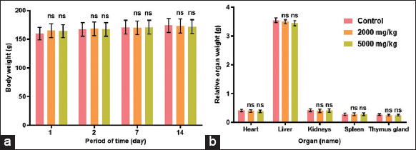 | Figure 4: Body weight (a) and relative organ weight (b) during acute toxicity test of Arctium tomentosum Mill. root extract. Data are expressed as mean ± standard deviation; n = 3, *P < 0.05, not significant (ns) compared to the control group. [Click here to view] |
28-day oral administration of the A. tomentosum Mill. root extract at the dose of 200 mg/kg did not induce vigorous signs of toxicity. No mortality, as well as abnormal signs in behavior, lesions on the skin, eyes, fur, and mucous membrane were observed. Similarly, to the acute test, no significant changes in the body weight and the relative weight of the organs (heart, liver, kidneys, spleen, and thymus gland) were indicated [Figure 5a and b].
 | Figure 5: Body weight (a) and relative organ weight (b) during sub-chronic toxicity test of Arctium tomentosum Mill. root extract. Data are expressed as mean ± standard deviation; n = 3, *P < 0.05, not significant (ns) compared to the control group. [Click here to view] |
The results of the blood analysis after 28-day sub-chronic administration of the extract are presented in Table 2.
Table 2: Blood cells and biochemical parameters of rats after 28-day oral administration of Arctium tomentosum Mill. root extract.
| Parameter | Control | 200 mg/kg |
|---|---|---|
| RBC, 106cells/μL | 6.10±0.6 | 5.30±0.7 |
| HGB, g/dL | 16.10±0.7 | 15.30±0.8 |
| HCT, % | 32.0±0.9 | 30.50±1.0 |
| MCV, fl | 60.0±1.0 | 59.20±1.5 |
| MCH, pg/cell | 30.3±1.1 | 28.9±1.6 |
| MCHC, g/dL | 51.20±1.0 | 49.70±1.3 |
| WBC, cells/μL | 5850±775 | 5600±850* |
| PLT, 103cells/μL | 815±115 | 857±125 |
| SGOT, IU/L | 145.0±2.5 | 157.0±3.7 |
| SGPT, IU/L | 35.1±1.7 | 38.5±2.0 |
| Creatinine, mg/dL | 0.5±0.0 | 0.6±0.0 |
| Total plasma protein, g/L | 6.5±0.0 | 6.1±0.0 |
| Urea, mg/dL | 46.8±2.0 | 48.2±2.2 |
Data are expressed as mean±SD, n=3, *P<0.05, compared to the control group, RBC: Red blood cells, HGB: Hemoglobin, HCT: Hematocrit, MCV: Mean corpuscular hemoglobin, MCHC: Mean corpuscular concentration, WBC: White blood cells, PLT: Platelets, SGOT: Serum Glutamic-oxaloacetic Transaminase, or Aspartataminotransferase, SGPT: Serum Glutamic pyruvic Transaminase, or Alaninaminotransferase
According to Table 2, a slightly decrease in WBCs count was found in the test group, compared to the control; however, it was remained within the normal range [39].
During microscopic investigation, no pathological changes in the structure of the liver [Figure 6a-c] and kidneys [Figure 7a-c] were observed.
 | Figure 6: Histologic aspect of the liver: (a) The control group, (b) the acute toxicity test group, and (c) the sub-chronic toxicity test group. There were no pathological changes in the structure and the shape of hepatocytes. Stain Hematoxylin and Eosin. Mag. ×400. [Click here to view] |
 | Figure 7: Histologic aspect of the kidneys: (a) The control group, (b) the acute toxicity test group, and (c) the sub-chronic toxicity test group. There were no pathological changes in the structure of the cortex and medulla. Stain H&E. Mag. ×400. [Click here to view] |
Overall, this study evaluates that the A. tomentosum Mill. root extract has no acute toxic effect at the doses of 2000 mg/kg and 5000 mg/kg, as well as no sub-chronic toxicity effect at the dose 200 mg/kg. It can be classified as practically nontoxic according to Hodge and sterner scale [40].
The studied dose for sub-chronic toxicity was chosen based on the previous research of Yaghoubi et al. (2018) [41]. In the group treated with 200 mg/kg of the extract over the period of 28 days, there were no significant changes in the body weight; however, a few toxic effects on lungs were observed. Hematological profile showed no significant difference between the control group and the group received the extract at the dose of 200 mg/kg for 28 days. Thus, the present study suggests a dose <200 mg/kg b.w. of the extract for the daily oral treatment. The results obtained here are in agreement with Yaghoubi et al. (2018) study of the plant of the same genus A. lappa L. Overall, in the presented in vivo adult Wistar rats appeared to tolerate A. tomentosum Mill. root extract at the acute dose of 5000 mg/kg b.w. well. The phytochemical composition of A. tomentosum Mill. root has not been widely analyzed so far, however, arctiin and arctigenin were found in its fruit extract [42]. Antioxidant compounds such as campesterol, squalene, lupeol, and tocopherols were found in the flowers and leaves [43,44]. Tannins are the most common in the root of the burdock. Since they exert inhibitory and cytotoxic activity on the tumor growth, they can cause some toxic effect on the liver and kidney in large doses [45].
4. CONCLUSION
The present study was conducted to evaluate the cytotoxic and toxic potential of the A. tomentosum Mill. root extract. The first objective was to study the cytotoxicity of the extract by MTT test. As a result, the 78–99% cell viability indicating the low cytotoxic effect was observed at the concentrations between 0.078 mg/mL and 0.001 mg/mL. The IC50 of the A. tomentosum Mill. root extract on the peripheral blood mononuclear cells was 0.091 mg/mL. The second objective was to study the acute and sub-chronic toxicity of the A. tomentosum Mill. extract in the adult Wistar rats. As a result, the extract showed no signs of toxicity at the doses of 2000 mg/kg and 5000 mg/kg b.w. and no death was observed in the first 24 h after oral administration of the extract. There were no significant changes in the body weight, physical state, and behavioral patterns between the control group and groups received the extract. In conclusion, the present work demonstrated that the A. tomentosum Mill. root extract is practically non-toxic and its LD50 is >5000 mg/kg. Sub-chronic administration of the extract at the dose of 200 mg/kg did not cause any obvious pathology in the organs of rats. Thus, the extract can be considered as safe in the limited doses.
5. AUTHORS’ CONTRIBUTIONS
All authors made substantial contributions to conception and design, acquisition of data, or analysis and interpretation of data; took part in drafting the article or revising it critically for important intellectual content; agreed to submit to the current journal; gave final approval of the version to be published; and agreed to be accountable for all aspects of the work. All the authors are eligible to be an author as per the International Committee of Medical Journal Editors (ICMJE) requirements/guidelines.
6. FUNDING
This research received no external funding.
7. CONFLICTS OF INTEREST
The authors report no financial or any other conflicts of interest in this work.
8. ETHICAL APPROVALS
The animal studies were approved by the Ethical Committee of JSC “Scientific Center for Anti-Infectious Drugs” No. 23/7 in accordance with the “Guide for the Care and Use of Laboratory Animals” and ARRIVE guidelines.
9. DATA AVAILABILITY
All the data is available with the authors and shall be provided upon request.
10. PUBLISHER’S NOTE
This journal remains neutral with regard to jurisdictional claims in published institutional affiliation.
REFERENCES
1. Ugwah-Oguejiofor CJ, Okoli CO, Ugwah MO, Umaru ML, Ogbulie CS, Mshelia HE, et al. Acute and sub-acute toxicity of aqueous extract of aerial parts of Caralluma dalzielii N. E. Brown in mice and rats. Heliyon 2019;5:e01179. [CrossRef]
2. Ibrahim JA, Muazzam I, Jegede I, Kunle O. Medicinal plants and animals sold by the Yan-Shimfidas of Sabo Wuse in Niger state, Nigeria. Afr J Pharm Pharmacol 2009;4:386-94. [CrossRef]
3. Skowro?ska W, Granica S, Dziedzic M, Kurkowiak J, Ziaja M, Bazylko A. Arctium lappa and Arctium tomentosum, sources of arctii radix:Comparison of anti-lipoxygenase and antioxidant activity as well as the chemical composition of extracts from aerial parts and from roots. Plants (Basel) 2021;10:78. [CrossRef]
4. Alhusaini A, Fadda L, Hasan IH, Ali HM, El Orabi NF, Badr AM, et al. Arctium lappa root extract prevents lead-induced liver injury by attenuating oxidative stress and inflammation, and activating Akt/GSK-3βsignaling. Antioxidants (Basel) 2019;8:582. [CrossRef]
5. Liu W, Wang J, Zhang Z, Xu J, Xie Z, Slavin M, et al. In vitro and in vivo antioxidant activity of a fructan from the roots of Arctium lappa L. Int J Biol Macromol 2014;65:446-53. [CrossRef]
6. Ghorat F, Azizkhani M, Naji S, Ranjbary AG, Doostishoar F. Histopathological evaluation of burdock (Arctium lappa) root hydroalcoholic extract on wound healing. Iran Red Crescent Med J 2017;19:e43788. [CrossRef]
7. Ferracane R, Graziani G, Gallo M, Fogliano V, Ritieni A. Metabolic profile of the bioactive compounds of burdock (Arctium lappa) seeds, roots and leaves. J Pharm Biomed Anal 2010;51:399-404. [CrossRef]
8. Wang D, B?d?rau AS, Swamy MK, Shaw S, Maggi F, da Silva LE, et al. Arctium species secondary metabolites chemodiversity and bioactivities. Front Plant Sci 2019;10:834. [CrossRef]
9. Knott A, Reuschlein K, Mielke H, Wensorra U, Mummert C, Koop U, et al. Natural Arctium lappa fruit extract improves the clinical signs of aging skin. J Cosmet Dermatol 2008;7:281-9. [CrossRef]
10. Strawa J, Wajs-Bonikowska A, Jakimiuk K, Waluk M, Poslednik M, Nazaruk J, et al. Phytochemical examination of woolly burdock Arctium tomentosum leaves and flower heads. Chem Nat Comp 2020;56:345-7. [CrossRef]
11. Ge L, Liu F, Hu Y, Zhou X. Qualitative and quantitative analysis of arctiin and arctigenin in Arctium tomentosum Mill. by high-performance thin-layer chromatography. JPC J Planar Chromat 2020;33:19-26. [CrossRef]
12. Bhatt NF, Gupta RC, Bansal Y. Secondary metabolites in Arctium lappa L.:Variation among plant parts and phenological stages. JPC J Planar Chromat 2019;32:461-5. [CrossRef]
13. Gao Q, Yang M, Zuo Z. Overview of the anti-inflammatory effects, pharmacokinetic properties and clinical efficacies of arctigenin and arctiin from Arctium lappa L. Acta Pharmacol Sin 2018;39:787-801. [CrossRef]
14. Yu HN, Zhu J, Pan WS, Shen SR, Shan WG, Das UN. Effects of fish oil with a high content of n-3 polyunsaturated fatty acids on mouse gut microbiota. Arch Med Res 2014;45:195-202. [CrossRef]
15. Fierascu RC, Georgiev MI, Fierascu I, Ungureanu C, Avramescu SM, Ortan A, et al. Mitodepressive, antioxidant, antifungal and anti-inflammatory effects of wild-growing Romanian native Arctium lappa L. (Asteraceae) and Veronica persica Poiret (Plantaginaceae). Food Chem Toxicol 2018;111:44-52. [CrossRef]
16. Mssillou I, Agour A, Hamamouch N, Lyoussi B, Derwich E. Chemical composition and in vitro antioxidant and antimicrobial activities of Marrubium vulgare L. ScientificWorldJournal 2021;2021:7011493. [CrossRef]
17. Xu Z, Ju J, Wang K, Gu C, Feng Y. Evaluation of hypoglycemic activity of total lignans from Fructus arctii in the spontaneously diabetic Goto-Kakizaki rats. J Ethnopharmacol 2014;151:548-55. [CrossRef]
18. Parasuraman S. Toxicological screening. J Pharmacol Pharmacother 2011;2:74-9. [CrossRef]
19. Hashemi SA, Abediankenari S, Ghasemi M, Azadbakht M, Yousefzadeh Y, Dehpour AA. The effect of fig tree latex (Ficus carica) on stomach cancer line. Iran Red Crescent Med J 2011;13:272-5.
20. Nemati F, Dehpouri AA, Eslami B, Mahdavi V, Mirzanejad S. Cytotoxic properties of some medicinal plant extracts from Mazandaran, Iran. Iran Red Crescent Med J 2013;15:e8871. [CrossRef]
21. Organization for Economic Co-operation and Development (OECD/OCDE). Test. No. 425:Acute Oral Toxicity:Up-and-Down Procedure;OECD Guidelines for the Testing of Chemicals. Paris, France:OECD;2008. 1-27. Available from:https://www.oecd-ilibrary.org/environment/test-no-425-acute-oral-toxicity-up-and-down-procedure_9789264071049-en [Last accessed on 2023 Mar 16].
22. Organization for Economic Co-operation and Development (OECD/OCDE). Test. No. 407:Repeated Dose 28-Day Oral Toxicity Study in Rodents;OECD Guidelines for the Testing of Chemicals. Paris, France:OECD;2018. 1-16. Available from:https://www.oecd-ilibrary.org/environment/test-no-407-repeated-dose-28-day-oral-toxicity-study-in-rodents_9789264070684-en [Last accessed on 2023 Mar 16].
23. Zhang S, Yang X, Coburn RA, Morris ME. Structure activity relationships and quantitative structure activity relationships for the flavonoid-mediated inhibition of breast cancer resistance protein. Biochem Pharmacol 2005;70:627-39. [CrossRef]
24. Romero-Benavides JC, Ortega-Torres GC, Villacis J, Vivanco-Jaramillo SL, Galarza-Urgilés KI, Bailon-Moscoso N. Phytochemical study and evaluation of the cytotoxic properties of methanolic extract from Baccharis obtusifolia. Int J Med Chem 2018;2018:8908435. [CrossRef]
25. Øverby A, Zhao CM, Chen D. Plant phytochemicals:Potential anticancer agents against gastric cancer. Curr Opin Pharmacol 2014;19:6-10. [CrossRef]
26. Srivastava S, Somasagara RR, Hegde M, Nishana M, Tadi SK, Srivastava M, et al. Quercetin, a natural flavonoid interacts with DNA, arrests cell cycle and causes tumor regression by activating mitochondrial pathway of apoptosis. Sci Rep 2016;6:24049. [CrossRef]
27. Umesalma S, Nagendraprabhu P, Sudhandiran G. Ellagic acid inhibits proliferation and induced apoptosis via the Akt signaling pathway in HCT-15 colon adenocarcinoma cells. Mol Cell Biochem 2015;399:303-13. [CrossRef]
28. Maxwell T, Lee KS, Kim S, Nam KS. Arctigenin inhibits the activation of the mTOR pathway, resulting in autophagic cell death and decreased ER expression in ER-positive human breast cancer cells. Int J Oncol 2018;52:1339-49. [CrossRef]
29. Maxwell T, Chun SY, Lee KS, Kim S, Nam KS. The anti-metastatic effects of the phytoestrogen arctigenin on human breast cancer cell lines regardless of the status of ER expression. Int J Oncol 2017;50:727-35. [CrossRef]
30. Lee KS, Lee MG, Kwon YS, Nam KS. Arctigenin enhances the cytotoxic effect of doxorubicin in MDA-MB-231 breast cancer cells. Int J Mol Sci 2020;21:2997. [CrossRef]
31. Das U. Essential fatty acids enhance free radical generation and lipid peroxidation toinduce apoptosis of tumor cells. Clin Lipidol 2011;6:463-89. [CrossRef]
32. Polavarapu S, Dwarakanath BS, Das UN. Differential action of polyunsaturated fatty acids and eicosanoids on bleomycin-induced cytotoxicity to neuroblastoma cells and lymphocytes. Arch Med Sci 2018;14:207-29. [CrossRef]
33. Li Y, Jiang F, Chen L, Yang Y, Cao S, Ye Y, et al. Blockage of TGFβ-SMAD2 by demethylation-activated miR-148a is involved in caffeic acid-induced inhibition of cancer stem cell-like properties in vitro and in vivo. FEBS Open Bio 2015;5:466-75. [CrossRef]
34. Brautigan DL, Gielata M, Heo J, Kubicka E, Wilkins LR. Selective toxicity of caffeic acid in hepatocellular carcinoma cells. Biochem Biophys Res Commun 2018;505:612-7. [CrossRef]
35. Pelinson LP, Assmann CE, Palma TV, da Cruz IB, Pillat MM, Mânica A, et al. Antiproliferative and apoptotic effects of caffeic acid on SK-Mel-28 human melanoma cancer cells. Mol Biol Rep 2019;46:2085-92. [CrossRef]
36. Li W, Guo Y, Zhang C, Wu R, Yang AY, Gaspar J, et al. Dietary phytochemicals and cancer chemoprevention:A perspective on oxidative stress, inflammation, and epigenetics. Chem Res Toxicol 2016;29:2071-95. [CrossRef]
37. Chen Z, Duan H, Tong X, Hsu P, Han L, Morris-Natschke SL, et al. Cytotoxicity, hemolytic toxicity, and mechanism of action of Pulsatilla Saponin D and its synthetic derivatives. J Nat Prod 2018;81:465-74. [CrossRef]
38. Podolak I, Galanty A, Sobolewska D. Saponins as cytotoxic agents:A review. Phytochem Rev 2010;9:425-474. [CrossRef]
39. Delwatta SL, Gunatilake M, Baumans V, Seneviratne MD, Dissanayaka ML, Batagoda SS, et al. Reference values for selected hematological, biochemical and physiological parameters of Sprague-Dawley rats at the Animal House, Faculty of Medicine, University of Colombo, Sri Lanka. Anim Model Exp Med 2018;1:250-4. [CrossRef]
40. Hodge A, Sterner B. Toxicity classes. In:Canadian Center for Occupational Health and Safety. Available form:https://www.ccohs.ca/oshanswers/chemicals/ld50.html [Last accessed on 2023 Mar 16].
41. Yaghoubi M, Mousavi Z, Rastegar T, Amin G. Safety assessment of Arctium lappa L. fruit extract in female wistar rats:Acute and repeated oral toxicity studies. Res J Pharmacogn 2019;6:39-48.
42. Zhou X, Zhang H, Ge L, Gong H, Tian S. Determination of arctiin and arctigenin contents in Arctium tomentosum Mill. By HPLC method. J Chem 2011;8:372-7. [CrossRef]
43. Skowro?ska W, Granica S, Dziedzic M, Kurkowiak J, Ziaja M, Bazylko A. Arctium lappa and Arctium tomentosum, sources of Arctii radix:Comparison of anti-lipoxygenase and antioxidant activity as well as the chemical composition of extracts from aerial parts and from roots. Plants (Basel) 2021;10:78. [CrossRef]
44. Strawa J, Jakimiuk K, Waluk M, Poslednik M, Nazaruk J, Tomczyk M. Phytochemical examination of wolly burdock Arctium tomentosum leaves and flower heads. Chem Nat Compd 2020;56:345-7. [CrossRef]
45. Chung KT, Wong TY, Wei CI, Huang YW, Lin Y. Tannins and human health:A review. Crit Rev Food Sci Nutr 1998;38:421-64. [CrossRef]