
1. INTRODUCTION
Lymphomas, the malignant transformation of the cells of the immune system, are broadly classified as Hodgkin’s lymphoma (HL) and non-Hodgkin’s lymphoma (NHL). Hodgkin’s lymphoma in most of the cases is known to involve altered B-cells [1]. Globally, in males and females, NHL is ranked the 10th and 12th most frequently occurring type of cancers, respectively. It involves the B-lymphocytes and a smaller portion of T-lymphocytes and natural killer cells. However, its classification is not easy as it requires a deep insight into the clinical features and genetic abnormalities [2]. With advances in areas of science such as genomics and molecular biology, chemotherapy and immunotherapy are promising in combating cancer; however, multidrug resistance and drug induced toxicity remains a major cause of concern. Plant-derived drugs, an alternative treatment to fight cancer, are the need of the hour as it has the potential to deliver highly efficient and non-toxic treatment [3]. The efficacy of anticancer agents depends on its ability to increase apoptosis, decrease cell proliferation, induce cell differentiation, modulate intracellular pathways, and inhibit activity of DNA topoisomerase and angiogenesis. Several plants such as Luffa aegyptiaca, Beta vulgaris, Capsicum frutescens, Solenostemma argel, and Colocasia antiquorum are known to have anticancer potentials against acute myeloid leukemia and lymphocyte leukemia [4]. Tragia involucrata Linn. belongs to the Family Euphorbiaceae and is widely distributed in South Asian countries. The various parts of this plant have tremendous ethnopharmacological importance, as it has been known to be used against inflammation, allergy, epilepsy, renal stones, asthma, bronchitis, vomiting, diarrhea, and numerous other ailments [5]. The present study aims to evaluate the apoptotic and tumor reduction potentials of T. involucrata ethanolic extract using lymphoma (DLA solid tumor) models.
2. MATERIALS AND METHODS
2.1. Plant Authentication and Extraction
The whole plant of T. involucrata Linn. was procured from Thrissur district, India (10.3469°N, 76.2074°E) and a voucher specimen (Accession number 17682) was submitted at Kerala Forest Research Institute, Kerala, India. The plant, after being cleaned thoroughly, was dried at 50°C in a hot air oven and powdered for extraction. The fine plant material was weighed (15 g) and extracted in accelerated solvent extractor (Thermo Scientific, Dionex 150) using (200 ml) Tragia ethanol extract (TME). A vacuum evaporator was used to dry the crude extracts and it was stored in a refrigerator at 4°C until further use.
2.2. Cell Lines and Animals
YAC-1 and DLA cell lines used for the study were procured from National Center for Cell Sciences, Pune and Amala Cancer Research Center, Thrissur, respectively. YAC-1 cell line was cultured in RPMI medium supplemented with FBS (10% v/v), streptomycin (100 μg/mL), and penicillin (100 U/mL) and incubated at 37°C in a CO2 incubator (Heracell 150i MD) which provided 5% vol CO2, 95% relative humidity, and a pH of 7.4. DLA cell line was maintained in the peritoneal cavity of mice. Swiss albino mice (25–30 g) belonging to both the sexes were procured from Small Animal Breeding Station, Kerala Veterinary, and Animal Science University, Thrissur, India. The animals were maintained in the animal house facility of Amala Cancer Research Center and standard conditions (24–28°C, 60–70% humidity, 12 h dark/light cycle) were provided. The animals were fed with standard mice feed from Sai Durga Feeds, Bangalore, India and water ad libitum. A prior approval from the Institutional Animal Ethics Committee (IAEC) (ACRC/IAEC/20(1)-P06) was obtained and the guidelines of the Committee for the Purpose of Control and Supervision of Experiments on Animals constituted by the Ministry of Environment and Forest, Government of India were strictly adhered to throughout the experiment.
2.3. Trypan Blue Exclusion Assay
The use of MTT assay was avoided in our study, as the plant extract contains diverse group of chemicals, and they may reduce the activity of MTT compound, giving false positive results for cell viability [6]. Cytotoxicity assay was conducted according to Strober (2015) [7] with minor modifications. Cells were seeded in six well plates with RPMI medium (1.0 × 10⁵ cells/well) and treated with different concentrations of extract at 37°C in a CO2 (5%) incubator. Trypan blue was added after the incubation and it was kept at room temperature for 3 min. The observations were made in a hemocytometer under a microscope (×20). The percentage was calculated using the formula:
 |
2.4. Gas chromatography–mass Spectrometry (GC–MS) Analysis
The ethanol extract (TME) was examined using the Shimadzu GC-MS instrument (model no: QP2010S). ELITE 5-MS column with 30 m × 0.25 mm (inner diameter) and 0.25 μm film thickness was used for performing GC. 1 μl was the injection volume and the temperature and pressure provided were 260°C and 61.5 kPa, respectively. The sample injections were done in the split mode and every 0.5 s over a range set with an m/z of 50–500 Da, the mass spectra was recorded. The initial oven temperature was 70.0°C for 2 min and then increased to 200°C at the rate of 10°C/min. The temperature was then maintained at 280°C for an additional 15 min. The computation of phytochemical components from GC peak areas was automatically done by the software. The library searches were done using NIST 11 and WILEY 8 and the comparison of the mass spectrum with the reference spectrum was done to identify exact names of fourteen unknown components.
2.5. Liquid chromatography–mass Spectrometry (LC–MS) Analysis
For detection of nonvolatile analytes in TME, Ultra Performance Liquid chromatography coupled to UPLC-Q-ToF-MS (Quadrupole time-of-flight mass spectrometry) was done. The Acquity UPLC system (Waters) with a TUV detector (J12 TUV750A), sample manager FTN (K12 SD1069G) a column chamber (J12 CHA730G), and a quaternary solvent manager (H12 QSM632A) was used. Reverse phased BEH C18 column (50 mm × 2.1 mm × 1.7 μm) with 0.3 mL/min flow rate was used for chromatographic separation. Mixture of water and acetonitrile with 0.1% formic acid was used as the mobile phase in a gradient mode. ESI ionization mode was used to study the samples and 10 μL of sample was injected. The m/z range scanned was between 50 and 1000. The dissolution gas flow was at the rate of 900 L/h at 350°C and collision energy ranged from 5 to 30 eV. For data acquisition, Masslynx software (4.1) was used.
2. 6. DLA-Induced Solid Tumor Model
The nontoxic doses were calculated after conducting acute and subacute toxicity studies. The doses for subacute toxicity studies were calculated as 1/5 (high dose) and 1/10 (low dose) of the nontoxic single oral dose (2g/kg b. wt) given in the acute toxicity studies. The subacute toxicity studies were then conducted with the low and high doses of 200 and 400 mg/kg b. Wt, respectively, for 28 days. After ensuring the safety of these doses, further antitumor studies were conducted using same dosage. Male Swiss albino mice (6–8 weeks old) weighing 25–30 g were sought into five groups (n = 6) for different treatments as follows. Group 1 and 2: Control (untreated tumor induced mice) and vehicle control (1% propylene glycol), respectively, Group 3: Standard (cyclophosphamide drug 10 mg/kg b. wt), and Group 4 and 5: TME low dose (200 mg/kg b. wt) and high dose (400 mg/kg b. wt), respectively. DLA cells were aspirated from the peritoneal cavity of tumor bearing mice and washed in PBS. The tumor was induced by intramuscularly injecting approximately 1 × 10⁶ of DLA cells in 100 μL of saline into the right hind limb of all animals. Twenty-four hours post-tumor cell inoculation, the treatment was initiated by oral administration of the extract and it continued for 10 consecutive days. The tumor growth was measured with a Vernier caliper, by measuring the diameter of its growth in two perpendicular planes. With an interval of 3 days between each reading, the readings were taken for 30 days. The tumor volume was calculated using the formula;
 |
where,
r1; minor radius and r2; major radius.
2.7. Statistical Analysis
The values are expressed as Mean ± SD. Student’s t-test was used to analyze P-value, thereby understanding the level of significance.
3. RESULTS
3.1. GC–MS Analysis
Identification and characterization of several compounds were possible by comparing the mass spectrum obtained for each with the reference spectrum. The major chemical compounds in ethanol extract identified are listed in Table 1. Figure 1 shows the peaks obtained in the GC–MS analysis.
Table 1: Compounds identified by GC-MS.
| Peak no | Retention time | Area (%) | Name | Molecular formula |
|---|---|---|---|---|
| 1 | 10.308 | 1.06 | Phenylacetaldehyde- diethylacetal | C12H18O2 |
| 2 | 16.41 | 6.61 | Neophytadiene | C20H38 |
| 3 | 17.313 | 1.29 | (E)-Phytol | C20H40O |
| 4 | 19.447 | 7.36 | Ethyl palmitate | C18H36O2 |
| 5 | 22.297 | 8.18 | Phytol | C20H40O |
| 6 | 23.358 | 4.94 | Ethyl linolate | C20H26O2 |
| 7 | 23.468 | 3.18 | Ethyl elaidate | C20H38O2 |
| 8 | 23.517 | 3.80 | Linolenic acid | C18H30O2 |
| 9 | 24.010 | 1.95 | Ethyl octadecanoate | C20H40O2 |
| 10 | 30.732 | 13.08 | 2-Ethylhexyl phthalate | C24H38O4 |
| 11 | 34.974 | 13.91 | Squalene | C30H50 |
| 12 | 40.139 | 11.21 | Vitamin E | C29H50O2 |
| 13 | 44.720 | 21.05 | Clionasterol | C29H50O2 |
| 14 | 47.183 | 4.41 | Viminalol | C30H50O |
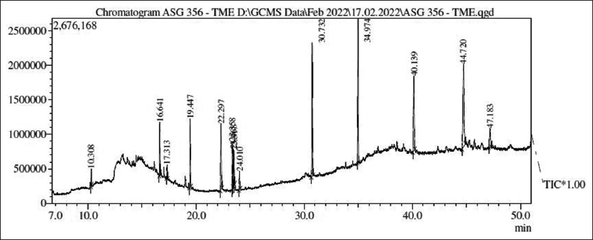 | Figure 1: Graph showing peaks in GC experiment. [Click here to view] |
3.2. LC–MS Analysis
Ten most abundant compounds were identified and the details are given in Table 2. The total ion chromatogram of TME in both positive and negative ion modes is shown in Figure 2.
Table 2: Compounds identified by LC-MS.
| Retention time | Name | Molecular Formula | m/z value |
|---|---|---|---|
| 4.310 | Agathisflavone | C30H18O10 | 537.082 |
| 4.153 | Loquatoside | C20H22O11 | 437.107 |
| 3.970 | Leufolin A | C30H28O12 | 579.150 |
| 3.813 | Quercetin | C15H10O7 | 303.049 |
| 3.805 | Echinacin | C30H26O12 | 577.133 |
| 3.257 | Apigetrin | C21H20O10 | 431.097 |
| 3.057 | Cynaroside | C21H20O11 | 447.092 |
| 2.926 | 1,2,36-tetrakis-O- galloyl-B-D-glucose | C34H28O22 | 787.095 |
| 2.883 | Isoquercetin | C21H20O12 | 465.104 |
| 2.674 | Corilagin | C27H22O18 | 633.072 |
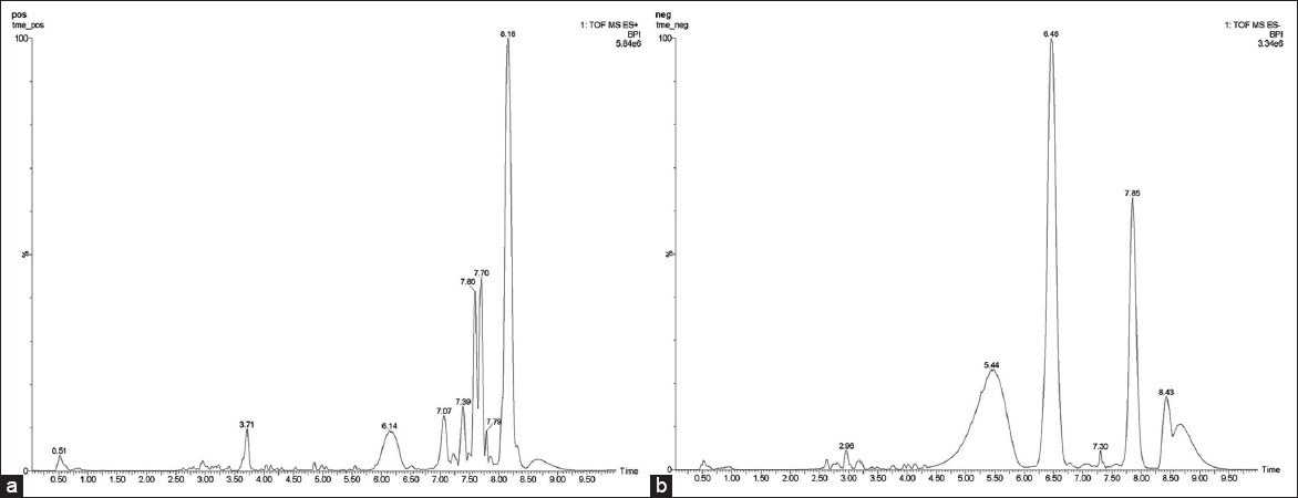 | Figure 2: LC-MS analysis of the extract. (a) Total ion chromatogram of TME in the positive ion mode. (b) Total ion chromatogram of TME in the negative ion mode. [Click here to view] |
3.3. Antiproliferation Effect of TME In Vitro
A cell proliferation assay was conducted to examine the growth inhibition exerted by TME on YAC-1 cells. The cells were treated for 12, 24, and 48 h, with different concentrations (5, 10, and 15 μg/mL). The potential of TME to induce cell death for a prolonged time at physiological temperature was revealed, and also, there were a concentration and time dependent reduction in the number of live cells [Figure 3].
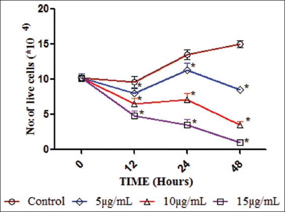 | Figure 3: Cell proliferation. Graph showing the number of live cells (YAC-1) at three different time points (12, 24, and 48 h) after treatment with extract (5, 10, and 15 μg/ml). Comparisons were made between vehicle controls with the treated groups separately at each time point. The symbol (*) represents statistical significance at P ≤ 0.05. [Click here to view] |
3.4. Antitumor Studies
The extract showed that no signs of toxicity in animals as no mortality or behavioral changes were observed. It also reduces cell proliferation in culture, which prompted us to evaluate its tumor reduction potential in the in vivo system. The tumor volume showed significant reduction (P ≤ 0.05) when compared to the vehicle control at the 30th day of tumor study [Figure 4]. Tumour tissue was further used for histopathological analysis that also showed correlating results [Figure S1, S2].
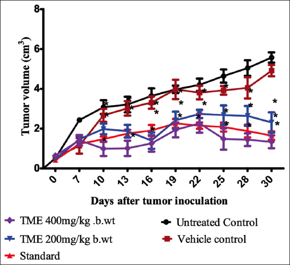 | Figure 4: Tumor volume. Graph showing the volume of DLA solid tumors in Swiss albino mice for 30 days. Untreated control, vehicle control, and standard (Cyclophosphamide) groups are included. Comparisons were made between vehicle control with the extract treated groups separately. The symbol (*) represents statistical significance at P ≤ 0.05. [Click here to view] |
4. DISCUSSION
Since aerial parts of T. involucrata were known to be traditionally used in few villages of Kerala, for treating tumors. A study was conducted earlier in ascites tumor models using ethyl acetate and hexane extracts of aerial parts of T. involucrata and a notable decrease in tumor burden and considerable increase in lifespan was observed [8]. The ethyl acetate and chloroform extracts of leaves of T. involucrata showed potent antiproliferative effect when tested against K562 (chronic myelogenous leukemia) cell lines [9]. Further, exploration of this activity was not conducted, and hence, here, we used the ethanol extract (TME) of T. involucrata to conduct an elaborate antitumor study. TME was effectively interfering with cell proliferation and has the ability to induce apoptosis in tumor cells.
The antiproliferative and proapoptotic changes in the tumor tissue can be attributed to several of the phytocompounds present in the extract that was revealed by GC-MS analysis. Neophytadiene detected in TME extracts are known to inhibit the nuclear factor kappa B (NF-κB) translocation to the nucleus and, thereby, significantly reduce the pro-inflammatory cytokine production [10]. In several types of cancer, especially, the ones with hematopoietic origin are known to have mutations in members of NF-κB protein family. Elevated NF-κB levels in tumor tissues promote increased accumulation of proinflammatory cytokines and this creates a protumorigenic microenvironment. Apart from this, the inhibition of NF-κB had inhibitory effects on tumor angiogenesis as it abolishes vascular endothelial growth factor production, a vital angiogenic factor [11]. Phytols detected in the extracts have shown antiproliferative effects and also induced apoptosis in lung cancer cell lines [12]. In HepG2 hepatocellular carcinoma cells, phytols that induced apoptosis were found to induce it through caspase 9/3 activation and also were found to inhibit EMT (epithelial mesenchymal transition) which plays a key role in tumor invasion [13]. Ethyl palmitate and ethyl linoleate present in the extract were also known to have inhibitory effects on NF-κB [14,15]. The NF-κB inhibition by linolenic acid is also well studied and was known to decrease the NO production in both breast and cervical cancer cell lines [16,17]. The inhibition of NF-κB/Rel induced apoptosis in murine B-cells [18]. The silencing of C1s (a protein of the complement system) reduced cell proliferation and viability in ccRcc (clear cell renal carcinoma) [19]. Clionasterol, a compound detected in the TME extract, was known to be a potent inhibitor of C1 protein [20]. Thus, it may play a major role in reducing cell proliferation. Viminalol was also known to possess anti leukemia properties [21].
The LC–MS analysis of the extract revealed several non-volatile compounds present in the extract with pharmacological importance. Most of the compounds detected through LC–MS belong to the flavonoid group of polyphenolic compounds with numerous potentials. Flavonoids are a well-known class of polyphenolic compounds, with immense potential to put a check on cell proliferation and invasiveness, induce apoptosis, arrest cell cycle, and modulate ROS-scavenging enzyme activities [22]. Agathisflavaone showed promising therapeutic potentials in docking studies conducted against important biomacromolecules with therapeutic importance. NF-κB is involved in cell survival and maintenance, and hence, inhibition of NF-κB signaling will lead to apoptosis of the cell. Agathisflavaone showed successful binding to NF-κB as well as cytokines IL-β and IL-10, in which the former is responsible for tumor invasion and metastasis and the latter for inhibiting the immune clearance of tumor cells. Therefore, agathisflavone can be considered a potential source of natural anticancer agent [23]. Quercetin, an important anticancer agent, has therapeutic effects in various types of cancer cells, with various target points. In lymphoma, it has a therapeutic role as they target the mitochondrial system, which is impaired in them. TNF-related apoptosis inducing ligand (TRAIL) is a cytokine that induces apoptosis by binding to its death inducing receptors TRAIL 1 and TRAIL 2. In resistant B-cell lymphoma cell lines, the TRAIL apoptosis mechanism was restored by quercetin. It also inhibited the expression of survivin (BIRC5) and myeloid cell leukemia 1 (Mcl-1), which would otherwise inhibit caspase 9/3. The inhibition of survivin by quercetin aided in caspase 9/3 activation and led to apoptosis. Similarly, the Mcl-1 proteasomal degradation resulted in the activation of BAX, thereby releasing cytochrome c from the mitochondria ultimately leading to apoptosis [24]. Apigetrin showed significant anticancer activity against gastric cancer cells (ASG), by inducing apoptosis through the extrinsic pathway, as the expression of FasL and death receptor 4 proteins were significantly elevated. This eventually led to increase in the caspase 3 and 8 resulting in Poly (ADP-ribose) polymerase cleavage and apoptosis [25]. Apigetrin also induced apoptosis in gastric cancer cells, through the intrinsic pathway by generating ROS, and changing the membrane potential of mitochondria. It was also noted to reduce the IL-6 mediated phosphorylation of signal transducer and activator of transduction (STAT) 3, which could augment the anti-apoptotic response. The upstream signal molecules Janus Kinase-2 and src of STAT 3 signaling pathway were also inactivated by apigetrin, thus exhibiting its strong apoptosis inducing potential [26]. A vital oncogenic determinant called Mesenchymal-Epithelial-Transition (MET) protein (the opposite of EMT) is known to regulate the occurrence, development and deterioration of tumors, and hence decrease in levels of MET will aid in inhibiting the activation of its downstream related pathway thus inhibiting cell survival. Cynaroside was known to enhance the ubiquitin degradation of MET in gastric cancer cells, thus proving to be a good anticancer agent [27]. Corilagin is a very potent anticancer molecule and targets various pathways in various types of cancers. Pheochromocytoma is a hormone secreting tumor of adrenal medulla. Corilagin was found to reduce NF-κB activation in this type of cancer. The NF-κB1 transcription factor has five members namely; Rel A (p65), Rel B, c-Rel, NF-κB1(p50), and NF-B2 (p52). It prevented the phosphorylation of RelA in a concentration dependent manner putting a check on NF-κB activation. In ovarian cancer cells, corilagin was found to interfere with the TGF-β signaling pathway. TGF-β was down regulated by corilagin and it thereby prevented the phosphorylation of its downstream signaling proteins such as suppressor of mothers against decapentaplegic 2, AK strain transforming, and extracellular regulated kinase, leading to cell cycle arrest and apoptosis. In cholangiocarcinoma, corilagin suppressed the expression of downstream target molecules of notch signaling pathway and thus led to apoptosis of the cell [28].
5. CONCLUSION
T. involucrata Linn. is an important ingredient in major Ayurvedic formulations called “Vidaryadi Kwatham Chuna” and “Vidyaryadi Ghrita” [29]. Several potent bioactive molecules were detected in the ethanol extract of T. involucrata. Many of these molecules have strong anticancer properties, and thus, the tumor reduction ability of the plant extract may be attributed to these biomolecules. The biomolecules may be individually contributing to the tumor reduction property or may be functioning in a synergistic manner. Thus, further studies are required to understand the exact molecules which impart the tumor reducing ability.
6. AUTHORS’ CONTRIBUTIONS
All authors made substantial contributions to conception and design, acquisition of data, or analysis and interpretation of data; took part in drafting the article or revising it critically for important intellectual content; agreed to submit to the current journal; gave final approval of the version to be published; and agree to be accountable for all aspects of the work. All the authors are eligible to be an author as per the international committee of medical journal editors (ICMJE) requirements/guidelines.
7. FUNDING
There is no funding to report.
8. CONFLICTS OF INTEREST
The authors declare no conflicts of interest with this manuscript.
9. ACKNOWLEDGMENTS
Authors like to thank Dr. Achuthan C Ragavamenon and Ms. Soorya Parathodi Illam of Amala Cancer Research Center, Amalanagar, Thrissur for providing necessary help and facilities for the animal experiments. Authors are also thankful to Rajiv Gandhi Center for Biotechnology for availing the flow cytometry service in their Laboratory. Madhuri Menon is a recipient of Christ College Research Fellowship (CCRF).
10. ETHICAL APPROVALS
All the animal experiments were conducted after getting approval from the Institutional animal Ethics Committee (IAEC) (ACRC/IAEC/20(1)-P06).
11. DATA AVAILABILITY
The authors confirm that the data supporting the findings of this study are available within the article and/or its supplementary materials.
12. PUBLISHER’S NOTE
This journal remains neutral with regard to jurisdictional claims in published institutional affiliation.
REFERENCES
1. Amini RM, Enblad G. Relationship between Hodgkin's and non-Hodgkin's lymphomas. Med Oncol 2003;20:211-20. [CrossRef]
2. Miranda-Filho A, Piñeros M, Znaor A, Marcos-Gragera R, Steliarova-Foucher E, Bray F. Global patterns and trends in the incidence of non-Hodgkin lymphoma. Cancer Causes Control 2019;30:489-99. [CrossRef]
3. Zou H, Li Y, Liu X, Wu Z, Li J, Ma Z. Roles of plant-derived bioactive compounds and related microRNAs in cancer therapy. Phytother Res 2021;35:1176-86. [CrossRef]
4. Gezici S, ?ekero?lu N. Current perspectives in the application of medicinal plants against cancer:Novel therapeutic agents. Anticancer Agents Med Chem 2019;19:101-11. [CrossRef]
5. Pallie MS, Perera PK, Kumarasinghe N, Arawwawala M, Goonasekara CL. Ethnopharmacological use and biological activities of Tragia involucrata L. Evid Based Complement Alternat Med 2020;2020:884?. [CrossRef]
6. Karaka?D, Ari F, Ulukaya E. The MTT viability assay yields strikingly false-positive viabilities although the cells are killed by some plant extracts. Turk J Biol 2017;41:919-25. [CrossRef]
7. Strober W. Trypan blue exclusion test of cell viability. Curr Protoc Immunol 2015;111:A3.B.1-A3.B.3. [CrossRef]
8. Chandrashekhar CG, Gopal M, Kumari NS. Antitumor activity of hexane and ethyl acetate extracts of Tragia involucrata. Int J Cancer Res 2011;7:267-77. [CrossRef]
9. Thomas R, Megha KB, Surya PH, Rosalin T, Varghese S, Elyas KK. Investigation on the biological attributes of Tragia involucrata Linn. using in vitro methods. J. Pharmacogn Phytochem 2021;10:398-404. [CrossRef]
10. Bhardwaj M, Sali VK, Mani S, Vasanthi HR. Neophytadiene from Turbinaria ornata suppresses LPS-induced inflammatory response in RAW 264.7 macrophages and Sprague Dawley rats. Inflammation 2020;43:937-50. [CrossRef]
11. Xia Y, Shen S, Verma IM. NF-kB, an active player in human cancers. Cancer Immunol Res 2014;2:823-30. [CrossRef]
12. Sakthivel R, Malar DS, Devi KP. Phytol shows anti-angiogenic activity and induces apoptosis in A549 cells by depolarizing the mitochondrial membrane potential. Biomed Pharmacother 2018;105:742-52. [CrossRef]
13. Kim CW, Lee HJ, Jung JH, Kim YH, Jung DB, Sohn EJ, et al. Activation of caspase-9/3 and inhibition of epithelial mesenchymal transition are critically involved in antitumor effect of phytol in hepatocellular carcinoma cells. Phytother Res 2015;29:1026-31. [CrossRef]
14. Saeed NM, El-Demerdash E, Abdel-Rahman HM, Algandaby MM, Al-Abbasi FA, Abdel-Naim AB. Anti-inflammatory activity of methyl palmitate and ethyl palmitate in different experimental rat models. Toxicol Appl Pharmacol 2012;264:84-93. [CrossRef]
15. Park SY, Seetharaman R, Ko MJ, Kim TH, Yoon MK, Kwak JH, et al. Ethyl linoleate from garlic attenuates lipopolysaccharide-induced pro-inflammatory cytokine production by inducing heme oxygenase-1 in RAW264. 7 cells. Int Immunopharmacol 2014;19:253-61. [CrossRef]
16. Kim KB, Nam YA, Kim HS, Hayes AW, Lee BM. a-Linolenic acid:Nutraceutical, pharmacological and toxicological evaluation. Food Chem Toxicol 2014;70:163-78. [CrossRef]
17. Deshpande R, Mansara P, Suryavanshi S, Kaul-Ghanekar R. Alpha-linolenic acid regulates the growth of breast and cervical cancer cell lines through regulation of NO release and induction of lipid peroxidation. J Mol Biochem 2013;2:6-17.
18. Wu M, Lee H, Bellas RE, Schauer SL, Arsura M, Katz D, et al. Inhibition of NF-kappaB/Rel induces apoptosis of murine B cells. EMBO J 1996;15:4682-90. [CrossRef]
19. Daugan M, Revel M, Russick J, Dragon-Durey MA, Gaboriaud C, Robe-Rybkine T, et al. Complement C1s and C4d as prognostic biomarkers in renal cancer:Emergence of noncanonical functions of C1s. Cancer Immunol Res 2021;9:891-908. [CrossRef]
20. Cerqueira F, Watanadilok R, Sonchaeng P, Kijjoa A, Pinto M, van Ufford HQ, et al. Clionasterol:A potent inhibitor of complement component C1. Planta Med 2003;69:174-6. [CrossRef]
21. Neto SF, Prada AL, Achod LD, Torquato HF, Lima CS, Paredes-Gamero EJ, et al. a-amyrin-loaded nanocapsules produce selective cytotoxic activity in leukemic cells. Biomed Pharmacother 2021;139:111656. [CrossRef]
22. Kopustinskiene DM, Jakstas V, Savickas A, Bernatoniene J. Flavonoids as anticancer agents. Nutrients 2020;12:457. [CrossRef]
23. Islam MT, Zihad SM, Rahman MS, Sifat N, Khan MD, Uddin SJ, et al. Agathisflavone:Botanical sources, therapeutic promises, and molecular docking study. IUBMB Life 2019;71:1192-200. [CrossRef]
24. Soofiyani SR, Hosseini K, Forouhandeh H, Ghasemnejad T, Tarhriz V, Asgharian P, et al. Quercetin as a novel therapeutic approach for lymphoma. Oxid Med Cell Longev 2021;2021:3157867. [CrossRef]
25. Kim SM, Preethi V, Ha SE, Kim HH, Kim JA, Kim GS. Apigetrin induces extrinsic apoptosis, autophagy and G2/M phase cell cycle arrest through Pi3k/Akt/Mtor pathway in Ags human gastric cancer cell. J Nutr Biochem 2020;83:108427. [CrossRef]
26. Sun Q, Lu NN, Feng L. Apigetrin inhibits gastric cancer progression through inducing apoptosis and regulating ros-modulated Stat3/Jak2 pathway. Biochem Biophys Res Commun 2018;498:164-70. [CrossRef]
27. Ji J, Wang Z, Sun W, Li Z, Cai H, Zhao E, et al. Effects of cynaroside on cell proliferation, apoptosis, migration and invasion through the MET/AKT/mTOR axis in gastric cancer. Int J Mol Sci 2021;22:12125. [CrossRef]
28. Gupta A, Singh AK, Kumar R, Ganguly R, Rana HK, Pandey PK, et al. Corilagin in cancer:A critical evaluation of anticancer activities and molecular mechanisms. Molecules 2019;24:3399. [CrossRef]
29. API. The Ayurvedic Pharmacopoeia of India, Ministry of Health and Family Welfare, Department of Ayush. Vol. 4. (Part-1);2001.
SUPPLEMENTARY INFORMATION
SUPPLEMENTARY METHODOLOGY
Histopathology (Ref. S1)
The tumor excised from the mice was subjected to histopathological analysis. 10% formalin was used as the fixative. Tissue processing and slide preparations for staining were conducted following the standard methods [S1]. The observations were made using (Leica 500M) a microscope (×40 magnification) and LAS EZ software was used to capture images.
TUNEL analysis was performed on the slides prepared from the extracted tumor mass of the vehicle controls and extract treated animals. TUNEL enzyme and TUNEL label mix with fluorescein dUTPs and dNTPs (Roche-SigmaAldrich) were used for staining. The slides immersed for 30 min in a mixture of 0.1 M Tris HCl, 3% BSA and 20% normal bovine serum at room temperature and pH 7.5 were rinsed twice in 1×PBS. Further, these slides were incubated at 37°C for 60 min in the dark after adding 50°μL of TUNEL mixture (45°μL TUNEL label + 5°μL TUNEL enzyme). This was followed by rinsing each slide thrice in PBS for 5 min and addition of 10°μL of propidium iodide (PI) to each slide for obtaining red fluorescence with green illumination. Rinsing in PBS followed and samples was observed under a Fluorescent Cell Imager (BIO RAD, Singapore) after adding a drop of PBS. The images were captured at a magnification of ×100.
SUPPLEMENTARY RESULTS
Histopathological Studies of Tumor Tissue
Several changes were seen in tumor tissue of treated groups when compared to the vehicle control [Figure S1]. Pleomorphic cells with hyperchromatic nuclei, mononucleated, and multinucleated giant cells with and high mitotic index were observed in the sections of untreated and vehicle controls, while sections from TME-treated groups showed fewer numbers of mitotic cells and degenerating cells with extensive areas of necrosis. Clear evidence for apoptotic changes was seen in tissues extracted from treated (TME 400 mg/kg b. wt) groups of animals when compared to untreated control groups [Figure S2]. In the TUNEL mix, the FITC-tagged dUTPs bind to free 3-OH’ of nicked DNA strands. Hence, green fluorescence (FITC) indicates the early to middle stage of apoptosis. The late apoptosis/necrosis is characterized by disrupted cell membrane integrity and hence red fluorescence (PI) is an indication of late apoptosis/necrosis as the PI easily traverses across the disrupted cell membrane.
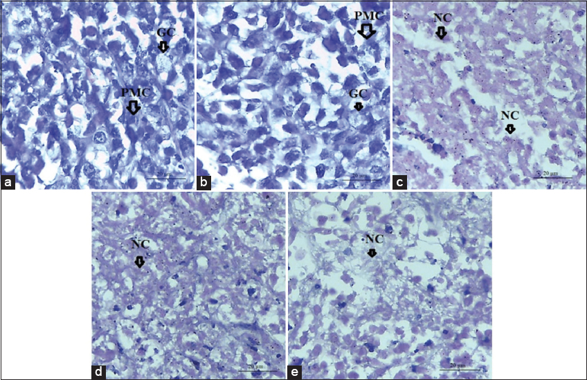 | Figure S1: Histopathology. H & E stained section of tumor extracted from (a); untreated control, (b); vehicle control, (c); cyclophosphamide treated, (d); TME 200 mg/kg b. wt, and (e); TME 400 mg/kg b. wt. The untreated and vehicle treated animals showed pleomorphic cells (PMC) and giant cells (GC) with hyperchromatic nuclei in the tumor tissues. The areas of necrosis marked as NC are shown in the treated groups as well as in the cyclophosphamide treated tissues sections. [Click here to view] |
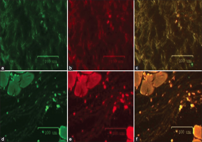 | Figure S2: TUNEL Analysis: (a-c) untreated control and (d-f) treated with TME 400 mg. In the untreated control group, no fluorescence was observed which shows that apoptosis has not taken place in them. Green fluorescence indicates early to middle stage of apoptosis with dUTP-FITC-positive cells. Red fluorescence indicates terminal stage apoptotic/necrotic cells with PI-positive fragmented nuclei. (c and f) are merged. In the treated groups, both green and red fluorescence was observed and it is an indication of early and late stages of apoptosis, respectively. [Click here to view] |
REFERENCE
S1. Feldman AT, Wolfe D. Tissue processing and hematoxylin and eosin staining. In: Histopathology. Germany: Springer; 2014. p. 31-43.