
1. INTRODUCTION
Begomovirus infection has attacked various crops in Indonesia, specifically the Solanaceae and Cucurbitaceae families [1,2]. It is also known as ssDNA virus and a member of the Geminivirus family with great diversity. According to the International Committee on Taxonomy of Viruses, the total species of Begomovirus is about 424. The infection in Cucurbitaceae and Solanaceae families often shows several symptoms such as stunting, mosaics, curls [2], yellow mosaics in eggplant’s leaves [1], green mosaics, and yellow mosaics in melon’s leaves [3]. In Solanaceae plants, particularly Capsicum annum L., Begomovirus tends to reduce productivity. A positive correlation was also reported between the presence of Begomovirus and plant growth inhibition such as stem diameter, the number of fruits, and mass of fruit [4].
Since Begomovirus is transmitted by whiteflies, higher infection cases are likely to occur. One of the popular species of whitefly is Bemisia tabaci, with a unique reproduction named arrhenotoky parthenogenesis, which enhances the yield. The higher the population, the greater the tendency of the virus to spread faster [5,6]. The type of Begomovirus inside the vector is circularly persistent, settling in a long period without any replication. Therefore, when B. tabaci carrying Begomovirus sucks the phloem sap, infection simultaneously spreads out and infects the plant [7].
Detection of Begomovirus in plant leaves through the polymerase chain reaction (PCR) method has been carried out on eggplant, melon, chili, watermelon, papaya, and pepper plants [2,8]. DNA extraction is an initial step in Begomovirus detection using PCR. A common simple extraction method to obtain DNA from insects is lysis buffer [9], while plant leaves DNA extraction can be performed using cetyltrimethylammonium bromide (CTAB) [10]. It is important to note that there are only a few reports on the use of lysis and CTAB buffers to detect Begomovirus. Therefore, this study aims to detect Begomovirus in B. tabaci and its host with a brief extraction method to predict Begomovirus incidence.
2. MATERIALs AND METHODS
2.1. Sample Collection and Identification
The symptomatic eggplant leaves and whitefly were collected in pairs at an eggplant plantation in Rejodani, Sleman, Yogyakarta, Indonesia, in October 2021. A total of 15–25 whitefly (code: s) were collected from the underside of yellow and mosaic leaves into a microtube containing 70% alcohol. In addition, whitefly was sampled using a respirator for the observations with light microscopy and stereo zoom. The whole body color and wings type were identified according to the manual identification book titled Borror and Delong’s Introduction to The Study of Insects [11]. Eggplant leaves were collected and stored in a zip lock with chill condition inside an icebox. Several codes were used as sample identifiers including yellow and yellow mosaic leaves with code of k and km, respectively.
2.2. Whitefly DNA Extraction
Whitefly DNA extraction was performed using lysis buffer [9], with several modifications such as dithiothreitol replacement with β-mercaptoethanol [12]. The samples were washed using a sterile ddH2O, then it was crushed simultaneously with the 100 μL lysis buffer solution containing 0.01 M Tris-HCl pH 8.0, 0.01 M NaCl, 0.005 M CaCl2, 0.002 M EDTA pH 8.0, 2% sodium dodecyl sulfate (SDS), β-mercaptoethanol, and 0.25 mg/mL of the Proteinase K. Furthermore, the crushed samples were incubated overnight at 55°C and centrifuged to produce supernatant and pellet. The supernatant was stored at –20°C as the DNA sample.
2.3. Solanum melongena DNA Extraction
CTAB buffer containing 2% CTAB, 0.1 M Tris-HCl pH 8.0, 1.4 M NaCl, 0.02 M EDTA pH 8.0, and 1% PVP [10] was heated at 60°C using a water bath. Afterward, 0.4 gr of S. melongena leaves were crushed in 2 mL of chilled CTAB buffer, and 10 μL RNase was added to the supernatant, while the microtube was inverted to obtain a completely dissolve sample. Since it was homogenous, the sample was incubated at 37°C for 10 min, then 2 μL β-mercaptoethanol was added and incubated at 65°C for 30 min in a water bath with microtubes inverted every 10 min. The sample was cooled at room temperature for 3 min, while 2 mL of 1:1 of phenol: Chloroform was added to the sample, and the microtube was inverted reverse for 3 min. The homogenous sample was centrifuged at 12000 rpm, 10 min, at 25°C, then chloroform was added in a ratio of 1:1 to the supernatant and centrifuged at 12000 rpm, 10 min, at 25°C. Furthermore, 3 M Na-acetate pH 5.2 up to 1/10 of the supernatant volume and cold isopropanol 1:1 were added to the supernatant. The samples were inverted and incubated for 1 h at –20°C and then centrifuged at 12000 rpm, 10 min, at 25°C to obtain the pellet. The DNA pellet was washed with 500 μL of 70% alcohol, then centrifuged at 12000 rpm, 10 min, at 25°C. The 70% alcohol was removed from the microtube and DNA pellet was air dry, while 30 μL of TE buffer was added and stored at –20°C.
2.4. Begomovirus Coat Protein (CP) Gene Amplification
A total of 2.5 μL DNA samples were dropped onto the Nanodrop surface and measured at 260, 280, and 230 nm for quantity and quality. The amplification of the Begomovirus’ CP gene was carried out by PCR. About 5 μL of 5x PCR master mix (Smobio), 1 μL of each 10 mM forward primer 5’CCNMRDGGHTGTGARGGNCC’3 and reverse primer 5’SVDGCRTGVGTRCANGCCAT’3 [13], 1 μL MgCl2, 9 μL ddH2O, and 8 μL of template DNA were added into the PCR tube. PCR reaction was performed with an initial denaturation at 95°C for 5 min, and 35 cycles of denaturation at 94°C for 30 s, annealing at 50°C for 30 s, and elongation at 72°C for 45 s. The last cycle of post elongation was performed at 72°C for 5 min. The amplicon was analyzed using 1% agarose gel electrophoresis and visualized using UV light.
3. RESULTS AND DISCUSSION
3.1. Symptoms of Begomovirus Infection
The symptoms of Begomovirus infection in eggplants are stunting, mosaic, yellow, and curling in the leaves [1,2]. The eggplant at the Rejodani area is an interlude plant to the chili garden with most of the plants receiving direct sunlight without any canopy. About 13 out of the 25 eggplant trees show symptoms of infection in the form of yellow and yellow mosaics leaves. Based on the observations, most of the plants have similar symptoms, yellow, and yellow mosaic [Figure 1a and b].
 | Figure 1: Begomovirus symptoms in S. melongena leaves and whitefly on the underside of leaves. (a) Yellow mosaic symptom; (b) Yellow symptom; (c and d) Whitefly; Bar=5 cm. [Click here to view] |
Whitefly was widely found on the underside of the eggplants’ leaves [Figure 1c and d], which were exposed to direct sunlight since it is stimulated by the green-yellow light spectrum reflected by the leaves [12]. Based on the results, Whitefly Bemisia spp. exists on a variety of plants, including chili as the main crop on this plantation. B. tabaci has a higher survival ability when living on eggplant plants than other hosts such as cucumber and pepper [14,15], also, it tends to lay eggs on dense trichomes leaves to attach to the body [6].
3.2. Whitefly Identification
Based on the results, the overall body length of the whitefly Bemisia spp. is ±0.6 mm, and it consists of the head/caput, thorax, and abdomen [Figure 2a and b]. The caput comprises antennae, eyes, and mouth, which each has seven segments, provides few ommatidia, and is a piercing-sucking type that cannot be observed using light microscope. On the thorax area, there are 3 pairs of legs with long femur and tibia, as well as two pairs of wings with a relatively equal wingspan and little venation. Based on microscopic observations, the prothoracic and mesothorax wings are the same size. Both wings are membranous, opaque white in color, and covered in white dust. Therefore, these insects can be classified into the Order Hemiptera, Aleyrodidae Family [14].
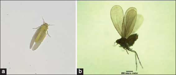 | Figure 2: Bemisia spp. observed using stereo zoom (a) and light microscope (4 × 10) (b) Bar= 200 µm. [Click here to view] |
According to Martin and Mound [16], there are ±41 species in the genus Bemisia, which can be identified based on the morphological characteristics of the pupa since the adult phase has an undistinguishable morphology. B. tabaci species are known as vectors of various plant viruses, including Begomovirus [17]. Whitefly Bemisia spp. in Rejodani eggplant plantation might belong to the B. tabaci species when it carries Begomovirus. Although there is no other supporting evidence, Trialeurodes ricini, and T. vaporariorum have been reported to carry Begomovirus [18-20]. The whole body and wings of Bemisia spp. is more oblong and slenderer than the whitefly genus Trialeuroides. Comparing the morphology, the species observed in Rejodani plantation were similar to Bemisia spp., specifically in its phenotypic body and wings.
3.3 Bemisia spp. and S. melongena DNA Extraction Using Lysis and CTAB Buffer
In general, DNA extraction consists of lysis, contaminant removal, precipitation, and washing. In this study, DNA extraction was conducted for Bemisia spp. using lysis buffer containing 0.01 M Tris-HCl pH 8.0, 0.01 M NaCl, 0.005 M CaCl2, 0.002 M EDTA pH 8.0, 2% SDS, β-mercaptoethanol, and 0.25 mg/mL of the Proteinase-K. Cell lysis was performed using micro pestle, while lysis buffer containing CaCl2 and SDS played a role in weakening cell membranes as well as dissolving labile fragments [21,22]. Furthermore, Tris and EDTA pH 8.0 maintained pH stability and bind ions in charge of maintaining the integrity of cell membranes [23,24]. The removal of contaminants such as the protein was carried out with Proteinase-K and β-mercaptoethanol. The protease enzyme was used to break peptide bonds while the sulfur bonds in proteins were separated using β-mercaptoethanol [25-27]. The DNA precipitation process was performed by Na+ from NaCl [24].
S. melongena DNA extraction was conducted with CTAB buffer. The detail CTAB procedure referred to Nugroho et al. [10] along with other modifications in sample weight, buffer volume per reaction, cold isopropanol, time, and temperature incubation of precipitation [10]. Cell lysis was performed by CTAB as an amphipathic cationic detergent [28]. Tris and EDTA were used to stabilize pH and assist the process of membrane lysis, as well as DNase inactivation [21,22]. The removal of RNA, protein, carbohydrates, tannins, polyphenols, lipids contamination from DNA was assisted by RNase, β-mercaptoethanol, Proteinase-K, PVP, phenol, and chloroform [24]. After removing the contaminant, Na-acetate and isopropanol precipitated the DNA for an hour at –20°C to obtain DNA pellet [28]. The washing step was carried out with 70% ethanol to reduce excess salt [23].
Based on these results, most of the DNA pellets were colored brownish indicating that PVP was not optimal enough in binding the polyphenols located in the vacuole leading to the oxidation of polyphenols which are then turned into brown [24,29]. Although the pellets were brownish, a higher density was obtained indicating that the extraction method with CTAB buffer, modification of 0.4 gr sample weight, 2 mL buffer in each reaction, precipitation with Na-acetate, and cold isopropanol for 1 h in a –20°C is suitable to produce a large amount of DNA.
Table 1 shows the quantity and purity of DNA, although Bemisia spp. samples, namely, S1 and S2, had poor purity, all samples had a high DNA concentration. According to a previous study, A260/A280 ratio near 1.8 indicates pure DNA [30]. Since the whitefly extraction method is quite brief with no separation of contaminant and washing step, poor quality of B. tabaci DNA was obtained. The overall Bemisia spp. in the S. melongena sample has slight purer DNA even though the quantity was low. This condition occurred due to the more complex extraction steps in S. melongena extraction.
Table 1: The quantity and purity of DNA.
| S. no. | Sample | DNA concentration (ng/µL) | DNA purity A260/A280 |
|---|---|---|---|
| 1 | S1 | 1632 | 1.764 |
| 2 | S2 | 1736 | 1.772 |
| 3 | S3 | 1484 | 1.828 |
| 4 | S4 | 1536 | 1.864 |
| 5 | K1 | 719.7 | 2.018 |
| 6 | K2 | 1749 | 1.885 |
| 7 | K3 | 816.4 | 1.985 |
| 8 | KM1 | 1855 | 1.971 |
S. melongena: Solanum melongena, S: Bemisia spp., K: S. melongena with yellowing leaves, KM: S. melongena with yellow mosaic leaves.
3.4. Begomovirus Detection
The amplification of Begomovirus CP gene is shown in Figure 3, and both Bemisia spp. and S. melongena samples were positive Begomovirus with an existing band in ±580 bp. According to Table 1, Bemisia spp. samples (S1, S2) collected from yellow leaves are not pure enough, but proper amplification occurred and produced a specific DNA band as demonstrated in Figure 3. The band of S4 sample appeared weak, fade but specific, in comparison, all S. melongena samples both yellow and mosaic leaves namely K1, K2, K3, and KM1 produced clear and fair band in ±580 bp. Begomovirus infection was observed from yellow and mosaic leaves, but the presence in Bemisia spp. cannot be observed morphologically. However, Bemisia spp. Samples, namely, S1, S2, S3, and S4 were collected in pairs with S. melongena leaves, indicating the probability of Begomovirus presence in Bemisia spp. is relatively high. When B. tabaci sucks infected leaves, the phloem sap along with Begomovirus will be sucked through the stylets. The virions pass through the esophagus, midgut (middle intestine) to hemolymph, and then they are translocated to the primary salivary glands (PSGs) of B. tabaci [31].
 | Figure 3: Amplification of coat protein gene in Bemisia spp. and S. melongena leaves in 1% agarose. M: 1kb marker, S: Bemisia spp., K: S. melongena with yellowing leaves, KM: S. melongena with yellow mosaic leaves, NC: Control. [Click here to view] |
The presence of Begomovirus on B. tabaci can be detected after four to seven hours of phloem suction. From the primary salivary glands, the infection spreads through the saliva when it makes contact with phloem [31]. As a vector, all Bemisia spp. detected positive are able to spread the virus by sucking the phloem sap. This virus remains stable inside the body due to the interaction of CP to GroEL from bacteria endosymbiont and heat-shocked protein 16 (HSP 16) from Bemisia spp. [31,32]. According to Rana et al. [32], these two proteins have a role in virus transmission. The presence of symbiotic bacteria that secretes GroEL protein might cause the virus to remain stable due to the interaction with the viral coat. Virions protected by GroEL can be released into the PSG [33], while the presence of heat-shocked protein 16 (HSP 16) or BtHSP16 Bemisia spp. serves as a chaperone in stabilizing other proteins. This interaction can prevent virus aggregation during transmission [31].
4. CONCLUSION
Based on the results, Begomovirus DNA in the whitefly Bemisia spp. can be extracted using lysis buffer, while in S. melongena, it can be extracted using CTAB buffer. Bemisia spp. on the lower leaves side of S. melongena was found as a potential vector to spread the Begomovirus infection in plantations.
5. ACKNOWLEDGMENTS
The authors are grateful to the technician assistant at FALITMA, Animal Physiology Laboratory, and Plant Physiology Laboratory, Faculty of Biology, Universitas Gadjah Mada. We are also grateful to Arief Muammar for supporting this study, Gilang Ari Saputra for guiding with several sampling locations, and Khoiruddin Anshori for guidance on how to use Leica DM750 ICC50E and ImageJ.
6. AUTHORS’ CONTRIBUTIONS
All authors made substantial contributions to conception and design, acquisition of data, or analysis and interpretation of data; took part in drafting the article or revising it critically for important intellectual content; agreed to submit to the current journal; gave final approval of the version to be published; and agreed to be accountable for all aspects of the work. All the authors are eligible to be an author as per the International Committee of Medical Journal Editors (ICMJE) requirements/guidelines.
7. FUNDING
This research received no specific grant from any funding agency in the public, commercial, or not-for-profit sectors.
8. CONFLICTS OF INTEREST
The authors report no financial or any other conflicts of interest in this work.
9. ETHICAL APPROVALS
Ethics approval is not required for this type of study in the country where the study was conducted.
10. DATA AVAILABILITY
Data sharing not applicable – no new data generated.
11. PUBLISHER’S NOTE
This journal remains neutral with regard to jurisdictional claims in published institutional affiliation.
REFERENCES
1. Kintasari T, Septariani DW, Sulandari S, Hidayat SH. Tomato yellow leaf curl Kanchanaburi virus Associated with yellow mosaic disease in eggplant in Java. J Fitopatol Indones 2013;9:127-31. [CrossRef]
2. Subiastuti AS, Hartono S, Daryono BS. Detection and identification of begomovirus infecting cucurbitaceae and solanaceae in Yogyakarta, Indonesia. Biodiversitas 2019;20:738-44. [CrossRef]
3. Haerunisa R, Suastika G, Damayanti TA. Identification of begomovirus associated with yellow mosaic disease on cucumber plant in West Java and Bali. J Hortik Indones 2019;7:9. [CrossRef]
4. Kesumawati E, Asdhani M. Correlation between Virus Attacks at the Several Phases of Growth with the Yield of Chili Pepper (
5. Hidayat P, Ludji R, Maryana N. Reproductive ability and life history of the whitefly
6. Indrayani IG, Sulistyowati E. Role of trichome density of cotton leaves to colonization of Bemisia tabaci Gennadius. J Penelitian Tanaman Ind 2011;11:101. [CrossRef]
7. Gilbertson RL, Batuman O, Webster CG, Adkins S. Role of the insect supervectors
8. Sutrawati M, Parwito P, Priyatiningsih P, Zarkani A, Sipriyadi S, Sariasih Y,
9. Modi A, Vergata C, Zilli C, Vischioni C, Vai S, Tagliazucchi G,
10. Nugroho K, Terryana RT, Lestari P. DNA extraction method for chili paper
11. Triplehorn C, Jhonson N. Borror and De Long's Introduction to the Study of Insects. 7th ed. Florence, KY:Thomson Brooks/Cole;2005.
12. Putri R. Detection of Begomovirus in Whitefly (
13. Revill PA, Ha CV, Porchun SC, Vu MT, Dale JL. The complete nucleotide sequence of two distinct geminiviruses infecting cucurbits in Vietnam. Arch Virol 2003;148:1523-41. [CrossRef]
14. Mound LA. Studies on the olfaction and colour sensitivity of
15. Mostafizur M, Shah R, Liu TX. Feeding experience of
16. Martin JH, Mound LA. An Annotated Check List of the World's Whiteflies(Insecta:
17. Hasegawa DK, Wenbo C, Yi Z, Navneet K, William MW, Alvin MS,
18. Idriss M, Abdallah N, Aref N, Haridy G, Madkour M. Biotypes of the castor bean whitefly
19. Sangeetha B, Malathi VG, Alice D, Suganthy M, Renukadevi P. A distinct seed-transmissible strain of tomato leaf curl New Delhi virus infecting Chayote in India. Virus Res2018;258:81-91. [CrossRef]
20. Fiallo-OlivéE, Pan, LL, Liu SS, Navas-Castillo J. Transmission of begomoviruses and other whitefly-borne viruses:Dependence on the vector species. Phytopathology 2020;110:10-7. [CrossRef]
21. Kamble SP, Fawade MM. A rapid and inexpensive one-tube genomic DNA extraction method from
22. Lever MA, Torti A, Eickenbusch P, Michaud AB, Šantl-Temkiv T, Jørgensen BB. A modular method for the extraction of DNA and RNA, and the separation of DNA pools from diverse environmental sample types. Front Microbiol 2015;6:476. [CrossRef]
23. Lade BD, Patil AS, Paikrao HM. Efficient genomic DNA extraction protocol from medicinal rich
24. Heikrujam J, Kishor R, Mazumder P. In:Oana-Maria, Balt?B, Awwad NS, editors. The Chemistry Behind Plant DNA Isolation Protocols. Biochemical Analysis Tools Methods for Bio-Molecules Studies. London:IntechOpen;2020. [CrossRef]
25. Neil DR, Guy S, editors. Saenger proteinase. In:Handbook of Proteolytic Enzymes. 3rd ed., Ch. 714. Cambridge, Massachusetts:Academic Press;2013. 3240-2. [CrossRef]
26. Flores-Gallegos AC, Delgado-García M, Ascacio-Valdés JA, Villareal-Morales S, Michel-Michel MR, Aguilar-González CN,
27. Jadhav K, Raja R, Natesan S. Chemistry of plant genomic dna extraction protocol. J Life Sci 2015;12:543-8.
28. Sari VK, Murti RH. An effective method for dna extraction of mature leaf of sapodilla (
29. Holderbaum DF, Kon T, Kudo T, Guerra MP. Enzymatic browning, polyphenol oxidase activity, and polyphenols in four apple cultivars:Dynamics during fruit development. Am Soc Hortic Sci 2010;45:1150-4. [CrossRef]
30. Hassan R, Husin A, Sulong S, Yusoff S, Farid J, Yahaya B,
31. Ohnesorge S, Bejarano ER. Begomovirus coat protein interacts with a small heat-shock protein of its transmission vector (
32. Rana VS, Singh ST, Priya NG, Kumar J, Rajagopal R. Arsenophonus GroEL interacts with CLCuV and is localized in midgut and salivary gland of whitefly
33. Czosnek H, Hariton-Shalev A, Sobol I, Gorovits R, Ghanim M. The incredible journey of Begomoviruses in their whitefly vector. Viruses 2017;9:273. [CrossRef]