
1. INTRODUCTION
Tissue engineering is an interdisciplinary field that connects biomedical engineering, mechanical engineering, clinical medicine, genetic engineering, and biotechnology. It is a complex process as tissue regeneration depends upon various factors such as maintenance of cell-cell interactions, surface properties, porosity, mechanical stability, solubility, and degradability of biomaterials used. Accidents and conditions, such as osteomyelitis, arthritis, anaemia, cancers, hereditary multiple exostoses, and hereditary bone marrow failure syndromes, cause severe damage to the bones cartilage and tissues[1–5]. With the increase in demand for bone grafts in organ transplantation and substitute surgery, the market value is expected to be 11.5 billion US dollars by the end of 2025 [6]. Tissue engineering is the development of porous scaffolds to provide a favourable environment for the regeneration, growth of tissues, and complex organs. Scaffolds are three-dimensional structures that are porous, fibrous and permeable biomaterials. It helps in the transport of body fluids, promotes cell-cell interaction, deposition of extracellular matrix, viability against various pathogens with minimum inflammation, and toxicity rate. In the 3D scaffolds fabrication technique, the materials are generally classified into chemical factors such as synthetic and natural polymers, hydrogels, metals and non-metals, composites, ceramics, and non-ceramics [7]. The development of biologically synthesized scaffolds depends on various factors such as pore sizes, interconnectivity between the pores, biodegradability, ability for cells to produce their extracellular matrix, machine limitation, biocompatibility, clinical status, instrumentation choice, good manufacture practices, and mechanical properties. In addition, the modifications considered during fabrication of bio-scaffolds are bio-mimicking with various cellular components, delivering of different bioactive molecules and ameliorative agents like antibiotics, cytokines, drugs, inhibitor, growth factors, proteins, which provide an anchorage for great importance in stacking to the scaffolds [8,9]. With the advancement in technology and various architectural demands, various approaches have been developed for the fabrication of scaffold materials.
Although the conventional methods are widely used and are evolving with decades, advanced prototyping techniques are also being adopted. The advanced techniques use high- end computer-aided designing software for the bio fabrication process with micro, macro, and nanoscale architecture [7,8]. With the recent development of biologically active agents, natural biopolymers are replacing synthetic polymers. The natural polymers are either plant-derived or animal-derived and are biodegradable, non-toxic, and provide customized pore sizes and renewability [10]. Moreover, the various types of natural bio-polymers play an important role in biocompatibility, influence cell behaviour, possess high surface area, with pre-existing vascular networks, porosity, and rapid biodegrading properties [11].
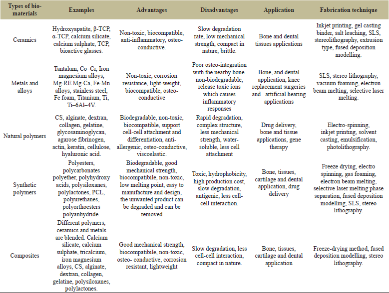 | Table 1: Type of materials used in scaffolds fabrication. [Click here to view] |
The biomaterials and fabrication methods are selected for processes depending upon the analysis and complexity of targeted tissues. Different type of materials used in scaffolds fabrication for various tissue engineering applications is compiled in the Table 1.
Recently, various studies reported on advancement in biomaterials fabrication technique and development of scaffolds from biomaterials which enhanced cell proliferation, cell viability, and printability without any stress [7,12–15]. Plant-based biomaterial such as plant proteins, lignin, polysaccharides, and plant extracts have various bioactive products that are useful in various restoration, regeneration and improvement of scaffolds fabrication. However, the synthetic and animal-derived biomaterials used in regeneration and restoration applications, have some disadvantages such as scarcity, expensiveness, high cell deaths, and poor biocompatibility [11].
This review describes different techniques of advanced rapid prototyping (RP) and conventional 3D fabrication for bio-scaffolds preparation. These techniques can create porous 3D structures with controlled mechanical possession, pore size, interconnected pores, and porosity. The paper summarizes research status of each of the methods and the opportunities and challenges are analyzed.
2. FABRICATION OF SCAFFOLDS BASED ON THEIR REQUIREMENTS
The scaffold fabrications and designing consider various properties, such as mechanical, biological, and physicochemical based on its feasibility and requirements. Additionally, the interconnectivity within the pores, shape, pore size, porosity, strength, and degradation rate are the important factors based on which the scaffolds fabrications depend. The 2D scaffolds technique possesses several advantages by providing higher resolution and accuracies with control over physical and chemical properties. The imaging and characterization are easier with automated lab facilities and high processing screening methodologies [16,17]. The 3D bio fabrication techniques for scaffolds design uses advanced bio-printing and bio-assembly methodology that include cells for the fabrication process in an automated manner [18]. It uses various types of computer-assisted designing software packages to create virtual cross-sections of cell-loaded matrices by consecutive layers formation with a computational fabrication process. The 3D scaffold designs are a form of sponges, foams, and meshes and can resist the external pressure caused by various factors such as different tissue interaction with the extracellular matrix, mechanical stiffness, rapid degradation, cell deaths, toxicity, and biocompatibility. Nevertheless, the designing of 3D scaffolds develops proper homogeneous mixture, cell-to-cell contacts, cell proliferation and cell attachment. The emerging 3D technology has revolutionized into 4D printing depending on the type of biomaterials and environmental factors [19]. However, the 4D printing technique is quite expensive as compared to other 3D printing technologies [20]. The scaffolds prints are prepared with advanced processing through multiscale finite element analysis and computational neuromusculoskeletal evaluation to obtain load-holding capacity, in vivo cyclic stress and biocompatibility [21].
Different types of scaffolds and fabrication techniques are used depending upon their biomaterials’ composition (Table 2). For example, in bone tissue engineering, nanofibrous scaffolds are used that mimics extracellular matrices and collagen fibres [32–34]. Gelatin and Fibrin are natural biopolymers that have been widely used in scaffold fabrication as it has high biodegradability and biocompatibility [35,36]. Alginate has been widely used for bone and cartilage tissue engineering and capable of scaffold-reinforcement and non-immunogenic property [37].
The design of scaffold for tissue engineering involves high interconnectivity within the nano-vascular networks of extracellular matrix, transport of oxygen, nutrient, and various soluble factors that are responsible for removing metabolic wastes [7,11]. Based on its complexity, construct materials source, geometrical distribution of structure and the process of fabrication technique, the 3D scaffolds are available in various forms Table 3.
3. DIFFERENT TYPES OD SCAFFOLD FABRICATION TECHNIQUES
The conventional tissue engineering scaffold production techniques include thermally induced phase separation (TIPS), fiber bonding, electrospinning, solvent casting and particulate leaching, membrane lamination, freeze-drying, and gas foaming [49]. The recent development in scaffold fabrication mainly comprises integration with computer-aided design (CAD) software through RP technology such as stereolithography, bio-plotting, solvent-based free forming, combination modelling technique, fused deposition modelling, 3D printing, and selective laser sintering (SLS) [8,9]. The techniques retain the ability to maintain pore structures, cell–cell interaction, reduction in mechanical instability, and control over mitigation of the cellular matrix. Advantages and disadvantages of different type scaffolds fabrication technique for tissue engineering applications are compiled in Table 4.
4. CONVENTIONAL METHODS
There are various conventional methods of scaffold fabrication (Fig. 1) and have been developed for drug delivery, 3D cell culture and tissue engineering. However, the conventional scaffold fabrication techniques sometimes remain incompatible as they deviate from the optimal environment for cell attachment, multiplication, and reproduction. In the case of skin tissue engineering, it forms a non-homogeneous structure and is limited to the only internal design of the scaffolds. Additionally, the technique is limited to manual intervention as it involves a time-consuming, multistage process that is labor intensive and requires high skill and experience. Conventional methods do not result in the regularity of pore shape, reproducibility and sufficient pore interconnectivity [9,49].
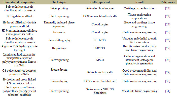 | Table 2: Different scaffolds fabrication for various tissue engineering applications. [Click here to view] |
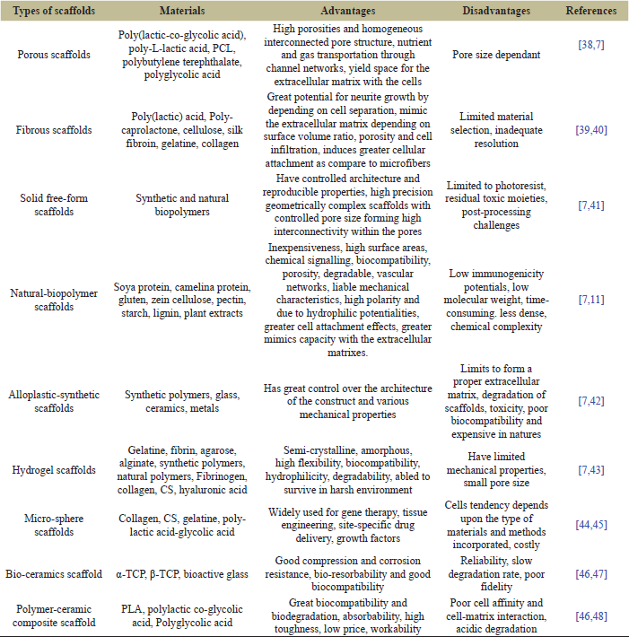 | Table 3: Various forms of 3D scaffolds. [Click here to view] |
4.1. Freeze-Drying
Freeze-drying, also known as lyophilization, is a widely utilized fabrication technique. In this technique, it freezes a synthetic polymer dissolved in selective solvent at a temperature between −20°C and −80°C, resulting in the solid solvent. The frozen sample then evaporated by sublimation with help of a lyophilizer to form a solid scaffold with various interconnected pores [68]. Soumya et al. [69] have successfully developed an osteoinductive herbal scaffold by freeze-drying. They blended medicinal plant extracts with natural biopolymers [O-carboxymethyl chitosan (CS) and alginate] by lyophilization process. Later, fabricated scaffolds with desirable properties for tissue engineering applications. The cytocompatibility studies on the developed composite scaffolds with human mesenchymal stem cells (MSCs) proved its biocompatible nature. The scaffolds fabricated with plant extract showed a remarkable difference in cell attachment and cell reproduction as compared to scaffolds fabricated without extract. The developed scaffold showed promising porosity and water absorption properties. The freeze-drying technique is mostly favorable for biomedical application as water solvents are used rather than other organic solvents for the fabrication of scaffolds but remain challenging for the fabrication of classified structured scaffold-like vascular systems used in biomedicine applications [70]. The cytotoxic solvents used for the mixing of the polymers consume high energy. The developed scaffold requires washing several times to remove the toxic solvents and hence declines cell death as well as triggers the irregularities in pore sizes for a longer time- span [71,72].
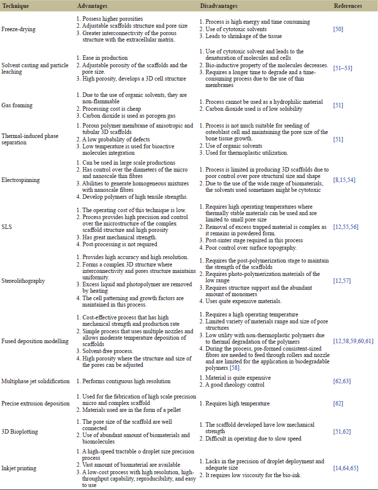 | Table 4: Advantages and Disadvantages of different types of scaffolds fabrication techniques for tissue engineering application. [Click here to view] |
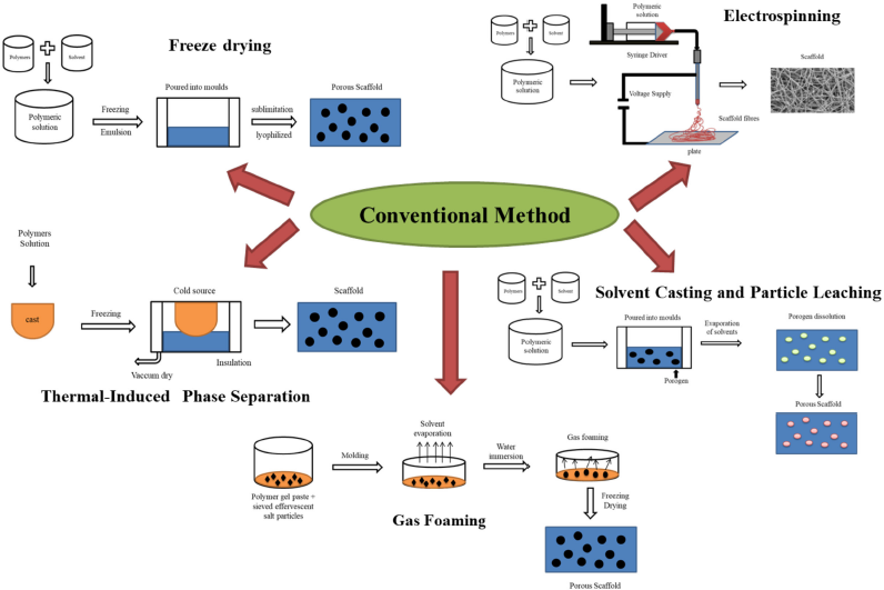 | Figure 1: Conventional methods of scaffold fabrication [66, 67] . [Click here to view] |
4.2. Solvent Casting and Particle Leaching
This method is generally used for Bone and cartilage tissue engineering applications [73]. In this technique, a highly volatile solvent solvates the polymer and cast in molds along with porogen. The solvents evaporate with a matrix consisting of salt particles and the formed composite matrix contains porogen and polymers. The matrix submerged in water allows salt leaching to develop scaffolds structure with high porosity. The solvent combined with uniformly distributed organic amalgams like glucose, gelatine microsphere and otherwise soluble inorganic salts like potassium chloride is selectively leached to get a certain pore size which is used as porogen to dissolve in the polymer solution [74]. The scaffolds prepared through this technique have a porosity of 50% to 90% and are of a low-cost process [75]. Zeng et al. [66] successfully fabricated and developed 3D porous polylactic acid (PLA) scaffold with high porosity using methods like phase separation, particle leaching techniques, vacuum-assisted solvent casting, and solvent extraction. In this process, the surface is modified with the help of a CS/osteogenic growth peptide (OGP) coating layer that can be potentially applied in bone, cartilage regeneration and tissue engineering purposes. The experiment proved that the higher the porosity of the PLA scaffolds developed, the lower the PLA solution concentration and the temperature. The hydrophilicity and mechanical properties of CS/OGP/PLA developed scaffold were higher as compared to the uncoated PLA scaffold.
4.3. Gas Foaming
Gas foaming utilizes natural cytostatic solvents and high temperature. In this method, the fabrication model uses inert gas foaming agents like methane, carbon dioxide, hydrogen, nitrogen to pressurize with a biodegradable polymer like fluoroform or water solvents until it reaches saturated conditions to form gas bubbles [76]. The gas foaming techniques and fused deposition methods are combined where, the porous PLA scaffolds dominate 1 to 10 μm micro pores sizes structures. Gas foaming process generates micropores (?10 μm) while in conventional 3D process, it is barely developed. The fused deposition method incorporates macro porosity through attached channels that have developed the saturation capabilities by reducing the time consumptions in the process. This technique achieves generally 85% porosity and 30 to 700 μm pore size for the developed structure which is sponge in nature [58]. Song et al. [67] developed tailored macro/micro-porosity architectures scaffolds by combined technology of gas foaming and fused deposition modelling process for applications in tissue engineering technology.
The PLA is blended with poly(vinyl alcohol) (PVA) to fabricate composite filaments by fused deposition modelling. After the fabrication process, the developed scaffolds were dominated by the gas foaming process to create micropores of size less than 10 μm. The outcomes of this process revealed that without further dense skin layers, interconnected pores attained micropores size of 2 to 10 μm. Hence the scaffolds developed have great potentialities for cartilage and bone tissue regeneration. Manavitehrani et al. [77] have studied polyester-based poly propylene carbonate (PPC)-starch bioscaffolds in tissue engineering technologies. In this process, to develop a porous scaffold, the bi-products are degraded and fabricated by gas-foaming technique with PPC blended with starch and bioglass particles. The pore sizes developed were ranged from 100 to 500 μm, with high pore interconnectivity.
4.4. Thermal-Induced Phase Separation (TIPS)
In the TIPS process, various polymeric solutions of solvent and non-solvent are demixed, quenched and consist of different polymeric phases. This demixing process takes place either by evaporation or extraction process, solvent is removed resulting in pores formation [74]. In this technique, the mixing of various types of selective solvent, additive blenders, and multicomponent polymer are used at low temperature for developing force separation process, where the homogeneous polymer solution at high-temperature environment settled down to induce phase separation by achieving variant polymeric phases [75,58]. The scaffold structures are attained, as the solvent gets eliminated by the freeze-drying process with relatively porous and nano-scale fibrous meshes. The thermoplastic crystalline polymer scaffolds are generally used for construction and low temperature is mostly used for blending bioactive materials with the fibrous scaffold [9]. In this method, the porosity of the fibres is achieved 98% higher than the surface to volume ratio of the scaffold constructed. Biswas et al. [78] have developed porous CS scaffold by using combined technology with mechanical foaming and thermal-induced phase separation technique and obtained 80% porosity, 2.6 to 25 kPa adjustable compacting parameters with 120 mm pore size. The scaffold showed great potential for tissue engineering and the foaming process incorporated by air bubbles, functioned as a mould for the macro-porous construct of the developed scaffold. In this technique, materials are limited to the fabrication process, inadequate resolution and very selective minimal materials are used in the phase separation process with uniform porous structures [70].
4.5. Electrospinning
Electrospinning is extensively used in nanofibers polymers and for scaffolds fabrication process from the selective solution by using electric current [79]. In this technique, the nanofiber scaffolds exhibit great porosity, high surface area, and biomimetic like natural extracellular matrix. During this process, the polymeric droplet is executed by the stress at the needle tip by using an electrically charged jet. Then at high voltage, the charging solvent gets dominated by an interaction between electrostatic repulsion and surface tension, where the spinneret droplets erupt and gets stretched by passing through grounded collector from the spinneret tip. Finally, the jet solidified into nanofibers as the solvent starts evaporating [80]. For the production of nanofibers, various parameters are followed such as surface tension, conductivity, flight distance, type and concentration of polymeric solvents, viscosity, spinneret diameter, interactions between the molecules, the electric current supplied, rate of flow, types of collector [81]. Yuan et al. [82] has successfully developed nanofibrous scaffolds with polyethylene oxide (PEO) and CS using the electrospinning fabrication technique. In this study, with the reduction of mass of CS and PEO from the scaffolds, degradation is exhibited. The bactericidal study shows stronger growth inhibition and cytotoxic effects. This fabrication technique is categorized into three depending on the types of manufacturing methods consisting of solution materials by changing electrospinning materials, setting up of collector by using liquid-assisted collectors with perfunctory setup, post-processing operation after electrospinning [8]. Hejazi and Mirzadeh [83] have developed 3D scaffold with electrospraying and electrospinning combined technique by using polycaprolactone (PCL). The 3D scaffold was fabricated using macro and nanofibres particle with optimized pore size, porosity, interconnectivity within the pores and biomimetic the extracellular matrix structure. Although electrospinning is widely used, there is various limitation such as distribution of sufficient homogeneous pores sizes, limited to some applications in biomedicine [70], and the solvent used is toxic and depended on various variables.
5. RAPID PROTOTYPING (RP)
Due to various limitations in the conventional methods, the use of RP technique was introduced to develop 3D porous scaffolds with great architecture advantages, higher porosity and interconnectivity within the pores. This process, also known solid free-form fabrication technique, is a strong fabricating tool and immensely used for the preparation of scaffolds in tissue engineering. The scaffolds are developed by using computer-aided designing tools to provide a perfect fit architectural structure with physio-chemical properties. In the RP process, closely attached materials are combined with powder, sheet or liquid and the fabrication process is employed by the addition of consecutive layers to produce 3D scaffolds utilizing the computer-generated models [84]. Initially, development takes place for a 3D volumetric computer model which is derived from yield information produced by surface digitizers or by clinical imaging frameworks. Later, the digital model extracted is stacked and fused on top of each other to make user-defined structures that statistically cut into layers with a consistent thickness [49]. The main advancement of these fabrication techniques is that they can maintain the porosity, pores shape and size of the scaffolds and have highly interconnected pore structure. It additionally empowers for development of patient-specific customizable scaffolds that are appropriate for tissues and organs designing technology [58]. There are various types of RP fabrication techniques for tissue engineering purposes (Fig. 2) such as fused deposition modelling, stereolithography, electron beam melting, 3D printing and SLS [9].
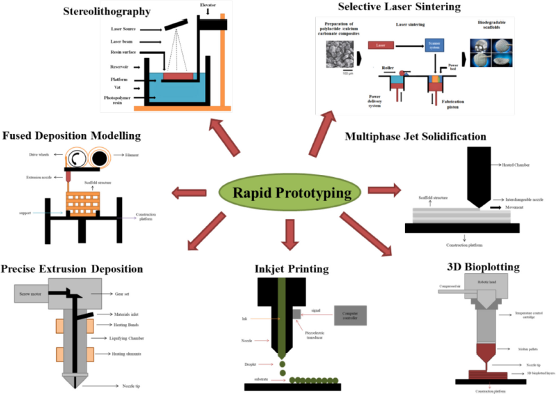 | Figure 2: Types of RP fabrication techniques for tissue engineering [85] . [Click here to view] |
5.1. Selective Laser Sintering (SLS)
SLS is a powder-based fabrication technique that uses laser technology to sinter powdered material such as polymers, ceramics, and metals. It is utilized for manufacturing and developing the scaffolds for fabrication in tissue engineering [9]. Patient-specific implants are developed with composite interconnectivity pore structures to advance bone ingrowth [86]. Although there are various process for the treatment and developing solutions to treat bone imperfections such as with titanium and polyetherketone ketone inserts as they are non-degradable but have the chance of getting several complications in future [85]. The productivity of this method is that it provides outstanding dominance over the microstructures of the developed scaffolds under various parameters like compositions of percentage by physically mixed polymers or by composite powder mixes to get the properties of the favoured scaffold [87]. Gayer et al. [85] have successfully developed and fabricated solvent-free polylactide-calcium carbonate composite scaffolds by using the SLS technique. In the cycle, four distinctive composite powders with about 75% (wt%) polylactide (PLLA and poly(D,L-lactic acid) and 25% (wt%) calcium carbonate (calcite) composite were set up by milling process dependent on Good Manufacturing Practice principles, where the four different grades of polylactide were selected for shelling the broad inherent viscosity range of 1.0–3.6 dl/g. The composite material with the most minimal inherent viscosity at (1.0 dl/g) showed the best processability by SLS process where the biaxial twisting quality of up to 75 MPa was accomplished. While the cell culture measures showed great feasibility of MG-63 osteoblast-like cells on the SLS exhibits. At last, the 3D scaffolds with great pore structure and interconnectivity with the pores were developed [87].
5.2. Stereolithography
Stereolithography technique is the process of developing solid 3D scaffolds based on structurally regulated solidification of the fluid resin by photo-polymerization measure. In this process, the printing of thin layers of ultraviolet curable material is done layer by layer using photolithography pattern [88]. The four main components in this process are UV laser for radiating resin, photosensitive liquid resin, transferable built platform, and dynamic mirror system [9]. The process starts by depositing the surface layer of photosensitive liquid resin with a UV laser and solidified to a characterized depth.
The initial layer is solidified and the platform is lowered down vertically layer by layer through photo-polymerization methods. The assisted platform is moved from the surface and the assembled layer is recoated with fluid resin. At that point, the subsequent layer is put over the principal layer where the process persistent until the 3D scaffold is created. Lastly, the uncured sap wiped out and the scaffolds are post cured under UV light with controlled exposure time, light intensity [9]. Farzan et al. [89] developed an elastic 3D-printed scaffold by stereolithography and solvent-free methods followed for the fabrication process using PCL and polyethylene glycol (PEG). The outcomes demonstrated that PEG-containing PUs had higher degradation rates, and the elasticity of PU/PCL/PEG was 1.4 and twice higher than that of PU/PEG and PU/PCL, separately. The 3D printed PU/PCL/PEG demonstrated high appropriateness in delicate tissue engineering during the scaffold development process. Elomaa et al. [90] developed photo-cross linkable PCL based resin utilizing solvent-free stereolithography methods. In this process, the photo-cross linkable macromers was set up by methacrylating three-outfitted oligomers alongside methacrylic anhydride where the porous scaffolds were developed by stereolithography technique using PCL resin. The ideal resin viscosity was formed during the curing process by heating the resin. Due to absence of solvent, the scaffolds developed provide the best fits with CADs as the material shrinkage do not take place. The interconnectivity of the pores was high in the photo-cross linkable biodegradable PCL resin and has a great potential for tissue engineering scaffolds [91].
6. EXTRUSION-BASED SYSTEM
Extrusion based techniques are the advanced fabrication technique because of various advantages like high resolution, drug-loading, cell-friendly environments, high physical properties, feasibility controlled over drop-on-demand high precision deposition [92]. This technique is mechanically accessible and are less expensive with correlation to other solid freeform manufacturing process. The extrusion-based system depends on material melting where computer-aided designing software is used and scaffold is formed layer after layer. This technique is categorized based on material melting methods and type, however, the heat method is commonly used, some of the other methods like premixed pastes and inks methods are also used [93]. The various process of the extrusion-based system includes fused deposition modelling, multiphase jet solidification, precise extrusion manufacturing, and 3D plotting.
6.1. Fused Deposition Modelling
It is a hot melting extrusion process wherein, the casting of the solid polymer takes place through a nozzle which is ejected and melted on the surface of 3D patterns by utilizing a controlled computer-aided tool for extrusion and deposition process [9]. In this process, the 3D scaffold is made up of layer-by-layer method, where multiple layers of adjacent microfilaments take places. In this technique, the tailoring of bone scaffolds is done with controlled cross-hatch microarchitecture depending on various printing parameters like printing temperature, infill angle, and layer thickness. It is then adjusted directly along with the use of readily available filaments and can be used in developing pharmacological products of good auxiliary properties with the least post-processing requirements. Kalita et al. [94] developed and fabricated 3D particulate-reinforced polymer-ceramic composites of polypropylene (PP) polymer and tricalcium phosphate (TCP) ceramic by high shear blending process with adjusted porosity. The PP-TCP composite fibers are prepared to utilize a solitary screw extruder followed by fused deposition to manufacture permeable structures. In this process, the strength of the composite was decreased as pore volume increased to identify through compression testing. The porous scaffolds developed were later characterized to use as bone grafts. Chen et al. [95] manufactured PVA/β-TCP composite scaffolds with a combination of solid-state shear milling and fused deposition methods. For the development of bioactivity and osteo-conductive properties, β-TCP bioceramic material is used as the fortifying filler while PVA as the polymer grid. The composite scaffolds formed demonstrated that β-TCP particles homogeneously scattered into the PVA grid with the help of solid-state shear processing. The outcomes firmly demonstrate the capability of the fabricated scaffolds in tissue engineering applications. The deposition takes place at low temperature though encapsulation of cells during the fabrication process was not allowed [59].
6.2. Multiphase Jet Solidification
In this technique, high thickness metallic and ceramics parts are created by utilizing low melting point compounds or powdered blender by compressing out through computational controlled spout to fabricate the part layer-by-layer [49]. The very important parts in this process are controlled with computational positioning arrangements and a warmed chamber with a stream and a pulling framework. The materials in this technique are stacked in the structure of material-binder mixture, powder, bar, and pellets which are heated in a reservoir system or by the processed chambers exceeding the melting point of the binder. Gradually throughout the process, the binder gets liquefied where the heated paste is propelled out through a heated jet spout and settled onto a controlled computational system [59]. Xiong et al. [96] developed and fabricated porous PLLA/TCP scaffolds through a computer-aided low-temperature deposition manufacturing process for bone tissue engineering applications. The developed scaffolds showed great porosity of 89.6% besides that scaffolds has controlled interconnectivity pores, great biocompatibility, great bone conductivity, and suitable in vivo biodegradable property which make them suitable for tissue engineering.
6.3. Precise Extrusion Deposition
The Precise Extrusion Deposition technique developed scaffolds with controlled structural adaptations and pore sizes that can provide strength, structural integrity, and microenvironment for tissue regeneration and tissue engineering [97]. This method is more applicable in the pharmaceutical field. PCL, one of the most widely used biodegradable polyester has a low melting point and are approved by the Food and Drug Administration with peculiar applications which are particularly utilized for longer run embeds and controlled drug delivery applications [97]. The precision extruding deposition process is the variations of fused deposition modelling technique, where without filament preparation the scaffolding material are directly deposited [98,99]. Shor et al. [100] has developed scaffolds using a precision extrusion deposition process by fabricating PCL-hydroxyapatite composite through computer-aided tissue engineering technique. In this study, the capability of the precision extrusion deposition fabrication process of the scaffolds with structural integrity, controlled microstructure, pore interconnectivity pore size, mechanical property is required for cartilage and bone tissue engineering have been characterized. The developed scaffolds formed 60%–70% porous and with 100% interconnectivity. This technique presented great advantages for high-precision on the micro-scale for complex scaffold fabrication as compared to the conventional fabrication of scaffold methods.
6.4. 3D Bioplotting
3D Bioplotting is one of the versatile and widely used techniques. It involves the mapping of a 3D moving head extruder which is controlled by the use of packed air to compel out fluid or glue-like plotting medium to produce 3D solid scaffolds. Then the characterization is done based on different compositions of material such as tailor-made internal structures, pore interconnectivity, and complex shapes. The scaffolds developed in this technique are fabricated layer-by-layer, where the plotting materials are stored in the form of a movable dispenser. Then with the help of a heating jacket and with nozzle, the gaseous tension is controlled into fluid plotting medium are plotted and maintained [59]. In this process, the polarity and density of the fluid medium are in a limited manner along with plotting material for prevention of gravity-induced structural collapse and to get temporary support structures [101]. Naghieh et al. [102] have developed alginate scaffolds by an indirect-bioprinting process that is categorized for nerve tissue engineering applications. These low-concentration alginate scaffolds are developed through the indirect-bioprinting process. It involves various methods that include printing a conciliatory system from gelatin, impregnating the structure with low focused alginate, and eliminating the gelatin system by incubation process, and shaping low-fixation alginate scaffolds. The mechanical and biological properties of the developed scaffolds are influenced by the accumulation of alginate and the disinfection method used which give a viable method of adjusting scaffold properties during the backhanded bioprinting measures. Gómez-Lizárraga et al. [103] have developed a 3D bioplotting process for scaffolds fabrication which is made up of a composite of PCL and ceramic micro-powder. The scaffold was manufactured in a cell grid structure. The scaffold developed had a porosity of 32% and a pore size of 323 μm and that indicated a potential use in tissue regeneration and tissue engineering applications.
6.5. Inkjet Printing
This technique is also known as drop-on-demand or electro-dynamic inkjet printing. It is a non-contact technique that is controlled by the physical properties depending on the jet dispensing process where ink droplets or liquid material are discharge into the substrate with predetermined patterns [88,97]. The liquid material is forced through the nozzle by pressurizing the stream of droplets to form the bio-ink solution which is then made up for cell culture medium or the hydrogel [104,64]. In this technique, print heads are provided by the printer that is connected to nozzles through ink chambers. The droplets stipulated are signaled in the form of the pulse from the electrostatic, thermal, and piezoelectric actuators which produce pressure in the liquid materials [104]. While the surface tension of liquid material gets surpass by the pressure formed in the nozzle orifice and finally ink droplet gets emitted out through the nozzle tip. Later through the nozzle, high voltage is applied to the generated substrate. The liquid material gets encircled by the meniscus which prises the ink with the substrate where the electrical power is constrained by surface pressure, that deposit on the substrate [97]. Generally, there are two different methods for this type of technique i.e. piezoelectric-actuated ink jet printing and thermal inkjet printing. Zamanifard et al. [105] have developed and fabricated natural polyhydroxybutyrate biocompatible polymer scaffolds by using electro-spun technology and computer-aided software. The modelling of scaffolds was based on data received from the software with response to the exterior and artificial neural matrix strategies. The various data generated are compared with experimental results. In this inkjet process, the natural polymer polyhydroxybutyrate was electrospun and bioscaffolds with high biocompatibility were developed of 224 to 360 nm breadth range. However, the final results confirmed that the scaffold developed by printing polyaniline nanoparticles and oxygen plasma methods with defined designs has a great effect on cell attachment and cell development. In addition, was non-harmful for human fibroblasts and is reasonable for considering the impact of electrical incitement on human fibroblasts. The developed scaffold exhibits various uses in nerve and tissue engineering technologies established by the degradation studies.
6.6. CAD Technology
The CAD technology makes scaffold fabrication a cheap, safe and time convenient by replacing conventional drawing board methods. The CAD process uses multidimensional coordinating system where three-dimensional data are generated digitally with the uses of noncontact 3D laser scanner and rapid-measuring technology. It is then viewed in broad array of perspectives and results are procured with the uses of RP process. The CAD based scaffolds (Table 5) are the advanced integrated fabrication techniques. The computer-aided designing software include AutoCAD [116], FreeForm Plus software [117], MathMod, Meshmixer, Netfabb, and Cura [107] and used libraries such as Visualization Toolkit (VTK), numpy and wxPython libraries that provide complexity geometric primitives [118]. AutoCAD (Autodesk) is a specialized CAD application to develop 2D and 3D model precisely with respect to its dimensions from micro to macro structures and arrangements with the pores sizes. Similarly poncad [119], meccad [120], solidwork [121] and blockscad [122] are associated with designing of scaffolds. Ansys Fluent [123] is to visualize the scaffold with respect to fluidic properties and Abaqus [124] applied in modelling and finite element analysis. VTK [125], is an open-source, freely available software system for visualization, processing of the image and developing three dimensional computer graphics. wxWidgets/ wxPython [126] is a freely available program that allows to develop highly graphical user interfaces. NumPy [127] is specialized program in data analysis and numerical calculation for large dimensions of data using array processing Python package.
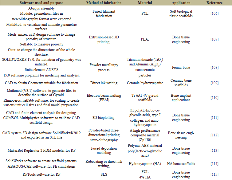 | Table 5: List of Software used in Scaffold design and fabrication. [Click here to view] |
7. CONFLICTS OF INTEREST
The authors report no financial or any other conflicts of interest in this work.
8. PUBLISHER’S NOTE
This journal remains neutral with regard to jurisdictional claims in published institutional affiliation.
9. CONCLUSION
Tissue engineering is a vast multidiscipline area with a wide range of applications. The fabrication of scaffolds is a very complex, dedicated, and sensitive process due to its various factors and parameters. Based on literature surveys, it can conclude that the RP techniques as an advanced fabrication technique that uses computer-aided software and tools for scaffold development in tissue engineering applications. The 3D scaffold should able to provide an innate cellular microenvironment and able to create great cellular interaction which plays a vital role in tissue engineering. Although, with recent progress in this research, many challenges have been overcome such as porosity, pore size, expensiveness, mechanical properties, suitable biomaterial selection, interconnectivity within the pores. Resolving these kinds of problems during the fabrication of 3D scaffold can lead to improved tissue regeneration, gene therapy, drug delivery, bone repairing, tissue-related various engineering processes.
REFERENCES
1. Baroli B. From natural bone grafts to tissue engineering therapeutics: brainstorming on pharmaceutical formulative requirements and challenges. J Pharm Sci 2009;98:1317–75. CrossRef
2. Wijewardena A, Vandervord E, Lajevardi SS, Vandervord J, Jackson CJ. Combination of activated protein C and topical negative pressure rapidly regenerates granulation tissue over exposed bone to heal recalcitrant orthopedic wounds. Int J Low Extrem Wounds 2011;10:146–51. CrossRef
3. Jimenez-Andrade, Miguel J, Mantyh WG, Bloom AP, Ferng AS, Geffre CP, et al. Bone cancer pain. Ann N Y Acad Sci 2010;1198:173. CrossRef
4. Duncan G, McCormick C, Tufaro F. The link between heparan sulfate and hereditary bone disease: finding a function for the EXT family of putative tumor suppressor proteins. J Clin Invest 2001;108:511–6. CrossRef
5. Bizzetto R, Bonfim C, Rocha V, Socié G, Locatelli F, Chan K, et al. Outcomes after related and unrelated umbilical cord blood transplantation for hereditary bone marrow failure syndromes other than Fanconi anemia. Haematologica 2011;96:134–41. CrossRef
6. Qasim M, Chae D, NY L. Advancements and frontiers in nano-based 3D and 4D scaffolds for bone and cartilage tissue engineering. Int J Nanomedicine 2019;14:4333–51.
7. Nikolova MP, Chavali MS. Recent advances in biomaterials for 3D scaffolds: a review. Bioact Mater 2019;4:271–92. CrossRef
8. Zhao P, Gu H, Mi H, Rao C, Fu J, Turng LS. Fabrication of scaffolds in tissue engineering: a review. Front Mech Eng 2018;13:107–19. CrossRef
9. Eltom A, Zhong G, Muhammad A. Scaffold techniques and designs in tissue engineering functions and purposes: a review. Adv Mater Sci Eng 2019;2019:3429527. CrossRef
10. Boni R, Ali A, Shavandi A, Clarkson AN. Current and novel polymeric biomaterials for neural tissue engineering. J Biomed Sci 2018;25:1–21. CrossRef
11. Iravani S, Varma RS. Plants and plant-based polymers as scaffolds for tissue engineering. Green Chem 2019;21:4839–67. CrossRef
12. Moreno Madrid AP, Vrech SM, Sanchez MA, Rodriguez AP. Advances in additive manufacturing for bone tissue engineering scaffolds. Mater Sci Eng C 2019;100:631–44. CrossRef
13. Sundar G, Joseph J, Prabhakumari C, John A, Abraham A. Natural collagen bioscaffolds for skin tissue engineering strategies in burns: a critical review. Int J Polym Mater Polym Biomater 2021;70:593–604. CrossRef
14. Bishop ES, Mostafa S, Pakvasa M, Luu HH, Lee MJ, Wolf JM, et al. 3-D bioprinting technologies in tissue engineering and regenerative medicine: current and future trends. Genes Dis 2017;4:185–95. CrossRef
15. Mabrouk M, Beherei HH, Das DB. Recent progress in the fabrication techniques of 3D scaffolds for tissue engineering. Mater Sci Eng C 2020;110:110716. CrossRef
16. Liu B, Li S, Hu J. Technological advances in high-throughput screening. Am J PharmacoGenom 2004;4:263–76. CrossRef
17. MacArron R, Banks MN, Bojanic D, Burns DJ, Cirovic DA, Garyantes T, et al. Impact of high-throughput screening in biomedical research. Nat Rev Drug Discov 2011;10:188–95. CrossRef
18. Melchels F, Malda J, Fedorovich N, Alblas J, Woodfield T. Organ printing. In: Ducheyne P, Healy KE, Hutmacher DW, Grainger DW, Kirkpatrick CJ (eds.). Comprehensive biomaterials, Italy, Rome, Elsevier, pp 587–606, 2011. CrossRef
19. Su JW, Tao X, Deng H, Zhang C, Jiang S, Lin Y, et al. 4D printing of a self-morphing polymer driven by a swellable guest medium. Soft Matter 2018;14:765–72. CrossRef
20. Gao B, Yang Q, Zhao X, Jin G, Ma Y, Xu F, et al. 4D bioprinting for biomedical applications. Trends Biotechnol 2016;34(9):746–56. CrossRef
21. Geris L. In vivo, in vitro, in silico: computational tools for product and process design in tissue engineering. In: Geris L. (ed.). Computational modeling in tissue engineering. Berlin, Heidelberg, Springer, pp 1–15, 2012. CrossRef
22. Cui X, Boland T, DD’Lima D, K Lotz M. Thermal inkjet printing in tissue engineering and regenerative medicine. Recent Pat Drug Deliv Formul 2012;6:149–55. CrossRef
23. Gautam S, Dinda AK, Mishra NC. Fabrication and characterization of PCL/gelatin composite nanofibrous scaffold for tissue engineering applications by electrospinning method. Mater Sci Eng C 2013;33:1228–35. CrossRef
24. Gong Y, He L, Li J, Zhou Q, Ma Z, Gao C, et al. Hydrogel-filled polylactide porous scaffolds for cartilage tissue engineering. J Biomed Mater Res - Part B Appl Biomater 2007;82:192–204. CrossRef
25. Markstedt K, Mantas A, Tournier I, Martínez Ávila H, Hagg D, Gatenholm P. 3D bioprinting human chondrocytes with nanocellulose-alginate bioink for cartilage tissue engineering applications. Biomacromolecules 2015;16:1489–96. CrossRef
26. Raman R, Bhaduri B, Mir M, Shkumatov A, Lee MK, Popescu G, et al. High-resolution projection microstereolithography for patterning of neovasculature. Adv Healthc Mater 2016;5:610–9. CrossRef
27. Bendtsen ST, Quinnell SP, Wei M. Development of a novel alginate-polyvinyl alcohol-hydroxyapatite hydrogel for 3D bioprinting bone tissue engineered scaffolds. J Biomed Mater Res Part A 2017;105:1457–68. CrossRef
28. Chen Z, Song Y, Zhang J, Liu W, Cui J, Li H, et al. Laminated electrospun nHA/PHB-composite scaffolds mimicking bone extracellular matrix for bone tissue engineering. Mater Sci Eng C 2017;72:341–51. CrossRef
29. Bhardwaj N, Kundu SC. Silk fibroin protein and chitosan polyelectrolyte complex porous scaffolds for tissue engineering applications. Carbohydr Polym 2011;85:325–33. CrossRef
30. Shamekhi MA, Rabiee A, Mirzadeh H, Mahdavi H, Mohebbi-Kalhori D, Eslaminejad M. Fabrication and characterization of hydrothermal cross-linked chitosan porous scaffolds for cartilage tissue engineering applications. Mater Sci Eng C 2017;80:532–42. CrossRef
31. Jiang L, Jiang Y, Stiadle J, Wang X, Wang L, Li Q, et al. Electrospun nanofibrous thermoplastic polyurethane/poly(glycerol sebacate) hybrid scaffolds for vocal fold tissue engineering applications. Mater Sci Eng C 2019;94:740–9. CrossRef
32. Li X, Xie J, Yuan X, Xia Y. Coating electrospun poly(ε-caprolactone) fibers with gelatin and calcium phosphate and their use as biomimetic scaffolds for bone tissue engineering. Langmuir 2008;24:14145–50. CrossRef
33. Li C, Vepari C, Jin HJ, Kim HJ, Kaplan DL. Electrospun silk-BMP-2 scaffolds for bone tissue engineering. Biomaterials 2006;27:3115–24. CrossRef
34. Zhang Y, Venugopal J R, El-Turki A, Ramakrishna S, Su B, Lim, C. T, et al. Electrospun biomimetic nanocomposite nanofibers of hydroxyapatite/chitosan for bone tissue engineering. Biomaterials 2008;29(32): 4314–22. CrossRef
35. Heo DN, Ko WK, Bae MS, Lee JB, Lee DW, Byun W, et al. Enhanced bone regeneration with a gold nanoparticle-hydrogel complex. J Mater Chem B 2014;2:1584–93. CrossRef
36. Steck E, Fischer J, Lorenz H, Gotterbarm T, Jung M, Richter W. Mesenchymal stem cell differentiation in an experimental cartilage defect: restriction of hypertrophy to bone-close neocartilage. Stem Cells Dev 2009;18:969–78. CrossRef
37. Chang SC, Tai CL, Chung HY, Lin TM, Jeng LB. Bone marrow mesenchymal stem cells form ectopic woven bone in vivo through endochondral bone formation. Wiley Online Libr 2009;33:301–8. CrossRef
38. Tran RT, Naseri E, Kolasnikov A, Bai X, Yang J. A new generation of sodium chloride porogen for tissue engineering. Biotechnol Appl Biochem 2011;58:335–44. CrossRef
39. Hejazian, Leila Beigom BE, Ghoroghi FM, Moradi F, Hejazian MB, Aslani A, et al. The role of biodegradable engineered nanofiber scaffolds seeded with hair follicle stem cells for tissue engineering. Iran Biomed J 2012;16:193.
40. Xia H, Chen Q, Fang Y, Liu D, Zhong D, Wu H, et al. Directed neurite growth of rat dorsal root ganglion neurons and increased colocalization with Schwann cells on aligned poly(methyl methacrylate) electrospun nanofibers. Brain Res 2014;1565:18–27. CrossRef
41. Turnbull G, Clarke J, Picard F, Riches P, Jia L, Han F, et al. 3D bioactive composite scaffolds for bone tissue engineering. Bioact Mater 2018;3:278–314. CrossRef
42. Gorth D, Webster T. Matrices for tissue engineering and regenerative medicine. In: Biomaterials for artificail organs, Woodhead Publishing Limited, Sawston, UK, pp 270–86, 2011. CrossRef
43. Hoffman AS. Hydrogels for biomedical applications. Adv Drug Deliv Rev 2012;64:18–23. CrossRef
44. Jaklenec A, Hinckfuss A, Bilgen B, Ciombor DM, Aaron R, Mathiowitz E. Sequential release of bioactive IGF-I and TGF-β1 from PLGA microsphere-based scaffolds. Biomaterials 2008;29:1518–25. CrossRef
45. Stephens D, Li L, Robinson D, Chen S, Chang HC, Liu RM, et al. Investigation of the in vitro release of gentamicin from a polyanhydride matrix. J Control release 2000;63:305317. CrossRef
46. Alaribe FN, Manoto SL, Motaung SCKM. Scaffolds from biomaterials: advantages and limitations in bone and tissue engineering. Biologia (Bratisl) 2016;71:353–66. CrossRef
47. Baino F, Novajra G, Vitale-Brovarone C. Bioceramics and scaffolds: a winning combination for tissue engineering. Front Bioeng Biotechnol 2015;3:202. CrossRef
48. Ge Z, Jin Z, Cao T. Manufacture of degradable polymeric scaffolds for bone regeneration. Biomed Mater 2008;3:22001. CrossRef
49. Hoque ME, Chuan YL, Pashby I. Extrusion based rapid prototyping technique: an advanced platform for tissue engineering scaffold fabrication. Biopolymers 2012;97:83–93. CrossRef
50. Perez-Puyana V, Jiménez-Rosado M, Romero A, Guerrero A. Polymer-based scaffolds for soft-tissue engineering. Polymers (Basel) 2020;12:1566. CrossRef
51. Chocholata P, Kulda V, Babuska V. Fabrication of scaffolds for bone-tissue regeneration. Materials (Basel) 2019;12:568. CrossRef
52. Sanz-Herrera J, García-Aznar J, Doblaré M. On scaffold designing for bone regeneration: a computational multiscale approach. Acta Biomater 2009;5: 219–29. CrossRef
53. Agrawal P, Singh RP, Sharma G, Mehata AK, Singh S, Rajesh CV, et al. Bioadhesive micelles of d-α-tocopherol polyethylene glycol succinate 1000: synergism of chitosan and transferrin in targeted drug delivery. Collo Surf B Biointer 2017;152:277–88. CrossRef
54. Kopp A, Smeets R, Gosau M, Kröger N, Fuest S, Köpf M, et al. Effect of process parameters on additive-free electrospinning of regenerated silk fibroin nonwovens. Bioact Mater 2020;5:241–52. CrossRef
55. Yuan S, Strobbe D, Li X, Kruth JP, Van Puyvelde P, Van der Bruggen B. 3D printed chemically and mechanically robust membrane by selective laser sintering for separation of oil/water and immiscible organic mixtures. Chem Eng J 2020;385:123816. CrossRef
56. Du Y, Liu H, Yang Q, Wang S, Wang J, Ma J, et al. Selective laser sintering scaffold with hierarchical architecture and gradient composition for osteochondral repair in rabbits. Biomaterials 2017;137:37–48. CrossRef
57. Creff J, Courson R, Mangeat T, Foncy J, Souleille S, Thibault C, et al. Fabrication of 3D scaffolds reproducing intestinal epithelium topography by high-resolution 3D stereolithography. Biomaterials 2019;221:119404. CrossRef
58. Abdelaal OAM, Darwish SMH. Review of rapid prototyping techniques for tissue engineering scaffolds fabrication. In: Characterization and development of biosystems and biomaterials, Springer, Berlin, Heidelberg, pp 33–54, 2013. CrossRef
59. Provaggi E, Kalaskar DM. 3D printing families: laser, powder, nozzle based techniques. In: 3D printing in medicine, Elsevier Inc, pp 21–42, 2017. CrossRef
60. Dhinakaran V, Kumar KM, Ram PB, Ravichandran M, Vinayagamoorthy M. A review on recent advancements in fused deposition modeling. Mater Today Proc 2020;27:752–6. CrossRef
61. Pu’ad NM, Haq RA, Noh HM, Abdullah HZ, Idris MI, Lee TC. Review on the fabrication of fused deposition modelling (FDM) composite filament for biomedical applications. In: Materials today: proceedings, Elsevier Ltd, pp 228–32, 2019. CrossRef
62. Yuan B, Zhou SY, Chen XS. Rapid prototyping technology and its application in bone tissue engineering. J Zhejiang Univ: Sci B 2017;18:303–15. CrossRef
63. Rey F, Barzaghini B, Nardini A, Bordoni M, Zuccotti GV, Cereda C, et al. Advances in tissue engineering and innovative fabrication techniques for 3-D-structures: translational applications in neurodegenerative diseases. Cells 2020;9:1636. CrossRef
64. Pati F, Gantelius J, Svahn HA. 3D bioprinting of tissue/organ models. Angew Chemie - Int Ed 2016;55:4650–65. CrossRef
65. Hulbert SF, Young FA, Mathews RS, Klawitter JJ, Talbert CD, Stelling FH. Potential of ceramic materials as permanently implantable skeletal prostheses. J Biomed Mater Res 1970;4:433–56. CrossRef
66. Zeng S, Ye J, Cui Z, Si J, Wang Q, Wang X, et al. Surface biofunctionalization of three-dimensional porous poly(lactic acid) scaffold using chitosan/OGP coating for bone tissue engineering. Mater Sci Eng C 2017;77:92–101. CrossRef
67. Song P, Zhou C, Fan H, Zhang B, Pei X, Fan Y, et al. Novel 3D porous biocomposite scaffolds fabricated by fused deposition modeling and gas foaming combined technology. Compos Part B Eng 2018;152:151–9. CrossRef
68. Roseti L, Parisi V, Petretta M, Cavallo C, Desando G, Bartolotti I, et al. Scaffolds for bone tissue engineering: state of the art and new perspectives. Mater Sci Eng C 2017;78:1246–62. CrossRef
69. Soumya S, Sajesh KM, Jayakumar R, Nair SV, Chennazhi KP. Development of a phytochemical scaffold for bone tissue engineering using Cissus quadrangularis extract. Carbohydr Polym 2012;87:1787–95. CrossRef
70. Lu T, Li Y, Chen T. Techniques for fabrication and construction of three-dimensional scaffolds for tissue engineering. Int J Nanomed 2013;8:337–50. CrossRef
71. Tomihata K, Ikada Y. In vitro and in vivo degradation of films of chitin and its deacetylated derivatives. Biomaterials 1997;18:567–75. CrossRef
72. Aranaz I, Gutiérrez M, Ferrer M, Del Monte F. Preparation of chitosan nanocomposites with a macroporous structure by unidirectional freezing and subsequent freeze-drying. Mar Drugs 2014;12:5619–42. CrossRef
73. Prasad A, Sankar MR, Katiyar V. State of art on solvent casting particulate leaching method for orthopedic scaffolds fabrication. In: Materials today: proceedings. Elsevier Ltd, pp.898–907, 2017. CrossRef
74. Aram E, Mehdipour-Ataei S. A review on the micro- and nanoporous polymeric foams: preparation and properties. Int J Polym Mater Polym Biomater 2016;65:358–75. CrossRef
75. Li Z, Xie M Bin, Li Y, Ma Y, Li JS, Dai FY. Recent progress in tissue engineering and regenerative medicine. J Biomater Tiss Eng 2016;6:755–66. CrossRef
76. Zhou C, Yang K, Wang K, Pei X, Dong Z, Hong Y, et al. Combination of fused deposition modeling and gas foaming technique to fabricated hierarchical macro/microporous polymer scaffolds. Mater Des 2016;109:415–24. CrossRef
77. Manavitehrani I, Le TYL, Daly S, Wang Y, Maitz PK, Schindeler A, et al. Formation of porous biodegradable scaffolds based on poly(propylene carbonate) using gas foaming technology. Mater Sci Eng C 2019;96:824–30. CrossRef
78. Biswas DP, Tran PA, Tallon C, O’connor AJ. Combining mechanical foaming and thermally induced phase separation to generate chitosan scaffolds for soft tissue engineering. J Biomater Sci Polym Ed 2017;28:207–26. CrossRef
79. Li D, Xia Y. Electrospinning of nanofibers: reinventing the wheel? Adv Mater 2004;16:1151–70. CrossRef
80. Gañán-Calvo AM, Dávila J, Barrero A. Current and droplet size in the electrospraying of liquids. Scaling laws. J Aerosol Sci 1997;28:249–75. CrossRef
81. Braghirolli DI, Steffens D, Pranke P. Electrospinning for regenerative medicine: a review of the main topics. Drug Discov Today 2014;19:743–53. CrossRef
82. Yuan TT, Jenkins PM, Digeorge Foushee AM, Jockheck-Clark AR, Stahl JM. Electrospun chitosan/polyethylene oxide nanofibrous scaffolds with potential antibacterial wound dressing applications. J Nanomater 2016;2016:6231040. CrossRef
83. Hejazi F, Mirzadeh H. Novel 3D scaffold with enhanced physical and cell response properties for bone tissue regeneration, fabricated by patterned electrospinning/electrospraying. J Mater Sci Mater Med 2016;27:1–17. CrossRef
84. Jahed E, Khaledabad MA, Almasi H, Hasanzadeh R. Physicochemical properties of Carum copticum essential oil loaded chitosan films containing organic nanoreinforcements. Carbohydr Polym 2017;164:325–38. CrossRef
85. Gayer C, Ritter J, Bullemer M, Grom S, Jauer L, Meiners W, et al. Development of a solvent-free polylactide/calcium carbonate composite for selective laser sintering of bone tissue engineering scaffolds. Mater Sci Eng C 2019;101:660–73. CrossRef
86. Taniguchi N, Fujibayashi S, Takemoto M, Sasaki K, Otsuki B, Nakamura T, et al. Effect of pore size on bone ingrowth into porous titanium implants fabricated by additive manufacturing: an in vivo experiment. Mater Sci Eng C 2016;59:690–701. CrossRef
87. Tan KH, Chua CK, Leong KF, Cheah CM, Cheang P, Bakar MA, et al. Scaffold development using selective laser sintering of polyetheretherketone-hydroxyapatite biocomposite blends. Biomaterials 2003;24:3115–23. CrossRef
88. Woodfield TBF, Morouço P, Levato R, et al. Biofabrication in tissue engineering, Elsevier, Netherlands, 2017; doi: 10.1016/B978-0-12-803581-8.10221-8
89. Farzan A, Borandeh S, Zanjanizadeh Ezazi N, Lipponen S, Santos HA, Seppälä J. 3D scaffolding of fast photocurable polyurethane for soft tissue engineering by stereolithography: Influence of materials and geometry on growth of fibroblast cells. Eur Polym J 2020;139:09988. CrossRef
90. Elomaa L, Teixeira S, Hakala R, Korhonen H, Grijpma DW, Seppälä JV. Preparation of poly(ε-caprolactone)-based tissue engineering scaffolds by stereolithography. Acta Biomater 2011;7:3850–6. CrossRef
91. Landers R, Mülhaupt R. Desktop manufacturing of complex objects, prototypes and biomedical scaffolds by means of computer-assisted design combined with computer-guided 3D plotting of polymers and reactive oligomers. Macromol Mater Eng 2000;282:17–21. CrossRef
92. Dimitrov D, Schreve K, De Beer N. Advances in three dimensional printing - State of the art and future perspectives. Rapid Prototyp J 2006;12:136–47. CrossRef
93. Vaezi M, Yang S. Freeform fabrication of nanobiomaterials using 3D printing. In: Rapid prototyping of biomaterials: techniques in additive manufacturing, Woodhead Publishing Limited, Sawston, UK, pp 41–92, 2014. CrossRef
94. Kalita SJ, Bose S, Hosick HL, Bandyopadhyay A. Development of controlled porosity polymer-ceramic composite scaffolds via fused deposition modeling. Mater Sci Eng C 2003;23:611–20. CrossRef
95. Chen G, Fan M, Wu J, Li N, Guo MQ. Antioxidant and anti-inflammatory properties of flavonoids from lotus plumule. Food Chem 2019;277:706–12. CrossRef
96. Xiong Z, Yan Y, Wang S, Zhang R, Zhang C. Fabrication of porous scaffolds for bone tissue engineering via low-temperature deposition. Scr Mater 2002;46:771–6. CrossRef
97. Zhang J, Vo AQ, Feng X, Bandari S, Repka MA. Pharmaceutical additive manufacturing: a novel tool for complex and personalized drug delivery systems. AAPS PharmSciTech 2018;19:3388–402. CrossRef
98. Hutmacher DW, Sittinger M, Risbud MV. Scaffold-based tissue engineering: Rationale for computer-aided design and solid free-form fabrication systems. Trends Biotechnol 2004;22:354–62. CrossRef
99. Wang F, Shor L, Darling A, Khalil S, Sun W, Güçeri S, et al. Precision extruding deposition and characterization of cellular poly-ε-caprolactone tissue scaffolds. Rapid Prototyp J 2004;10:42–9. CrossRef
100. Shor L, Güçeri S, Wen X, Gandhi M, Sun W. Fabrication of three-dimensional polycaprolactone/hydroxyapatite tissue scaffolds and osteoblast-scaffold interactions in vitro. Biomaterials 2007;28:5291–7. CrossRef
101. Landers R, Pfister A, Hübner U, John H, Schmelzeisen R, Mülhaupt R. Fabrication of soft tissue engineering scaffolds by means of rapid prototyping techniques. J Mater Sci 2002;37:3107–16. CrossRef
102. Naghieh S, Sarker M, Abelseth E, Chen X. Indirect 3D bioprinting and characterization of alginate scaffolds for potential nerve tissue engineering applications. J Mech Behav Biomed Mater 2019;93:183–93. CrossRef
103. Gómez-Lizárraga KK, Flores-Morales C, Del Prado-Audelo ML, Álvarez-Pérez MA, Piña-Barba MC, Escobedo C. Polycaprolactone- and polycaprolactone/ceramic-based 3D-bioplotted porous scaffolds for bone regeneration: a comparative study. Mater Sci Eng C 2017;79:326–35. CrossRef
104. Derby B. Inkjet printing of functional and structural materials: fluid property requirements, feature stability, and resolution. Annu Rev Mater Res 2010;40:395–414. CrossRef
105. Zamanifard M, Khorasani MT, Daliri M, Parvazinia M. Preparation and modeling of electrospun polyhydroxybutyrate/polyaniline composite scaffold modified by plasma and printed by an inkjet method and its cellular study. J Biomater Sci Polym Ed 2020;31:1515–37. CrossRef
106. Liu H, Ahlinder A, Yassin MA, Finne-Wistrand A, Gasser TC. Computational and experimental characterization of 3D-printed PCL structures toward the design of soft biological tissue scaffolds. Mater Des 2020;188:108488. CrossRef
107. Cai Z, Liu Z, Hu X, Kuang H, Zhai J. The effect of porosity on the mechanical properties of 3D-printed triply periodic minimal surface (TPMS) bioscaffold. Bio-Design Manuf 2019;2:242–55. CrossRef
108. Kashan JS, Ali SM. Modeling and simulation for mechanical behavior of modified biocomposite for scaffold application. Ing e Investig 2019;39:63–75. CrossRef
109. Roberge J, Norato J. Computational design of curvilinear bone scaffolds fabricated via direct ink writing. CAD Comput Aided Des 2018;95:1–13. CrossRef
110. Ataee A, Li Y, Fraser D, Song G, Wen C. Anisotropic Ti-6Al-4V gyroid scaffolds manufactured by electron beam melting (EBM) for bone implant applications. Mater Des 2018;137:345–54. CrossRef
111. Uth N, Mueller J, Smucker B, Yousefi AM. Validation of scaffold design optimization in bone tissue engineering: finite element modeling versus designed experiments. Biofabrication 2017;9:015023. CrossRef
112. Asadi-Eydivand M, Solati-Hashjin M, Fathi A, Padashi M, Osman NA. Optimal design of a 3D-printed scaffold using intelligent evolutionary algorithms. Appl Soft Comput J 2016;39:36–47. CrossRef
113. Makowski P, Ku? W. Optimization of bone scaffold structures using experimental and numerical data. Acta Mech 2015;227:139–49. CrossRef
114. Entezari A, Zhang Z, Chen J, Li Q. Optimization of bone tissue scaffolds fabricated by robocasting technique. 11th world congress on structural and multidisciplinary optimisation , Sydney, Australia, pp 1–6, 2015.
115. Dias MR, Guedes JM, Flanagan CL, Hollister SJ, Fernandes PR. Optimization of scaffold design for bone tissue engineering: a computational and experimental study. Med Eng Phys 2014;36:448–57. CrossRef
116. Liu Z, Takeuchi M, Nakajima M, Hu C, Hasegawa Y, Huang Q, et al. Three-dimensional hepatic lobule-like tissue constructs using cell-microcapsule technology. Acta Biomater 2017;50:178–87. CrossRef
117. Leberfinger AN, Jones CM, Mackay DR, Samson TD, Henry CR, Ravnic DJ. Computer-aided design and manufacture of intraoral splints: a potential role in cleft care. J Surg Res 2021;261:173–8. CrossRef
118. Dinis JC, Morais TF, Amorim PHJ, Ruben RB, Almeida HA, Inforçati PN, et al. Open source software for the automatic design of scaffold structures for tissue engineering applications. Procedia Technol 2014;16:1542–7. CrossRef
119. Poncad, 2021. Available via http://www.meccad.net/software-cad-3d-scaffolding
120. Meccad, 2021. Available via http://https://www.meccad.net
121. Solidwork, 2021. Available via https://www.solidworks.com
122. Blockscad, 2021. Available via https://www.blockscad3d.com/editor
123. AnsysFluent, 2021. Available via https//www.ansys.com
124. Abaqus, 2021. Available via https//www3ds.com/products-services/simulia/products/abaqus/
125. VTK, 2021. Available via http//www.vtk org
126. WxPython, 2021. Available via https//www.wxpython.org
127. NumPy, 2021. Available via https://numpy.org/