
1. INTRODUCTION
Increasing environmental pollution throughout the world, particularly aquatic pollution, has become one of the global problems of various toxins, such as heavy metals and toxic chemicals, when released into water bodies without proper treatment is most prevalent in developing countries. Such toxic metals and chemicals and their indiscriminate use resulted in problems with contaminants and polluted the marine environment, which threatened the survival of aquatic organisms like fish.
In recent years, the consumption of fish worldwide has increased at the same time as a growing concern for their nutritional and therapeutic benefits [1]. In addition to its significant source of protein, it also contains a rich content of essential minerals, vitamins, and unsaturated fatty acids. In the assessment of pathways, toxic heavy metals that entered into the human body produced fatal poisoning with contaminated fish, prawn, and shrimp in the human diet. Two main ways in which heavy metals penetrate the aquatic food chain are through direct water intake and through food in the digestive tract, and through non-dietary routes through permeable membranes such as muscles and gills [2].
Such heavy metals, due to their special properties such as long half-life, bioaccumulation, biomagnification in the food chain, and non-biodegradability, are harmful to aquatic organisms and also to their users, who may suffer from tremendous health problems and the threat of life when exposed to these heavy metals [3]. Fish has more economic importance and is quite prone to toxins discharged into aquatic ecosystems [4]. Due to their prevalence and toxicity, heavy metal contamination in aquatic ecosystems poses a serious environmental hazard.
Aquatic animals have the ability to accumulate heavy metals in their tissues through absorption along the gill layer, lungs, liver, and intestinal walls to levels higher than those of the environment [5]. Non-essential heavy metals such as Cadmium (Cd), Mercury (Hg), and Lead (Pb) have no known essential role in living organisms; they exhibit significant toxicity even at very low levels of exposure and are considered to be major threats to all forms of life, especially human health [6].
The bioaccumulation of metal toxicants depends on the availability and the persistence of the contamination in water, food, and physicochemical properties of the toxicants. The levels of heavy metals in fish have been extensively studied over the last several decades. Evidence has shown that the degree of accumulation of heavy metals in fish depends on metal types, fish species, and tissues, respectively [7,8]. Nevertheless, fish are comparatively at the bottom of the aquatic food chain; thus, heavy metals can usually be absorbed from food, water, and sediment [9,10]. This present investigation evaluated the level of heavy metal concentration in edible fish species (Mugil cephalus), which appears to have great economic and ecological value at the coastal area of the Nellore. Fishes are known for their ability to store heavy metals in their muscles and the body's different organs. Bioaccumulation is the net build-up of liquid substances in the aquatic organism as a result of increased absorption and sluggish discharge of substances [11].
2. MATERIALS AND METHODS
2.1. Fish Collection
Brackish water fish flathead grey mullet (M. cephalus) ranging from 10 to 14 cm in length and weighing between 120 and 145 g were collected from the brackish water canal located near the thermal power plants in Nelaturu coastal village, Nellore district of Andhra Pradesh (Fig. 1). The collected fish samples were carried to the laboratory with an artificial aeration system. We found that the selected fish used for this study was identified as male observed during the dissection time, because the selected fishes are in immature stage so that it is difficult to identify the sex of fish by physical observation. In mature stages of the mullets, sex can be easily determined by the gonad secretion color formed after pressure at the abdominal part of the fish: can identify yellow organ with visible eggs as female and milky white sperm as male.
 | Figure 1: Map showing the sample (M. cephalus) collection site of the brackish water canal near Nelaturu coastal village in Nellore coast of Andhra Pradesh in India. [Click here to view] |
2.2. Description of the Sampling Site
The new sampling site Nelaturu area was fast-growing with thermal power station and Port trades Iron ore, coal, fertilizers, edible oils, natural gas, and so on. Coal and iron ore activities are developed around the coast and cultivating shrimp. Thermal power stations located in the village of Nelaturu near the coast. This sampling station is one of the main marine fishing areas carried out by the fishing community along the coast. Considering all the factors involved, the samples collected from Nelaturu for this study were taken.
2.3. Fish Conditioning
The fish are acclimatized to laboratory conditions for 5 days with 15 ppt (ppt-parts per thousand) salinity and maintained the water temperature at 28°C ± 2°C. The water exchanged (10%) in every 2 days and they were fed with formulated fish feed (2% body weight) and removed uneaten feed, waste materials from the experimental tanks (20 liters). The water quality parameters such as dissolved oxygen, salinity, and pH were maintained constantly in both control and experimental tanks.
2.4. Range Finding Test
The fishes were exposed to lead acetate (Pb (C2H3O2)2) at several selected concentrations of experimental groups of 15. For 96 hours, fish are exposed to different levels of lead acetate in the experimental tank. The test medium was not replaced during the study and no food was given to research animals. Mortality values were calculated at periods of 0, 24, 48, 72, and 96 hours.
2.5. Behavior Observation
The behavioral changes of the fish before and after exposure to the toxic metal Lead (Pb) were monitored. Physiological responses, such as rapid opercular activity and regular air intake, have been observed during the initial stages of exposure, after which it has become intermittent. During the test, the dead fish are removed from the tank every 12 hours.
2.6. Acute Toxicity Test
According to the results obtained from the range-finding test, acute toxicity tests were performed to measure 96 hours-LC for lead acetate in acute toxicity tests [35, 40, 45, 50, 55, and 60 ppm]. Mortality was reported after 24, 48, 72, and 96 hours, and LC50 values and its confidence limits [95%] were calculated based on the 96 hours LC50 values of lead acetate chosen for the present investigation as a sub-lethal concentration, fish mortality was observed at 12 hour intervals and dead fish were removed from the experimental tanks and slightly dried.
2.7. Bioaccumulation Assay
Dead fish collected from acute toxicity studies were dissolved, and their tissue samples, namely, gills, liver, and muscle, were isolated for heavy metal analysis, and then tissue samples were rinsed with distilled water and bloated with blotting paper. These have been digested in HNO3 and HClO4 (3:1 V/V) by putting in the flasks on the hot plate until a transparent solution has been obtained (S.M.E.W.W., APHA 1989). After this processing, the samples are cooled, distilled, screened, and then measured for metal concentration using the Atomic Absorption Spectrophotometer (Analyst-400 Perkin Elmer, USA). Calibration standards for the metal have been established by serial dilution solutions with reagent grade distilled water and established standards have been followed by samples.
2.8. Statistical Analyses
Mean values of 96 hours LC50 and lethal concentrations were determined for each mixture treatment at 95% confidence intervals using the Probit analysis method [12] with the aid of the MINITAB software program. The data obtained from this experiment were statistically analyzed using the computer MICROSTAT kit. Results on different parameters are statistically analyzed using the Analysis of Variance (ANOVA) and Factorial Design (RCBD) results. The mean values were calculated using the Tuckey Student Newman–Keul test and the p < 0.05 value was acknowledged as statistically significant.
3. RESULTS AND DISCUSSION
The results were observed that the mean values of heavy metal lead concentrations were recorded in control and experimental groups at different time intervals (10, 20, and 30 days) of exposure period and presented in Table 2 and corresponding percent changes in Figure 1. It is clear from the results that the average lead concentrations were significantly higher (p < 0.05; Table 2) in the experimental group than from controls in different intervals of exposure duration. It is also clear that the magnitude of increase in metal (Pb) accumulation in different tissues of fish was much greater in 30 days of exposure.
 | Table 1: Atomic absorption spectroscopy condition. [Click here to view] |
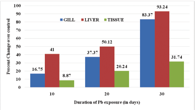 | Figure 2: Percent change in lead (Pb) accumulation in different tissues of fish M. cephalus. [Click here to view] |
The accumulation of lead has been found in various tissues (Liver, Muscle, and Gill) of M. cephalus exposed to sub-lethal concentration of lead acetate in different intervals of 10, 20, and 30 days of the exposure period. The significant (p < 0.05) changes were observed in the bioaccumulation of lead concentrations in different tissues of fish M. cephalus. The metal concentrations are found to be the maximum at 30 days of the sub-lethal exposure period in all tissues. The accumulation of lead is gradually increased from 10 to 30 days in the liver, gills, and muscle tissue of M. cephalus exposed to sub-lethal concentration of lead. The mean lead accumulation in the muscle tissues was 0.071, 0.162, and 0.254 μg/g, whereas in gills, 0.134, 0.299, and 0.671 μg/g, and in the liver, 0.328, 0.401, and 0.746 μg/g for 10, 20, and 30 days exposure periods, respectively (Table 2). The lead accumulation exhibits the maximum level in the liver (0.46 μg/g) followed by gill (0.672 μg/g) and muscle (0.254 μg/g) for a period of 30 days sub-lethal exposure (Fig. 1). Among the tissues of fish, liver shows a greater percent increment in metal accumulation compared with gill and muscle (Fig. 1).
3.1. Behavioral and Morphological Changes of Fish
Experimental fish samples showed different behavioral changes at different concentrations of lead. The type, frequency, and length of behavioral change increased with increased concentrations. In all of the procedures, the fish was hyperactive and attempted to escape from the procedure in the tank during the first hours of activity, in lead increased concentration tanks were observed fish swim near the surface and also observed respiratory difficulty, fishes attempted to breathe at the surface, their active movements increased due to toxicity, fish tried to escape from the tank, and observed sudden swimming motions also increased high mucus secretion. The anal fin and surrounding areas of the body were found bloody spots with high slime formation.
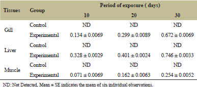 | Table 2: Concentration of lead (Mean ± S.E) in different tissues of fish M. cephalus exposed to sub-lethal concentration. [Click here to view] |
The present investigation, the highest level of the lead accumulation was found in the liver when compared to other tissues, followed by gill and muscle tissues of M. cephalus, when exposed to sub-lethal concentration of lead in different time intervals. These findings showed that lead acts as a cumulative poison in the liver and it is the prime site of metal accumulating or binding in the fishes.
The bioaccumulation of heavy metals in fish can be considered as an index of metal pollution in the aquatic environment. It is the capacity of organisms to concentrate a food chain and water element or compound at a level higher than that of their environment. The bioaccumulation of mixtures of metals may demonstrate a competitive, anti-competitive, or non-competitive inhibition, and various combinations of these may exhibit an increase in metal uptake [13].
Metals enter into the fish body by polluted food and water, after they accumulate in kidneys, liver, gills, muscles, skin, fins, heart, scales, gut, and brain [14]. However, the pattern of metal bioaccumulation in different fish tissues differed significantly at p < 0.05 during the current investigations, and it is found that liver and gills were deposited at higher levels. Lead (Pb), whereas muscles showed that there is least tendency for the accumulation of metal. Most of the research studies show that the kidney, liver, and gills are the main target organs for the metal accumulation. Similar results have been reported [15] with different metals such as Cd, Hg, Pb, Cr, and As in different tissues of fishes.
In this study, the organs of the fish M. cephalus shown heavy metal lead(Pb) concentration followed the order: liver > gill > muscle tissue. The liver plays a significant role in the processing, detoxification, transmission, and conversion of toxins and also serves as an active site for adverse effects caused by pollutants in animals. The synthesis of metallothionein (metal-binding protein) is caused by an elevated level of heavy metals. The disparity in the accumulation of metals in different fish tissues may be due to these metal-binding proteins (metallothionein) [16]. Previous studies have shown that the higher accumulation of Pb in the liver, body, intestine, gills, and freshwater fish muscles of Catla Catla. Similar results have also been recorded in the kidneys of fish Cyprinus carpio accumulated a higher amount of metals compared to the liver [17]. Fish gill surface is negatively charged, therefore it provides the binding site for the positively charged metals explained [18]. When part of routine control of environmental pollutants, muscle is a big tissue of concern because it is eaten by humans. Muscle is not an active tissue for the aggregation of metals like other tissues, this least potential may be due to low levels of protein binding in the muscles [15]. Water organisms exposed to heavy metals from run-off water continue to accumulate in their bodies, but fish are more frequently affected than other species [19].
4. CONCLUSION
Aquatic contamination caused by heavy metals has created a serious problem all over the world due to the ongoing dumping of waste materials from household waste and industrial waste into aquatic habitats. They can reach a dangerous level and pose a serious threat to the health of all aquatic organisms, as well as to humans. The biomonitoring approach allows us to predict the potential risk of persistent pollutants (heavy metals) and to formulate “safe” levels of these bioaccumulative chemicals with a genotoxic potential. Acute and chronic effects of individual metals (Pb, Co, and Cd) have been widely studied in various species of fish as these metals are important components of industrial and municipal effluents. Knowledge of the concentration of toxic metals in fish is important for management, such as the threat of using fish as part of the diet and the development of pollution control strategies. The result of the present investigation reveals that a time-dependent lead accumulation in different tissues of fishes exposed to different periods of sub-lethal concentration of lead. These findings extend for future studies on the evaluation of lead accumulation tendency in relation to the eco-toxicological examining program for pollution risk assessment.
REFERENCES
1. Sunlu U. Egemen, Babaran, A. The red mullet Mullus arbatus (Linnaeus, 1758) as an Indicator for heavy metal pollution in Izmir Bay (Turkey). In 36th CIESM Congress Proceedings, Monte-Carlo, Monaco, 2001, vol. 36, p 166.
2. Oliveira Ribeiro CA, Vollaire Y, Sanchez-Chardi A, Roche H. Bioaccumulation and the effects of organochlorine pesticides PAH and heavy metals in the eel (Anguilla anguilla) at the Camargue Nature Reserve, France. Aqua Toxicol 2005;74:53–69. CrossRef
3. De Forest KV, Brix WJ. Adams. Assessing metal bioaccumulation in aquatic environments: the inverse relationship between bioaccumulation factors, trophic transfer factors and exposure concentration. Aquat Toxicol 2007;84:236–46. CrossRef
4. Bhattacharya AK, Mandal SN, Das SK. Heavy metal accumulation in water sediment and tissues of different edible fishes in upper stretch of gangetic West Bengal. Trends in Applied Science Research 2008;3:61–8. CrossRef
5. Annabi A, Said K, Messaoudi I. Cadmium: bioaccumulation, histopathology and detoxifying mechanisms in fish. Am J Res Commun 2013;1:60–79.
6. Eisler R. Cadmium hazard to fish, wildlife and invertebrates: a synoptic review. US Fish Wildl Serv Biol Rep 1985;85:1–30.
7. Korkmaz Gorur F, Keser R, Akcay N, Dizman S. Radioactivity and heavy metal concentrations of some commercial fish species consumed in the Black Sea Region of Turkey. Chemosphere 2012;87:356–61. CrossRef
8. Petrovic Z, Teodrorovic V, Dimitrijevic M, Borozan S, Beukovic M, Milicevic M. Environmental Cd and Zn concentration in liver and kidney of erupean hare from different Serbian region: age and tissue difference. Bull Environ Contamin Toxicol 2013;90:203–7. CrossRef
9. Yilmaz F, Ozdemir N, Demirak A, Tuna AL. Heavy metal levels in two fish species Leuciscus cephalus and Lepomis gibbosus. Food Chem 2007;100:830–5. CrossRef
10. Zhao S, Feng C, Quan W, Chen X, Niu J, Shen Z. Role of living environments in the accumulation characteristics of heavy metals in fishes and crabs in the Yangtze River Estuary, China. Mar Pollut Bull 2012;64:1163–71. CrossRef
11. Vinodhini R, Narayanan M. Bioaccumulation of heavy metals in organs of freshwater fish Cyprino carpio (common carp). Int J Environ Sci Tech 2008;5(2):179–82. CrossRef
12. Ezeonyejiaku CD, Obiakora MO. Toxicological studies of single action of zinc on Tilapia specie (Tilapia nilotica). J Anim Feed Res 2011;1:139–43.
13. Eneji IS, Ato RS, Annune PA. Bioaccumulation of heavy metals in fish (Tilapia zilli and Clarias gariepinus) organs from River Benue, North-Central Nigeria. Pak J Anal Environ Chem 2011;12:25–31.
14. Jabeen G, Javed M, Azmat H. Assessment of heavy metals in the fish collected from the river Ravi, Pakistan. Pak Vet J 2012;32:107–11.
15. Squadrone S, Prearo M, Brizio P, Gavienelli S, Pellegrino M, Scanzio T, et al. Heavy metals distribution in muscle, liver, kidney and gill of European catfish (Silurus glanis) from Italian Rivers. Chemosphere 2013;90:358–65. CrossRef
16. Canli M, Atli G. The relationships between heavy metal (Cd, Cr, Cu, Fe, Pb, Zn) levels and the size of six Mediterranean fish species. Environ Pollut 2003;121:129–36. CrossRef
17. Ahmed MS, Bibi S. Uptake and bioaccumulation of water borne lead (Pb) in the fingerlings of a freshwater cyprinid. Catla catla L J Anim Pl Sci 2010;20:201–7.
18. Playle RC. Using multiple metal-gill binding models and the toxic unit concept to help reconcile multiple-metal toxicity results. Aquat Toxicol 2004;67:359–70. CrossRef
19. Henry F, Amara R, Courcot L, Lacouture D, Bertho ML. Heavy metals in four fish species from the French coast of the eastern English Channel and southern bight of the North Sea. Environ Int 2004;30:675–83. CrossRef