
1. INTRODUCTION
Today, biopolymers are in great demand for their various applications and extracellular polymeric substance (EPS) production by microorganisms is of great concern now-a-days. [1]. EPSs are secondary metabolites secreted by bacteria and accumulate over the cell surface [2]. The main components of extracellular polymeric substance are polysaccharides and proteins but also include some other macro-molecules [3]. Many microbial EPSs have gum like properties [4]. These microbial extracellular polymeric substances are highly soluble in water but many EPSs are insoluble and not easily separated from the cells [5].
Due to physicochemical and rheological properties, the extracellular polymeric substance is industrially important as they are used in textiles, food, cosmetics, agriculture, packaging industries, and pharmaceutical industries [6,7]. The EPS-producing bacteria have a good capability to convert nutrient into EPS [8]. It mainly consists of two types of groups, i.e., homopolysaccharides and heteropolysaccharides [9]. Some of the microbial exopolysaccharides have commercial applications, such as dextran, xanthan, pullulan, gellan, and hyaluronic acid [10]. The EPS also have been reported to be generally recognized as safe compounds [11]. It also has a great role in the field of bioremediation, among them, are sorption of heavy metals from wastewater [12,13], metabolizing and mineralizing of persistent organic pollutant [14] and improving activated sludge settleability [15]. In the medical field, the extracellular polymeric substance has a defensive role against desiccation, phagocytosis, antibiotics, helps to lower the cholesterol levels and prebiotic potential [16], used in oral, ophthalmic, and nasal drug formulations [10] and anticancer drugs [17]. There are many microorganisms reported earlier from different genera responsible for the production of EPS as a secondary metabolite. Among bacteria, Lactobacillus plantarum KX041 [7], Bacillus sp. MC3B-22, and Microbacterium sp. MC3B-10 [13], Leuconostoc mesenteroides [18], Sphingomonas paucimobilis [19], Kocuria rosea ZJUQH [8], Alteromonas infernus [20], and Escherichia coli [21]. Many fungi have also been reported for EPS production that include Fusarium equiseti ANP2 [22], Schizophyllum commune IBRC-M 30213 [23], Fusarium solani DO7 [24], Cordyceps militaris [25], etc. By keeping all the facts in mind and its vast application in various fields, the present research work was based on isolation and characterization of EPS-producing bacteria.
2. MATERIALS AND METHODS
2.1. Bacterial Cultures and Media Used
All the microorganisms used in this study were isolated from biofilms formed over the slides dipped in wastewater, and Salmonella, E. coli, Klebsiella sp., Pseudomonas sp., Staphylococcus aureus were used as test microorganisms for antimicrobial activity test. Different types of growth media were used viz. Nutrient agar medium (Peptone, 5 g/l; Beef extract, 3 g/l; Sodium chloride, 5 g/l; pH: 7.2) and casein hydrolysate agar medium (Sodium nitrate, 2 g; Dipotassium hydrogen phosphate, 1 g/l; Magnesium sulfate, 0.5 g/l; Potassium chloride, 0.5 g/l; Carboxymethyl cellulose, 5 g/l; and Peptone, 2 g/l; pH: 7). Each medium was first adjusted to appropriate pH and the agar–agar was added @2% whenever the solid medium was required before being sterilized by autoclaving at 121°C for 15 minutes.
2.2. Isolation of Biofilm Forming Bacteria
Glass slides were sterilized with 1N HCl and washed with distilled water. The sterilized glass slides were dipped into wastewater at different sites in Rohtak, Haryana for 20 days. After completion of 20 days, slides were rinsed with distilled water to remove debris and loosely attached bacteria [26]. The surfaces of the slides were scraped and suspended in 10 ml of sterilized normal saline. Bacterial cultures were isolated using the serial dilution technique. The agar plates were incubated at 37°C for 72 hours. Bacterial colonies having different morphology were selected and purified on nutrient agar plates [27].
2.3. Screening of EPS-producing Bacteria
Screening of EPS-producing bacteria was carried out on a morphological basis and formation of strings [28]. The colony characteristics, viz., shape, color, and polysaccharide production were observed on agar medium.
2.4. Production and Extraction of EPS
The EPS-producing colonies of each isolate were inoculated in appropriate media for the highest production of EPS. The one loopful inoculum was inoculated in 100-ml broth media and then incubated at 37°C for proper growth and production of EPS. The EPS extraction was carried out using the modified protocol of Subramanian et al. [26]. The bacterial broth culture was centrifuged at 10,000 rpm for 30 minutes at 4°C. The supernatant was precipitated with 97% chilled ethanol. The Precipitated EPS was collected by centrifugation at 10,000 rpm for 30 minutes. The dry weight of the extracted EPS was measured.
.png) | Table 1: Sites of wastewater sample collection and the selected EPS producing isolates. [Click here to view] |
2.5. Microscopic Studies and Biochemical Characterization of Isolates
The studies were carried out microscopically by different staining methods that involved simple staining and Gram staining. Biochemical tests were carried out as per the method given by Cappuccino and Sherman [29] with 24 hours old cultures. The various biochemical tests involved for the present study were starch hydrolysis, casein hydrolysis, cellulose hydrolysis, urease, Indole, Methyl Red, Voges-Proskauer and Citrate tests (IMVIC) citrate utilization test, etc.
2.6. Antimicrobial Activity of EPS-producing Isolates
The antimicrobial activity of isolates was studied by using agar well diffusion method. The test was performed according to a procedure developed by Rajoka et al. [30]. Twenty milliliters of the melted agar medium was poured in petriplate and 100 μl of 24 hours. Old broth culture of the test microorganism was spread on each plate. When the inoculums got absorbed in media, wells were cut on the surface using borer. The supernatant of isolates was poured into the well against different test microorganisms and incubated at 37ºC for 24–48 hours. After incubation, the plates were observed for a clear zone of inhibition around the well and diameter was measured.
3. RESULTS AND DISCUSSION
3.1. Isolation and Screening of Biofilm-forming Bacteria
Total of five samples of biofilms were collected from five different sites (Table 1). Bacteria from these samples were isolated on nutrient agar media amended with glucose (1.0%). In all, 50 representative bacteria were isolated, purified, and maintained as pure cultures. A similar method of isolation has also been used by Dhanya et al. [27] and isolated Pseudoalteromonas sp. from sea water. The EPS-producing bacteria are involved in the formation of biofilms, which in turn mediates close association of bacteria to abiotic and biotic surfaces [31]. According to another study by Gu et al. [8], EPS production is directly related to salt tolerance by K. rosea ZJUQH. Stress conditions are always preferable for EPS production [32].
3.2. Screening of EPS-producing Colonies
Out of 50 isolates, 21 isolates were screened for EPS production. The method used for the selection of EPS-producing microorganisms was based on their slimy colony and string formation by touching with inoculating loop on the growth medium (Table 1; Fig. 1). The EPS-producing colonies were mucoid in shape and produced a large amount of yellow pigmentation (Fig. 1C and 1D). The mucoid colonies have a glossy and slimy appearance on agar media were the primary criteria for selection of EPS-producing bacteria [33]. Earlier, the similar property of string formation and the mucoid colony has been reported by Bacosa et al. [34] and Rühmann et al. [28].
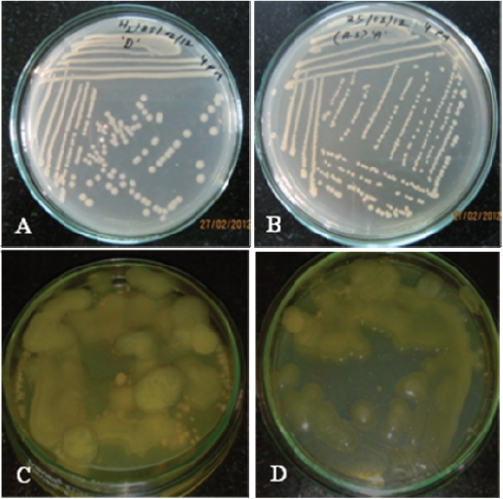 | Figure 1: Bacterial isolates purified colonies (Plate A) H2D isolate and (Plate B) A.SA isolate. Plate (C) and (D) are the bacteria producing yellow pigmentation after overproduction of EPS during preservation. [Click here to view] |
 | Figure 2: Production of EPS by different bacterial isolates. [Click here to view] |
3.3. Extraction of Exopolysaccharide (EPS)
All the 21 isolated microorganisms were used for EPS production and extraction using casein hydrolysate broth medium. On fourth day strain, ASB4 showed the highest EPS productivity, i.e., 17.2 g/l (Fig. 2). The concentration of EPS produced by 21 different bacterial strains varied from 1.2 to 17.2 g/l. Out of 21 isolates, nine were best EPS producers (ASA1, ASA2, ASA3, ASA4, ASA5, H2C6, H2E7, H2F8, and H2E9) and were further selected for EPS extraction. The strain ASB4 showed the highest productivity of 17.2 g/l, whereas the EPS produced by the isolates ASB5, ASA2, ASA1, and ASA3 was 8.4, 7.4, 7.0, and 5.8 g/l, respectively. The rest of the isolates showed average production (1.2–4.6 g/l) of EPS. In another study reported earlier, the production of exopolysaccharide was 4.65 mg/ml by isolate VS2B [35]. The comparative study revealed that isolate ASB4 was the highest producer of EPS. The EPS concentration from Bacillus subtilis was found in the range from 3.5 to 5.5 g/l [36].
3.4. Microscopic Studies of Isolates
Among nine isolates, six were rod-shaped and three were small rods. Five out of nine isolates were Gram-positive and four were Gram-negative (Table 2). The highest EPS-producing strain ASB4 was Gram-positive and rod-shaped. The bacterial isolate ASA1 was a small rod and Gram-positive, while the strain ASA2 was a small rod and Gram-negative. The strain ASB4 was found to be rod-shaped and gram-positive, whereas the isolate H2E9 was rod-shaped and Gram-negative. Earlier, it has been reported that most of the EPS-producing bacteria were Gram-positive and rod-shaped. Although, EPS producing bacteria has earlier been identified as Gram positive, Gram negative and maybe coccus or rods [37].
3.5. Biochemical Characterization of Isolates
Biochemical characterization showed that most of the bacterial isolates showed amylase positive except ASBB, H2E7, H2FB, and H2E9 (Table 2), and all of the isolates showed cellulase, casein hydrolysis, and urease test as negative, while most of the isolates were V-P, indole, and citrate utilization test positive. Six of the nine isolates were methyl red negative and three were positive. In our study, the strains ASA1, ASA2, ASA3, ASB5, and H2C6 (Table 2) showed a positive test for starch hydrolysis, i.e., these strain produced amylase enzyme. Amylase is the enzyme that hydrolyzes starch and forms simple glucose. The strains H2C6, H2E7, and H2E9 showed methyl red positive, whereas ASA1, ASA2, ASA3, ASB4, ASB5, H2C6, and H2F8 showed Voges–Proskauer test positive (Table 2). The isolates ASA1, ASA2, ASA3, ASB4, and ASB5 showed indole production test positive. The strains ASA1, ASA2, ASA3, ASB4, and ASB5 utilized sodium citrate as a carbon source, while the isolates H2C6, H2E7, H2F8, and H2E9 showed negative results for citrate utilization test. The unknown bacterial sample can be identified using biochemical tests as each bacterium has its own metabolic pathway. The biochemical tests have earlier been carried out to analyze the ability of bacteria to use enzymes and degrade amino acids, lipids, carbohydrates, and proteins [38]. It has been reported that EPS-producing bacteria having oxidase, protease, and starch hydrolysis as positive but catalase negative [39,40].
 | Table 2: Morphological, microscopic, and biochemical characterization of isolates. [Click here to view] |
3.6. Antimicrobial Activity by Bacterial Isolates
The test microorganisms, such as Salmonella sp., E. coli, Klebsiella sp., Pseudomonas sp., and S. aureus, were used in this study (Table 3; Fig. 3). The determination of the antimicrobial effect was carried out by the agar well diffusion method. In this study, strains ASA3, ASB5, H2C6, H2E7, H2F8, and H2E9 inhibited the growth of Salmonella sp. and the strain H2E7 inhibited the growth of E. coli and Klebsiella sp., whereas the strains ASA3 and H2F8 showed inhibition against Klebsiella sp. and E. coli, respectively (Table 3; Fig. 3). However, none of the isolates showed growth on Pseudomonas sp. and S. aureus plates. Antimicrobial activity reached the maximum after 48 hours of incubation of strains H2E7 and H2F8 against Salmonella sp. (Table 4). The highest antimicrobial activity shown by H2C6, H2E7, and H2E9 strains were 1.6, 1.4, and 1.4 cm against Salmonella sp. (Table 4), whereas the lowest antimicrobial activity was 1.0 and 1.1 cm by ASA3 strain against Salmonella sp. and Klebsiella sp. The strains H2C6, H2E7, and H2F8 showed the highest antimicrobial activity against Salmonella sp. Similar study on antimicrobial activity against some microorganisms has earlier been carried out by researchers by using EPS-producing bacteria [41,42]. It was reported that most of the EPS-producing bacteria effective against the Gram-positive bacterium, especially B. subtilis and M. tetragenus and their effectiveness depends on the concentration of culture filtrate and incubation time [43]. But in the present study, we found that EPS-producing strains were effective against Gram-negative bacteria, especially Salmonella sp., E. coli, and Klebsiella sp. but not against Gram-positive bacteria. Various factors are responsible for the antimicrobial activity of exopolysaccharides, including molecular weight, composition, and the chelating activities [44]. In our study, the strains ASA3, ASB5, H2C6, H2E7, H2F8, and H2E9 showed antimicrobial activity against Salmonella sp. (Table 3; Fig. 3A–C). Two strains H2E7 and H2F8 showed antimicrobial activity against E. coli (Fig. 3D), and the strains ASA3 and H2E7 showed antimicrobial activity against Klebsiella sp. (Table 3; Fig. 3E and F).
 | Table 3: Antimicrobial activity of EPS producing isolates. [Click here to view] |
 | Figure 3: Plate (A) strain ASA3, plate (B) ASB5 and H2C6, and plate (C) H2E7, H2F8, and H2E9 showed antimicrobial activity against Salmonella sp. In plate (D), strain H2E7 and H2F8 showed antimicrobial activity against E. coli. In plate (E) and (F), strain A.SA3 and H2E7 showed antimicrobial activity against Klebsiella sp. [Click here to view] |
 | Table 4: Antimicrobial activity (inhibition zone diameter in centimeter) of the selected isolates against Salmonella sp., E. coli, and Klebsiella sp. [Click here to view] |
4. CONCLUSION
In the present study, an attempt was made to isolate efficient EPS-producing bacteria from wastewater samples collected from different sites. A total of 21 bacterial isolates were used to EPS extraction and nine of them were efficient EPS producer. These isolates were checked for antimicrobial activity. On fourth day strain, ASB4 showed the highest EPS productivity, i.e., 17.2 g/l. Further studies are needed to study the application of produced exopolysaccharides at the industrial level.
REFERENCES
1. Delbarre-Ladrat C, Sinquin C, Lebellenger L, Zykwinska A, Colliec-Jouault S. Exopolysaccharides produced by marine bacteria and their applications as glycosaminoglycan-like molecules. FrontChem 2014;2:1–15.
2. Ates O. Systems biology of microbial exopolysaccharides production. Front Bioeng Biotechnol 2015;3:1–16.
3. Limoli DH, Jones CJ, Wozniak DJ. Bacterial extracellular polysaccharides in biofilm formation and function. Microbiol Spectr 2015;3(3):1–30.
4. Kaur V, Bera MB, Panesa PS, Kumar H, Kennedy JF. Welan gum: microbial production, characterization, and applications. Int J Biol Macromol 2014;65:454–61.
5. Casillo A, Lanzetta R, Parrilli M, Corsaro MM. Exopolysaccharides from marine and marine extremophilic bacteria: structures, properties, ecological roles and applications. Mar Drugs 2018;16:1–34.
6. Majee SB, Avlani D, Biswas GP. Rheological behavior and pharmaceutical applications of bacterial exopolysaccharides. J Appl Pharm Sci 2017;7:224–32.
7. Wang X, Shao C, Liu L, Guo X, Xu Y, Lü X. Optimization, partial characterization and antioxidant activity of an exopolysaccharide from Lactobacillus plantarum KX041. Int J Biol Macromol 2017;103:1173–84.
8. Gu D, Jiao Y, Wu J, Liu Z, Chen Q. Optimization of EPS production and characterization by a halophilic bacterium, Kocuria rosea ZJUQH from Chaka Salt Lake with response surface methodology. Molecules 2017;22:1–19.
9. Yildiz H, Karatas N. Microbial exopolysaccharides: resources and bioactive properties. Process Biochem 2018;72:41–6.
10. Moscovici M. Present and future medical applications of microbial exopolysaccharides. Front Microbiol 2015;6:1–11.
11. Kanmani P, Lim ST. Synthesis and structural characterization of silver nanoparticles using bacterial exopolysaccharide and its antimicrobial activity against food and multidrug resistant pathogens. Process Biochem 2013;48:1099–106.
12. Gupta P, Diwan B. Bacterial exopolysaccharide mediated heavy metal removal: a review on biosynthesis, mechanism and remediation strategies. Biotechnol Rep 2017;13:58–71.
13. Camacho-Chab J, Castañeda-Chávez M, Chan-Bacab M, Aguila-Ramírez R, Galaviz-Villa I, Bartolo-Pére P, et al. Biosorption of cadmium by non-toxic extracellular polymeric substances (EPS) synthesized by bacteria from marine intertidal biofilms. Int J Environ Res Public Health 2018;15:1–11.
14. Edwards SJ, Kjellerup BV. Applications of biofilms in bioremediation and biotransformation of persistent organic pollutants, pharmaceuticals/personal care products, and heavy metals. Appl Microbiol Biotechnol 2013;97:9909–21.
15. Lin YM, Wang L, Chi ZM, Liu XY. Bacterial alginate role in aerobic granular bioparticles formation and settleability improvement. SepSci Technol 2008;43:1642–52.
16. Maeda H, Zhu X, Suzuki S, Suzuki K, Kitamura S. Structural characterization and biological activities of an exopolysaccharide kefiran produced by Lactobacillus kefiranofaciens WT-2BT. J Agric Food Chem 2004;52:5533–8.
17. Escárcega-González CE, Garza-Cervantes JA, Vázquez-Rodríguez A, Morones-Ramíre JR. Bacterial exopolysaccharides as reducing and/or stabilizing agents during synthesis of metal nanoparticles with biomedical applications. Int J Polym Sci 2018;1–15.
18. Nwodo U, Green E, Okoh A. Bacterial exopolysaccharides: functionality and prospects. Int J Mol Sci 2012;13:14002–15.
19. Osmalek T, Froelich A, Tasarek S. Application of gellan gum in pharmacy and medicine. Int J Pharma 2014;466:328–40.
20. Zykwinska A, Tripon-Le Berre L, Sinquin C, Ropartz D, Rogniaux H, Colliec-Jouault S, et al. Enzymatic depolymerization of the GY785 exopolysaccharide produced by the deep-sea hydrothermal bacterium Alteromonas infernus: Structural study and enzyme activity assessment. Carbohydr Polym 2018;188:101–7.
21. Borgersen Q, Bolick DT, Kolling GL, Aijuka M, Ruiz-Perez F, Guerrant RL, et al. Abundant production of exopolysaccharide by EAEC strains enhances the formation of bacterial biofilms in contaminated sprouts. Gut Microbes 2018;9:264–78.
22. Prathyusha AMVN, Mohana SG, Bramhachari PV. Chemical characterization and antioxidant properties of exopolysaccharides from mangrove filamentous fungi Fusarium equiseti ANP2. Biotechnol Rep 2018;19:1–8.
23. Mohammadi A, Shojaosadati SA, Tehrani HJ, Mousavi SM, Saleh T, Khorasani AC. Schizophyllan production by newly isolated fungus Schizophyllum commune IBRC-M 30213: optimization of culture medium using response surface methodology. Ann Microbiol 2018;68:47–62.
24. Zeng YJ, Yang HR, Wang HF, Zong MH, Lou WY. Immune enhancement activity of a novel polysaccharide produced by Dendrobium officinale endophytic fungus Fusarium solani DO7. J Funct Foods 2019;53:266–75.
25. Wang CC, Wu JY, Chang CY, Yu ST, Liu YC. Enhanced exopolysaccharide production by Cordyceps militaris using repeated batch cultivation. J Biosci Bioeng 2018;127:499–505.
26. Subramanian SB, Yan S, Tyagi RD, Surampalli RY. Extracellular polymeric substances (EPS) producing bacterial strains of municipal wastewater sludge: isolation, molecular identification, EPS characterization and performance for sludge settling and dewatering. Water Res 2010;44:2253–66.
27. Dhanya BE, Chandra M, Rekha PD. Isolation and identification of exopolysaccharide producing bacteria from Someshwar beach of Dakshina Kannada, Mangalore. Pharma Innovation J 2018;7:382–6.
28. Rühmann B, Schmid J, Sieber V. Methods to identify the unexplored diversity of microbial exopolysaccharides. Front Microbiol 2015;6:1–8.
29. Cappuccino JC, Sherman N. Negative staining. In: Microbiology: a laboratory manual. 3rd edition, Benjamin/Cummings, Redwood City, CA, pp 27–8, 1992.
30. Rajoka MSR, Jin M, Haobin Z, Li Q, Shao D, Jiang C, et al. Functional characterization and biotechnological potential of exopolysaccharide produced by Lactobacillus rhamnosus strains isolated from human breast milk. LWT 2018;89:638–47.
31. Vu B, Chen M, Crawford R, Ivanova E. Bacterial extracellular polysaccharides involved in biofilm formation. Molecules 2009;14:2535–54.
32. Sandhya V, Ali SZ. The production of exopolysaccharide by Pseudomonas putida GAP-P45 under various abiotic stress conditions and its role in soil aggregation. Microbiology 2015;84:512–9.
33. Fusconi R, Godinho MJL. Screening for exopolysaccharide-producing bacteria from sub-tropical polluted groundwater. BrazJ Biol 2002;62:363–9.
34. Bacosa HP, Kamalanathan M, Chiu MH, Tsai SM, Sun L, Labonté JM, Quigg A. Extracellular polymeric substances (EPS) producing and oil degrading bacteria isolated from the northern Gulf of Mexico. PLoS One 2018;13:1–19.
35. Chaudhari R, Hajoori M, Suthar M, Desai S. Isolation, screening and characterization of marine bacteria for exopolysaccharide production. Biosci Discov 2017;8:643–9.
36. Razack SA, Velayutham V, Thangavelu V. Medium optimization for the production of exopolysaccharide by Bacillus subtilis using synthetic sources and agro wastes. Turk J Biol 2013;37:280–8.
37. Nicolaus B, Kambourova M, Oner ET. Exopolysaccharides from extremophiles: from fundamentals to biotechnology. Environ Technol 2010;31:1145–58.
38. Harley JP. Laboratory exercises in microbiology. 7th edition, McGraw-Hill Companies, New York, NY, 2008.
39. Al-Nahas MO, Darwish MM, Ali AE, Amin MA. Characterization of an exopolysaccharide-producing marine bacterium, isolate Pseudoalteromonas sp. AM. Afr J Microbiol Res 2011;5:3823–31.
40. Savadogo A, Cheik AT, Ouattara P, Savadogo W. Identification of exopolysaccharides-producing lactic acid bacteria from Burkina Faso fermented milk samples. Afr J Biotechnol 2004;3:189–94.
41. Chikkanna A, Ghosh D, Kishore A. Expression and characterization of a potential exopolysaccharide from a newly isolated halophilic thermotolerant bacteria Halomonas nitroreducens strain WB1. Peer J 2018;6:1–18.
42. Ghalem BR. Antioxidant and antimicrobial activities of exopolysaccharides from yoghurt starter. Adv Biochem 2017;5:97–101.
43. Li X, Nielsen L, Nolan C, Halverson LJ. Transient alginate gene expression by Pseudomonas putida biofilm residents under water-limiting conditions reflects adaptation to the local environment. Environ Microbiol 2010;12:1578–90.
44. Rabea EI, Badawy MET, Stevens CV, Smagghe G, Steurbaut W. Chitosan as antimicrobial agent: applications and mode of action. Biomacromolecules 2003;4:1457–65.