Research Article | Volume 13, Issue 1, January, 2025

Screening and characterization of thermostable protease-producing bacteria isolated from slaughter site soil samples from Southern India

Nafila Puthiya Punnarath Anjana Meppat Anitha Subash Servin Wesley Ponraj Shobana Arunachalam   

Open Access   

Published:  Nov 15, 2024

DOI: 10.7324/JABB.2024.193529
Abstract

The current study was isolated and identified Bacillus cereus from an animal slaughter site in the South Indian state of Kerala, as a potent additive to detergents for enhancing stain removal efficacy. Identifying the isolates as B. cereus was confirmed through microbial biomass production, biochemical tests, protease-specific activity, and sequencing of the 16S rRNA gene. Following the growth condition optimization, the protease enzyme was extracted from the biomass, exhibiting a protease activity of 72.212 ± 0.235 U/mL. Calcium, magnesium, and sodium positively influenced protease activity under the tested conditions and were negatively impacted by zinc, copper, and iron. The polar solvent dimethyl sulfoxide (DMSO) supported enzyme activity, whereas the non-polar solvents hexane and chloroform completely inhibited it. The highest protease-specific activity of the crude extract following dialysis was 62.997 U/mg. The purified fractions of Sephadex G-100 and DEAE-Sephadex A50 anion exchange columns showed the highest enzyme-specific activity of 193.652 U/mg and 324.004 U/mg, respectively. The addition of crude protease to a generic detergent significantly enhanced its efficacy in removing fabric stains from cotton, polyester, and silk materials. Therefore, the isolated serine protease demonstrates significant potential for industrial applications.


Keyword:     Soil isolation Protease activity Bacillus cereus Purification Stain removal Destaining


Citation:

Punnarath NP, Meppat A, Subash A, Ponraj SW, Arunachalam S. Screening and characterization of thermostable protease-producing bacteria isolated from slaughter site soil samples from Southern India. J App Biol Biotech. 2025;13(1):159-168. http://doi.org/10.7324/JABB.2024.193529

Copyright: Author(s). This is an Open Access article distributed under the terms of the Creative Commons Attribution-NonCommercial-ShareAlike license.

HTML Full Text

INTRODUCTION

Protease enzymes are essential for hydrolyzing peptide bonds within polypeptide chains composed of amino acids. They are ubiquitous across organisms, including plants, animals, and microorganisms, and are known for their specificity and selectivity in modifying proteins. Recent decades have seen revolutionary advancements in biocatalysis, integrating multidisciplinary technologies, and leveraging natural enzymatic reactions. Approximately one-third of the total enzyme production in industries worldwide is currently occupied by proteases. Approximately 60% of proteases used in industries are economically produced by microorganisms, including bacteria, molds, and yeasts that are responsible for generating around 40% of the chemicals used in various industries today [1-4] Proteases are engineered to function under physiological conditions and are frequently utilized to catalyze a wide range of organic transformations. However, in the context of biocatalysis, the focus is on employing proteases as effective process catalysts under specific and tailored environmental conditions.

In industry, the proteases isolated from thermophiles are much preferred as they tend to be stable at higher temperatures in industrial manufacturing processes. This leads to considerably accelerated reaction rates, increased solubility of non-gaseous reactants and products, and diminished susceptibility to microbial contamination by mesophiles [5]. Proteases used in industrial applications, such as in detergents and leather processing, must demonstrate stability at high temperatures. The broad-spectrum application of such enzymes in chemicals, food, pharmaceuticals, paper, textiles, and others primarily owes their resilience to denaturation and exceptional operational stability at elevated temperatures [6,7].

The industrial demand for enzymes that exhibit stability and activity in non-aqueous media is increasing more than ever, primarily due to their expanding utility in organic synthesis [8]. In the field of peptide synthesis, the application of proteases is currently limited due to their specificity and susceptibility in the presence of organic solvents, as numerous reactions occur in organic media [9]. The identification of microorganisms with thermostable and non-aqueous stable proteases also requires a focus on the optimization of microbial protease production [10]. Several factors include nutritional requirements and physical parameters, including pH, temperature, aeration, and agitation. These factors, when optimized for the culturing and cultivation processes, can lead to optimal protease production [11].

The present study aimed to identify suitable bacterial strains from soil samples that can deliver protease that is thermostable and possesses optimal activity in the presence of organic solvents and salt stress.


2. MATERIALS AND METHODS

2.1. Soil Study Area

Soil samples were collected near a meat processing facility in the Malappuram District of Kerala, India. This region has a humid tropical wet climate, with temperatures ranging from over 30°C during the monsoon season to up to 40°C in the summer.

2.2. Soil Collection and Isolation of Bacteria

Soil samples were collected randomly from a depth of 15 cm, placed in polystyrene bags, and stored at room temperature. The samples were enriched in 1% casein broth and incubated at room temperature for 24 h. One gram/mL of the enriched sample was diluted in 9 ml of sterile distilled water, and then serially diluted up to 10-5. Each dilution (0.1 ml) was spread on solid nutrient agar and incubated at 37°C for 48 h. Morphologically distinct colonies were selected and maintained on nutrient agar slants at 4°C for further analysis. The inoculation concentration for all experiments was maintained at 1% (0.5 mL of enriched seed culture in 50 mL of basal medium).

2.3. Screening and Selecting Protease-Producing Bacteria

2.3.1. Morphological screening

The selected isolates’ colony morphology was meticulously documented, considering factors such as colony size, color, texture, shape, and surface appearance. Pure cultures were preserved on nutrient agar slants and utilized for subsequent analyses.

2.3.2. Biochemical characterization

Gram staining and motility tests were performed on bacterial isolates to assess cellular structure and motility. Protease activity was evaluated using gelatin and casein hydrolysis tests. Preliminary bacterial identification followed the IMViC series of biochemical tests, as described in Bergey’s Manual of Systematic Bacteriology (Sneath et al., 1986), including lactose fermentation, methyl red, Voges-Proskauer, citrate utilization, urease, catalase, and hydrogen sulfide production.

2.3.3. Gelatin hydrolysis test

The bacterial pure colonies were inoculated on gelatin agar media, containing peptone (0.1% w/v), NaCl (0.5% w/v), agar (2.0% w/v), and gelatin (1% w/v) adjusted to neutral pH. These plates were incubated at 37°C for 24 h. A well-defined, clear zone of gelatin hydrolysis indicated protease activity. Colonies identified with wider gelatin clear zones were selected for subsequent experimental investigations.

2.3.4. Casein hydrolysis test

Bacterial colonies were inoculated on nutrient agar with 1% casein and incubated at 37°C for 24 h. Colonies forming clear zones were selected for further study. Isolates with prominent hydrolysis zones on both gelatin and casein were chosen for quantitative analysis. These colonies were repeatedly cultured on nutrient agar and preserved on nutrient agar slants at 4°C for future use.

2.3.5. Protease assay

Protease production began by incubating screened colonies for 120 h in an enrichment medium containing beef extract (0.15% w/v), peptone (0.5% w/v), NaCl (0.5% w/v), and glucose (0.5% w/v). Enriched cells were harvested by centrifugation at 10,000 rpm for 15 min at 4°C, and the supernatant was precipitated with 40% ammonium sulfate. The precipitate was redissolved in 20 mM Tris buffer for analysis.

For the protease assay, 1% β-casein was used as the substrate, and tyrosine was used as the standard. The Folin-Ciocalteau reagent quantified tyrosine by measuring absorbance at 660 nm using a Shimadzu UV 2401 spectrophotometer. The enzyme activity was calculated using the following formula:

For all experiments, the inoculation concentration remained consistent at 1%, constituted by 0.5 mL of enriched seed culture in 50 mL of basal medium. Initially, a single bacterial colony underwent inoculation into 5 mL of nutrient broth and was subjected to overnight incubation. Subsequently, 1% of this cultured broth (0.5 mL) was transferred to 50 mL of fresh nutrient broth and incubated for duration of 16-18 hours. Following cell harvesting, the culture underwent resuspension in phosphate-buffered saline (PBS), and the absorbance at 600 nm was determined. Subsequent dilution with PBS was performed to attain an optical density (OD600) of 1.0. This 1% inoculum served as the basis for enzyme production.

2.3.6. 16S rRNA analysis

Genomic DNA was extracted using the chloroform-isoamyl alcohol method from cells grown in 1.5 mL broth cultures. A 20-ng aliquot of genomic DNA from each isolate was amplified for 16S rRNA genes using universal bacterial primers 8f (5′-AGAGTTTGATCATGGCTCAG) and 1492r (5′-CGGTTACCTTGTTAC000000GACTT) [12-14]. PCR amplification employed the EmeraldAmp® GT PCR Master Mix. The sequencing PCR reaction was conducted in a final volume of 20 µl, and samples were purified as described by Chen et al. Sanger sequencing was performed by AgriGenome Labs Pvt. Ltd., Cochin, Kerala, IN, and sequences were compiled using appropriate software.

A BLAST search (https://blast.ncbi.nlm.nih.gov/Blast.cgi) identified similar sequences in the National Center for Biotechnology Information database. Phylogenetic analyses were conducted using MEGA software (https://www.megasoftware.net/), with sequence alignment by ClustalW and phylogenetic tree generation via the maximum likelihood method.

2.4. Optimization of Growth Conditions

2.4.1. Effect of time on protease production

The rate of protease production concerning time was tested with the 24 h growth isolate inoculation into a basal medium containing the following components: 1% glucose, 0.5% casein, 0.55% yeast extract, 0.2% KH2PO4, 1% Na2CO3, 0.2% MgSO4•7H2O, with a pH of 7. The inoculated medium was then incubated at 37°C for 72 h. One ml sample following 12 h of incubation was collected by 30 min of centrifugation at 5000 rpm to assess protease activity.

2.4.2. Effect of temperature on the production of protease

The effect of temperature on the production of protease was tested across a range of elevated temperatures, spanning from 25°C–50°C. Distinct cultures were incubated at temperatures of 25°C, 37°C, 40°C, 45°C, and 50°C, at pH 7.0 for 36 h, with continuous agitation at 150 rpm. Following incubation, the centrifugal pellet from the culture was observed for protease activity.

2.4.3. Effect of pH on the production of protease

Protease production based on the effects of pH was tested following incubation of bacterial samples on distinct basal media with pH ranging as 3.0, 5.0, 7.0, 9.0, and 11.0 (adjusted with 1 M NaOH or HCl), incubated at 37°C for 36 h and an agitation rate of 150 rpm. Protease activity was determined following the incubation period.

2.4.4. Effect of carbon source on the production of protease

The role of carbon sources in optimal bacterial growth and protease production was tested by inoculating samples into distinct basal media containing glucose, sucrose, maltose, xylose, galactose, and starch. The cultures were incubated at 37°C for 36 h with an agitation rate of 150 rpm.

2.4.5. Effect of nitrogen source on the production of protease

The effect of the nitrogen source on the production was tested with organic (yeast extract, peptone, and casein, each at 1% w/v) and inorganic nitrogen (ammonium sulfate ((NH4)2SO4), potassium nitrate (KNO3), and ammonium chloride (NH4Cl), each at 1% w/v) compounds added to the inoculation basal media. The cultures were incubated at 37°C for 36 h with an agitation rate of 150 rpm.

2.4.6. Effect of salt concentration on the production of protease

Various concentrations of NaCl, ranging from 0.0M to 1.0M, were introduced into the protease production medium. The cultures were incubated at 37°C for 36 h with an agitation rate of 150 rpm. Subsequently, the crude protease activity of these cultures was tested.

2.5. Enzyme Production and Characterization

2.5.1. Cultivation of bacteria in enzyme production media

Bacteria were cultured in a protein production medium at 37°C for 36 h with continuous agitation at 150 rpm. After incubation, dense bacterial growth was observed. The medium was filtered with Whatman® filter paper to remove bacterial biomass, then centrifuged at 5000 rpm and 4°C for 30 min to eliminate residual particles. The clarified filtrate was collected for further processing.

2.5.2. Ammonium sulfate precipitation

The filtrate obtained after centrifugation was subjected to ammonium persulfate precipitation. The supernatant was subjected to a gradual increase in ammonium sulfate concentration to reach 80% saturation while stirring at 4°C in an ice bath. Following a 30-min equilibration period, the protein precipitate was collected through centrifugation for 15 min at 15,000 rpm, maintaining a temperature of 4°C. The resulting pellet was re-suspended in 10 mL of ice-cold 0.2 M Tris–HCl buffer at pH 7. The protein, now dissolved in the buffer, underwent dialysis.

2.5.3. Membrane dialysis

The re-dissolved crude enzyme was placed in a cellulose dialysis membrane secured with knots. The membrane was submerged in 0.2 M Tris-HCl buffer (pH 7), with the buffer replaced hourly. After dialysis, the membrane was punctured, and the contents were transferred to a beaker for further purification.

2.5.4. Gel filtration and ion exchange chromatography

Protease was isolated using a Sephadex G-100 gel chromatography column with a 0.02 M phosphate buffer (pH 7.0) at a flow rate of 0.5 mL/min. Fractions were collected and analyzed at 280 nm. The enzyme sample was then loaded onto a DEAE-Sephadex A50 anion exchange column and eluted with 0.02 M Tris-HCl buffer (pH 8.5) using a 0 to 2 M sodium chloride gradient. Protease-active fractions were detected and combined for further use.

2.5.5. Protein quantification

Protein quantification was performed according to Lowry’s method, utilizing bovine serum albumin as a standard reference [15]. Purified protein (0.5 mL) was diluted to 1 mL with 0.1 N NaOH. Subsequently, 5.0 mL of alkaline copper reagent and 0.5 mL of Folin’s reagent were added, thoroughly mixed, and incubated at room temperature for 30 min. Absorbance was measured at 670 nm using a spectrophotometer, and protein concentration was determined using a standard graph generated from BSA.

2.5.6. SDS-PAGE and Zymogram

The protein samples’ purity was assessed using SDS-PAGE (Sodium Dodecyl Sulfate-Polyacrylamide Gel Electrophoresis) following Laemmli’s method (1970) on a 12% cross-linked polyacrylamide gel. Coomassie brilliant blue (CBB) staining was employed for visualizing protein bands, with molecular weight compared to a standard molecular marker ranging from 200 to 10 kDa [25].

Protease activity on SDS-PAGE was analyzed via zymography on a 12% SDS polyacrylamide gel with 0.1% copolymerized casein as a substrate and 10µl/well of buffer-diluted isolated protease. After electrophoresis, the gel underwent three consecutive one-hour washes in deionized water to remove SDS. Subsequently, the gel was incubated for 15 h at 37°C with gentle shaking in an activation buffer comprising 50 mM Tris-HCl and 10 mM CaCl2 (pH 8.0). CBB was used for staining and destaining the gel, with the protease activity appearing as a clear, colorless zone against the blue background.

2.6. Protease Characterization

2.6.1. Effect of pH on protease activity and stability

The isolated protease enzyme was incubated in 0.1 M potassium phosphate buffer at pH 5, pH 6, pH 7, pH 8, pH 9, and pH 10. The impact on enzyme activity was assessed over a 20-min incubation period, during which the residual enzymatic activity was measured using established protease assay techniques, as described earlier. To assess the enzyme’s stability, a pre-incubation period of 12 h was implemented, and subsequently, the residual enzymatic activity was determined using the established protease assay methods as described in section 2.3.3.

2.6.2. Effect of temperature on protease activity and stability

The protease enzyme was dissolved in 0.1 M potassium phosphate buffer and incubated at distinct temperatures at 20°C, 30°C, 40°C, 50°C, 60°C, and 70°C with 1% β-casein. The enzyme-substrate mixture was incubated for 20 min, followed by a protease assay.

2.6.3. Effect of salt concentration on protease activity and stability

The impact of different NaCl concentrations on the crude protease extract was examined by introducing NaCl into the reaction mixture to achieve final concentrations of 0%, 3%, 6%, 9%, and 15%. Enzyme activities were evaluated under conditions that were optimal for both pH and temperature, based on results from sections 2.6.1 and 2.6.2.

2.6.4. Effect of divalent ions on the protease activity

The influence of various metal cations, including calcium ions (Ca²+), magnesium ions (Mg²+), manganese ions (Mn²+), ferrous ions (Fe²+), and zinc ions (Zn²+), on the crude protease activity was tested. These metal cations were added to the reaction mixture at final concentrations of 1 and 5 millimolars (mM). In each case, enzyme activities were evaluated under conditions that matched the optimal pH and temperature, based on results from sections 2.6.1 and 2.6.2.

2.6.5. Effect of organic solvents on protease activity

The protease enzyme was subjected to a pre-incubation period of 4 h at 50°C in the presence of various organic solvents (hexane, chloroform, methanol, and dimethyl sulfoxide (DMSO)). The enzyme activity without any organic solvent (the control) was considered 100% to calculate the relative activity in percentage terms.

2.6.6. Enzyme characterization

To determine the class of protease produced by the potent isolate, specific inhibitors for different protease classes were employed. The enzyme was incubated with each inhibitor at a concentration of 5 mM for 2 h at 37°C, followed by the measurement of residual activity under standard protease assay conditions. The inhibitors used included phenylmethyl sulfonyl fluoride (PMSF) for serine proteases, β-mercaptoethanol (β-ME) for cysteine proteases, EDTA for metalloproteases, and aprotinin for aspartate proteases.

2.7. Stain Removal Efficacy of Protease Enzyme

The study assessed the protease enzyme’s efficacy as a detergent additive for stain removal on 5x5 cm pieces of silk, polyester, and white cotton fabrics stained with soy sauce and egg yolk. Each fabric type underwent three test groups: a control with stained samples, test group 1 with samples washed with varying volumes of wheel detergent (5 g/mL), and test group 2 with samples washed with a combination of wheel detergent (5 g/mL) and protease enzyme. Test group 1 for egg yolk stains was treated with 1000 µL of detergent, while test group 2 received a combination of 500 µL detergent and 500 µL protease enzyme. For soy sauce stains, test group 1 was treated with 300 µL of detergent, and test group 2 received 300 µL of detergent along with 300 µL of protease enzyme. The stained fabrics were dried at 50ºC for 3 h, followed by the application of detergent and enzyme, and a five min incubation at room temperature. Visual examination and microscopic images were utilized for efficacy assessment and reflectivity studies.

2.8. Statistical Analysis

All experimental data were collected in triplicates and analyzed using the analysis of variance (ANOVA) test, followed by a post-hoc Tukey HSD test.


3. RESULTS AND DISCUSSION

3.1. Isolation and Characterization of Protease-Producing Bacteria

The soil sampled from Malappuram District, Kerala, India, demonstrated loamy to clayey textures with notable gravel content [Figure 1A]. Employing sterile techniques, the soil underwent enrichment, serial dilutions, and subculturing on nutrient agar, resulting in the growth of eight distinct colonies with diverse morphological characteristics [Figures 1A,B].

Figure 1: Isolation of potent protease producing bacteria from soil sample. (A) Soil sample, (B) 1X solution of the soil sample, (C) Colonies from different serial dilutions from soil sample solution, (D) Isolated pure colonies from serial dilution, (E) Gelatin hydrolysis of isolated samples, (F) Casein hydrolysis of isolated samples, (G) Mass production of potent protease producers.


[Click here to view]

Enzymatic index (EI) analysis for gelatin and casein hydrolysis revealed significantly higher protein hydrolysis efficiency in isolates one, four, and seven compared to others [Figures 1C-F; Tables 1 and Figures 1E,G, Table 3].

Table 1: Hydrolysis results – Gelatin & Casein.

IsolatesGelatin hydrolysisCasein hydrolysis
Diameter of zone of degradation (mm)Enzymatic index (Diameter of zone of degradation (mm) / Colony diameter)Diameter of zone of degradation (mm)Enzymatic index (Diameter of zone of degradation (mm)/Colony diameter)
Isolate 116 mm2.2817 mm2.125
Isolate 2----
Isolate 314 mm1.7519 mm1.58
Isolate 419 mm2.1118 mm1.38
Isolate 515 mm1.2517 mm1.54
Isolate 6----
Isolate 717 mm1.8816 mm1.6
Isolate 8----

Table 2: Proteolytic activity of the isolates.

IsolatesConcentration of tyrosine released(mg/ml)Enzyme activity (U/mL)
Isolate 120.030 ± 0.0836.850 ± 0.08
Isolate 415.056 ± 0.0627.698 ± 0.08
Isolate 713.025 ± 0.1123.963 ± 0.09

Table 3: Biochemical characterization of the Isolate 1.

TestsIsolate 1
Indole test–ve
Methyl red test–ve
Voges proskauer test–ve
Citrate utilization test+ve
Urease production test–ve
Triple sugar iron agar testK/K no H2S production
Sugar fermentation
Glucose+ve
Lactose–ve
Sucrose+ve
Maltose+ve
Oxidase test–ve
Catalase test+ve

Morphological and microscopic examination of isolate one exhibited large, feathery, gray, granular, spreading, and opaque colonies with irregular margins [Figures 4B,C].

Figure 2: Enzyme production study. (A) Standard curve for tyrosine used for calculating enzyme activity; Optimization of enzyme production, (B) incubation time, (C) temperature, and (D) pH.


[Click here to view]
Figure 3: Effect on protease production by, (A) Sodium chloride, (B) Standard curve for tyrosine estimation, (C) carbon sources, (D) nitrogen sources.


[Click here to view]
Figure 4: Biochemical characterization of bacterial isolates, (A) pure culture of isolate one, (B) Gram staining, (C) IMViC test, (D) Catalase test, (E) Oxidase test.


[Click here to view]

Soil harbors a rich diversity of microorganisms, influenced by its composition and environmental conditions. Previous research worldwide has isolated various soil microorganisms with potential applications in antibacterial compounds, pigments, and industrial enzymes [16,17]. In our study, we isolated eight distinct bacterial strains from soil samples, each exhibiting unique morphological and biochemical characteristics. In contrast, a study conducted in Antarctica [18,19] successfully isolated seventy-five strains for alkaline protease production, highlighting the versatility of soil microorganisms across different environments. Identifying bacterial strains based on morphology alone can be challenging, as variations may arise from differential gene expression. Gene expression influences the synthesis of specific proteins, impacting cell structure and function, and leading to observable morphological differences.

Biochemical tests, including indole, methyl red, Voges Proskauer, Simmons citrate, catalase, oxidase, urease, and carbohydrate fermentation tests (glucose, sucrose, maltose, and lactose), aided in the preliminary identification of the isolated bacterial strain as belonging to the Bacillus species. Utilizing multivariate techniques [Figures 4D, E] offers a comprehensive approach to bacterial identification, with each method having its own strengths and weaknesses [20,26]. These tests evaluate metabolic activities such as substrate utilization and enzyme production, providing insights into the physiological characteristics of the bacterial isolates.

3.2. 16S rRNA Analysis

Isolate one, identified as Bacillus cereus, and was subjected to gene-based screening using 16S ribosomal RNA sequences with universal primers. Sanger sequencing of the 16S rRNA sequences revealed the highest identity (99.92%) with the P1 strain of B. cereus, ranging from 650 to 800 bases. The phylogenetic analysis included this strain along with nine other similar sequences [24]. The resulting phylogenetic tree [Figure 6C] displayed the P1 strain as the outgroup, indicating a more distantly related group. Two main clades branched from a common ancestor: rRNA strains A-D and E-H. Within these clades, A and B, as well as G and H, were closely related. Notably, strains I and H exhibited a distance of 0.016 (0.05 + 0.011) generic changes. Isolate one, identified as B. cereus, demonstrated superior enzyme activity in gelatin and casein degradation compared to other isolates.

Figure 5: Protease purification and enzyme activity, (A) Bacterial biomass production media, ammonium sulfate precipitation of protease crude extract, and membrane dialysis purification, (B) Enzyme activity of various fractions of from sephadex G-100, (C) Enzyme activity of various fractions from DEAE-Sephadex A50 anion exchange column.


[Click here to view]
Figure 6: Protease purification validation by, (A) SDS-PAGE, and (B) Zymogram Assay, (C) Phylogenetic tree of the protease-producing bacteria based on 16S rRNA gene sequence blast and sequence alignment results.


[Click here to view]

3.3. Optimization of Growth Conditions

Protease production from B. cereus was optimized across multiple growth parameters, as illustrated in Figure 3].

The isolates displayed varying strengths in protease production, with distinct enzymatic activities observed in each. Bacillus species’ bacterial isolates have shown effectiveness in extracellular protease production, consistent with previous studies by Alnahdi and Verma [21,22]. Notably, the isolate demonstrated a higher ratio of biomass production and alkaline protease activity, suggesting its potential for larger-scale commercial applications.

3.4. Enzyme Characterization

B. cereus was cultured in production media under optimized conditions, as described in section 3.4 [Figure 5A]. Following incubation, a crude enzyme unit was obtained, and subsequent downstream purification techniques were applied to isolate the protease enzyme. Evaluation of the crude enzyme’s protease activity using the Folin-Ciocalteau reagent at 660 nm revealed an activity of 72.212 ± 0.235 U/ml.

The purity of the isolated fractions was assessed through SDS-PAGE, which showed a single band corresponding to a 40 kDa protein weight, consistent with the standard protein marker. Zymogram staining confirmed proteolytic activity against a blue background, aligning with positions on the gel corresponding to the 40 kDa marker observed in SDS-PAGE [Figures 6A,B].

Peak protease activity of the eluted fractions from Sephadex-G 100 chromatography was observed in the range of 58 to 71 [Figure 5B]. These specific fractions (58 to 71) were collected and further eluted in a DEAE-Sephadex A-50 column. The protease activity of the eluted product was most prominent in fractions 37 to 54, with the 47th peak exhibiting the highest efficacy [Figure 5C].

Protein concentration was monitored at each purification step using Lowry’s method, demonstrating an increase in purification fold alongside enhanced enzyme activity (U/mL) [Table 5]. The DEAE-Sephadex A-50 column exhibited the highest enzyme activity, which was notably comparable to that of Sephadex-G 100.

Table 4: Identification of the potent isolate.

IsolateSequenceIdentityAccession ID
1GATTGGATTA AGAGCTTGCT CTTATGAAGT
TAGCGGCGGA CGGGTGAGTA ACACGTGGGT
AACCTGCCCA TAGGACTGGG ATAACTCCGG
GAAACCGGGG CTAATACCGG ATAACATTTT
GAACCGCATG GTTCGAAATT GAATGGCGGC
TTCGGCTGTC ACTTATGGAT GGACCCGCGT
CGCATTAGCT AGTTGGTGAG GTAA CGGCTC
ACGAAGGCAA CGATGCGTAG CCGACCTGAG
AGGGTGATCG GCCACACTGG GACTGAGACA
CGGCCCAGAC TCCTACGGGA GGCAGCAGTA
GGGAATCTTC CGCAATGGAC GAAAGTCTGA
CGGAGCAACG CCGCGTGAGT GATGAAGGCT
TTCGGGTCGT AAAACTCTGT TGTTAGGGAA
GAACAAGTGC TAGTTGAATA AGCTGGCACC
TTGACGGTAC CTAACCAGAA AGCCACGGCT
AACTACGTGC CAGCAGCCGC GGTAATACGT
AGGTGGCAAG CGTTATCCGG AATTATTGGG
CGTAAAGCGC GCGCAGGTGG TTTCTTAAGT
CTGATGTGAA AGCCCACGGC TGAACCGTGG
AGGGTCATTG GAAACTGGGA GACTTGAGTG
CAGAAGAGGA AAGTGGAATT CCATGTGTAG
CGGTGAAATG CGTAGAGATA TGGAGGAACA
CCAGTGGCGA AGGCGACTTT CTGGTCTGTA
ACTTACACTG AGGCGCGAAA GCGTGGGGAG
CAAACAGGAT TAGATACCCT GGTAGTCCAC
GCCGTAAACG ATGAGTGCTA AGTGTTAGAG
GGTTTCCGCC CTTTAGTGCT GAAGTTAACG
CATTAAGCAC TCCGCCTGGG GAGTACGGCC
GCAAGGCTGA AACTCAAAGG AATTGACGGG
GGCCCGCACA AGCGGTGGAG CATGTGGTTT
AATTCGAAGC AACGCGAAGA ACCTTACCAG
GTCTTGACAT CCTCTGAAAA CCCTAGAGAT
AGGGCTTCTC CTTCGGGAGC AGAGTGACAG
GTGGTGCATG GTTGTCGTCA GCTCGTGTCG
TGAGATGTTG GGTTAAGTCC CGCAACGAGC
GCAACCCTTG ATCTTAGTTG CCATCATTAA
GTTGGGCACT CTAAGGTGAC TGCCGGTGAC
AAACCGGAGG AAGGTGGGGA TGACGTCAAA
TCATCATGCC CCTTATGACC TGCGCTACAC
ACGTGCTACA ATGGACGGTA CAAAGAGCTG
CAAGACCGCG AGGTGGAGCT AATCTCATAA
AACCGTTCTC AGTTCGCATT GTAGGCTGCA
ACTCGCCTAC ATGAAGCTGG AATCGCTAGT
AATCGCGGA CAGCATGCCG
CGGTGAATAC
Bacillus cereus p1OP727433

Table 5: Purification table for enzyme extracted from isolate 1.

Purification stepVolume (ml)Protein concentration (mg/ml)Total protein (mg)Enzyme activity (U/ml)Total activity (U)Specific activity (U/mg)Yield (%)Purification fold
Culture filtrate2002.002 ± 0.020400.34260.009 ± 0.01212001.829.979100.001.000
Dialysis1001.146 ± 0.013114.62872.212 ± 0.0097221.262.99760.1682.101
Sephadex-100200.898 ± 0.03217.964173.939 ± 0.0223478.787193.65228.9866.460
DEAE-Sephadex A-50100.838 ± 0.0238.383271.620 ± 0.0172716.203324.00422.63210.808

The crude enzyme was effectively inhibited by phenylmethylsulfonyl fluoride (PMSF, 5 mM), indicating the presence of serine proteases in the strain’s enzyme. Conversely, EDTA, a metalloprotease inhibitor, did not inhibit enzyme activity, confirming the absence of metalloproteases. The enzyme retained activity levels of 265.09 U/mL when preincubated with 5 mM EDTA. Similar activity levels were observed with β-mercaptoethanol (278.26 U/mL) and aprotinin (238.753 U/mL), further supporting its classification as a serine protease [Table 6].

Table 6: Characterization of protease enzyme class.

Class of proteaseInhibitor usedEnzyme activity (U/mL)
Serine proteasePMSF0.2633 ± 0.048
Cysteine proteaseβ-mercaptoethanol278.26 ± 0.022
MetalloproteaseEDTA265.09 ± 0.060
Aspartate proteaseAprotinin238.75 ± 0.010

PMSF: phenylmethylsulfonyl fluoride; EDTA: Ethylenediaminetetraacetic acid.

The isolated enzymes were tested for stability and activity under varying conditions of temperature, pH, metal ions, and organic solvents [Figure 8]. The maximum enzyme activity was observed at 37°C (135.825 ± 0.240 U/mL), decreasing significantly at 60°C (58.73 ± 0.522 U/mL). The optimal pH for enzyme activity was 7.0 (67.59 ± 0.0167 U/mL). Serine protease inhibitors (serpins) inhibit proteases through a conformational change.

Figure 7: Stain Removal Test: Egg yolk stain (A-I), soy sauce stain (J-R) on white cotton, polyester and silk respectively. 500 µL and 300 µL detergent alone wash on egg yolk stain (B, E, and H) and soy sauce (K, N, and Q). Detergent and protease enzyme combination wash (500:500 µL and 300:300 µL) on egg yolk (C, F, and I) and soy sauce (L, O, and R). Microscopic surface image of all the test samples are labeled in their respective lowercases.


[Click here to view]
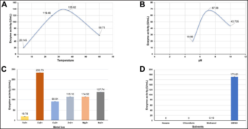
Figure 8: Effect of enzyme activity (U/mL) under varying conditions. (A) Temperature, (B) pH, (C) Presence of metal ions, (D) solvents.


[Click here to view]

Protease activity was notably influenced by the presence of divalent metal ions [Figure 8C]. Calcium ions (Ca2+) significantly enhanced protease activity, as indicated by the highest concentration of released tyrosine and enzyme activity. Magnesium (Mg2+) and sodium (Na2+) also increased protease activity, albeit to a lesser extent. Conversely, zinc (Zn2+), copper (Cu2+), and iron (Fe2+) resulted in lower enzyme activity compared to calcium, magnesium, and sodium. These results suggest that specific divalent metal ions, particularly calcium, positively influence protease activity under the tested conditions.

In both hexane and chloroform, the tyrosine release and enzyme activity were zero, indicating that these nonpolar solvents do not support protease activity [Figure 8D]. This lack of activity suggests that proteases are not stable or active in nonpolar environments. Conversely, methanol, a polar solvent, showed minimal protease activity. Dimethyl sulfoxide (DMSO), a polar aprotic solvent, exhibited higher tyrosine release and enzyme activity, suggesting that DMSO supports protease activity better than the other solvents tested.

The purified enzymes combined with detergent effectively removed egg yolk and soy sauce stains, as demonstrated by stain washing and microscopic imaging [Figure 7]. The enzyme-detergent combination showed superior stain removal efficacy compared to detergent alone. Reflectivity studies indicated that the enzyme-detergent combination significantly reduced residual stains on and between the fibers, resulting in smoother and looser fiber arrangements. In contrast, detergent alone left some stain residues.

The effectiveness of enzyme-detergent mixtures in stain removal aligns with previous studies showing that alkaline proteases from Bacillus species excel in stain removal and have potential in the enzyme-detergent market as effective cleaning agents. The demand for organic solvent-tolerant proteases in the industrial sector is high due to their application in organic solvent-based reactions. The thermostability of these proteases further enhances their suitability for industrial applications, particularly in laundry detergent formulations.

Overall, the present study establishes the higher attributes of the isolated B. cereus serine protease in comparison to prevalent microbial proteases, specifically emphasizing its thermostability, pH stability, catalytic efficiency, inhibitor and solvent resistance, and production yield. Particularly noteworthy is the exceptional thermostability of our protease, which sustains high activity levels (72.212±0.235 U/mL) even under elevated temperatures, presenting a significant advantage for industrial processes operating at high temperatures [11]. This characteristic contrasts with the common observation of reduced stability in many Bacillus proteases under similar conditions [6-8]. Furthermore, our protease exhibits sustained high activity across a wide pH range, outperforming the narrower pH tolerances of other Bacillus proteases, thus rendering it applicable across diverse industrial sectors such as wastewater treatment and food processing [6]. Additionally, our enzyme displays elevated specific activity and efficient hydrolysis (62.997 U/mg), resulting in diminished enzyme requirements in comparison to other Bacillus proteases [19, 23, 27, 28]. Its resilience against inhibitors and compatibility with polar solvents like DMSO further augment its utility in industrial contexts where enzyme inhibition poses a prevalent challenge. Moreover, the substantial production yield of our protease facilitates scalable production, rendering it economically viable and significantly amplifying detergent efficacy on various fabric substrates, thereby conclusively establishing its superiority over extant microbial proteases.


4. CONCLUSION

The present study highlights the isolation of B. cereus from the slaughter site soil samples in Malappuram District of Kerala state, India. The isolation and screening led to the identification of eight potent protease-producing bacterial colonies capable of degrading casein and gelatin. Based on the screening, one out of the eight was selected for a parameter optimization study related to growth and enzyme production. Morphological and biochemical assays revealed the screened isolate as a Bacillus species. Based on the 16S rRNA sequencing methods, the strain was subsequently identified as B. cereus. The enzyme from B. cereus was initially extracted in crude form and later purified via ion exchange chromatography. The enzyme activity of all the crude and purified enzyme solutions was studied under various physical and chemical parameters, revealing the industry favorable functioning. The stain removal efficacy of a known detergent on cotton, polyester, and silk against egg-yolk and soy sauce revealed potential for use in detergents. The isolated B. cereus and its protease can be recommended for further study on protease research work in enzyme-based industries.


5. AUTHOR CONTRIBUTIONS

All authors made substantial contributions to conception and design, acquisition of data, or analysis and interpretation of data; took part in drafting the article or revising it critically for important intellectual content; agreed to submit to the current journal; gave final approval of the version to be published; and agree to be accountable for all aspects of the work. All the authors are eligible to be an author as per the international committee of medical journal editors (ICMJE) requirements/guidelines.


6. FINANCIAL SUPPORT AND SPONSORSHIP

The authors declare that no funds, grants, or other support were received during the preparation of this manuscript.


7. CONFLICTS OF INTEREST

The authors report no financial or other conflicts of interest in this work.


8. ETHICAL APPROVALS

This study does not involve experiments on animals or human subjects.


9. DATA AVAILABILITY

All the data presented in this article is available on request from the authors.


10. USE OF ARTIFICIAL INTELLIGENCE (AI)-ASSISTED TECHNOLOGY

The authors confirm that there was no use of artificial intelligence (AI)-assisted technology for assisting in the writing or editing of the manuscript and no images were manipulated using AI.


11. PUBLISHER’S NOTE

All claims expressed in this article are solely those of the authors and do not necessarily represent those of the publisher, the editors and the reviewers. This journal remains neutral with regard to jurisdictional claims in published institutional affiliation.


REFERENCES

1.  Sloma A, Rudolph CF, Rufo GA, Sullivan BJ, Theriault KA, Ally D, et al. Gene encoding a novel extracellular metalloprotease in Bacillus subtilis. J Bacteriol. 1990;172:1024-9. [CrossRef]

2.  Rao MB, Tanksale AM, Ghatge MS, Deshpande VV. Molecular and biotechnological aspects of microbial proteases. Microbiol Mol Biol Rev. 1998;62:597-635. [CrossRef]

3.  Adinarayana K, Ellaiah P, Prasad DS. Purification and partial characterization of thermostable serine alkaline protease from a newly isolated Bacillus subtilis PE-11. AAPS PharmSciTech. 2003;4:E56. [CrossRef]

4.  Subba Rao C, Sathish T, Ravichandra P, Prakasham RS. Characterization of thermo- and detergent stable serine protease from isolated Bacillus circulans and evaluation of eco-friendly applications. Process Biochem. 2009;44:262-8. [CrossRef]

5.  Do Nascimento WCA, Leal Martins ML. Production and properties of an extracellular protease from thermophilic Bacillus sp. Brazilian J Microbiol. 2004;35:91-6. [CrossRef]

6.  Raveendran S, Parameswaran B, Ummalyma SB, Abraham A, Mathew AK, Madhavan A, et al. Applications of microbial enzymes in food industry. Food Technol Biotechnol. 2018;56:16-30. [CrossRef]

7.  Naveed M, Nadeem F, Mehmood T, Bilal M, Anwar Z, Amjad F. Protease—a versatile and ecofriendly biocatalyst with multi-industrial applications: an updated review. Catal Lett. 2021;151:307-23. [CrossRef]

8.  Gupta A, Khare SK. Enhanced production and characterization of a solvent stable protease from solvent tolerant pseudomonas aeruginosa PseA. Enzyme Microb Technol. 2007;42:11-6. [CrossRef]

9.  Kumar D, Bhalla TC. Microbial proteases in peptide synthesis: approaches and applications. Appl Microbiol Biotechnol. 2005;68:726-36. [CrossRef]

10.  Sellami-Kamoun A, Haddar A, Ali NEH, Ghorbel-Frikha B, Kanoun S, Nasri M. Stability of thermostable alkaline protease from Bacillus licheniformis RP1 in commercial solid laundry detergent formulations. Microbiol Res. 2008;163:299-306. [CrossRef]

11.  Abusham RA, Rahman RNZRA, Salleh A, Basri M. Optimization of physical factors affecting the production of thermo-stable organic solvent-tolerant protease from a newly isolated halo tolerant Bacillus subtilis strain Rand. Microb Cell Fact. 2009;8:1-9. [CrossRef]

12.  Weisburg WG, Barns SM, Pelletier DA, Lane DJ. 16S ribosomal DNA amplification for phylogenetic study. J Bacteriol. 1991;173:697-703. [CrossRef]

13.  Schumann P. E. Stackebrandt and M. Goodfellow (Editors), Nucleic Acid Techniques in Bacterial Systematics (Modern Microbiological Methods). J Basic Microbiol. 1991;31:479–80. [CrossRef]

14.  Voytek MA, Ward BB. Detection of ammonium-oxidizing bacteria of the beta-subclass of the class Proteobacteria in aquatic samples with the PCR. Appl Environ Microbiol. 1995;61:2811. [CrossRef]

15.  Shen C-H. Quantification and analysis of proteins. In Shen C-H, ed. Diagnostic Molecular Biology. Academic Press;2023:231-57. [CrossRef]

16.  Luang-In V, Yotchaisarn M, Saengha W, Udomwong P, Deeseenthum S, Maneewan K. Protease-producing bacteria from soil in Nasinuan Community Forest, Mahasarakham Province, Thailand. Biomed Pharmacol J. 2019;12:587-95. [CrossRef]

17.  Gaete A, Mandakovic D, González M. Isolation and identification of soil bacteria from extreme environments of chile and their plant beneficial characteristics. Microorganisms. 2020;8:1213. [CrossRef]

18.  Wery N, Gerike U, Sharman A, Chaudhuri JB, Hough DW, Danson MJ. Use of a packed-column bioreactor for isolation of diverse protease-producing bacteria from Antarctic soil. Appl Environ Microbiol. 2003;69:1457-64. [CrossRef]

19.  Masi C, Parthasarathi N. Production and process optimization of protease using various bacterial species-a review. Recent Trends Biotechnol Chem Eng. 2014;6:4268-75.

20.  Franco-Duarte R, Cernáková L, Kadam S, Kaushik KS, Salehi B, Bevilacqua A, et al. Advances in chemical and biological methods to identify microorganisms—from past to present. Microorganisms. 2019;7:130. [CrossRef]

21.  Alnahdi HS. Isolation and screening of extracellular proteases produced by new Isolated Bacillus sp. J Appl Pharm Sci. 2012;2:2915. [CrossRef]

22.  Verma A, Singh H, Anwar MS, Kumar S, Ansari MW, Agrawal S. Production of thermostable organic solvent tolerant keratinolytic protease from Thermoactinomyces sp. RM4: IAA production and plant growth promotion. Front Microbiol. 2016;7:1189. [CrossRef]

23.  Williams CM, Richter CS, MacKenzie JM, Shih JCH. Isolation, identification, and characterization of a feather-degrading bacterium. Appl Environ Microbiol. 1990;56:1509. [CrossRef]

24.  Chen L, Cai Y, Zhou G, Shi X, Su J, Chen G, et al. Rapid sanger sequencing of the 16S rRNA gene for identification of some common pathogens. PLoS One. 2014;9:88886. [CrossRef]

25.  He F. Laemmli-SDS-PAGE. Bio-Protocol. 2011;1:e80. [CrossRef]

26.  Mazotto AM, Lage Cedrola SM, Lins U, Rosado AS, Silva KT, Chaves JQ, et al. Keratinolytic activity of Bacillus subtilis AMR using human hair. Lett Appl Microbiol. 2010;50:89-96. [CrossRef]

27.  Keshapaga UR, Jathoth K, Singh SS, Gogada R, Burgula S. Characterization of high-yield Bacillus subtilis cysteine protease for diverse industrial applications. Braz J Microbiol. 2023;54:739-52. [CrossRef]

28.  Marathe SK, Vashistht MA, Prashanth A, Parveen N, Chakraborty S, Nair SS. Isolation, partial purification, biochemical characterization and detergent compatibility of alkaline protease produced by Bacillus subtilis, Alcaligenes faecalis and Pseudomonas aeruginosa obtained from sea water samples. J Genet Eng Biotechnol. 2018;16:39-46. [CrossRef]

Reference

1. Sloma A, Rudolph CF, Rufo GA, Sullivan BJ, Theriault KA, Ally D, et al. Gene encoding a novel extracellular metalloprotease in Bacillus subtilis. J Bacteriol. 1990;172:1024-9. https://doi.org/10.1128/JB.172.2.1024-1029.1990

2. Rao MB, Tanksale AM, Ghatge MS, Deshpande VV. Molecular and biotechnological aspects of microbial proteases. Microbiol Mol Biol Rev. 1998;62:597-635. https://doi.org/10.1128/MMBR.62.3.597-635.1998

3. Adinarayana K, Ellaiah P, Prasad DS. Purification and partial characterization of thermostable serine alkaline protease from a newly isolated Bacillus subtilis PE-11. AAPS PharmSciTech. 2003;4:E56. https://doi.org/10.1208/pt040456

4. Subba Rao C, Sathish T, Ravichandra P, Prakasham RS. Characterization of thermo- and detergent stable serine protease from isolated Bacillus circulans and evaluation of eco-friendly applications. Process Biochem. 2009;44:262-8. https://doi.org/10.1016/J.PROCBIO.2008.10.022

5. Do Nascimento WCA, Leal Martins ML. Production and properties of an extracellular protease from thermophilic Bacillus sp. Brazilian J Microbiol. 2004;35:91-6. https://doi.org/10.1590/S1517-83822004000100015

6. Raveendran S, Parameswaran B, Ummalyma SB, Abraham A, Mathew AK, Madhavan A, et al. Applications of microbial enzymes in food industry. Food Technol Biotechnol. 2018;56:16-30. https://doi.org/10.17113/FTB.56.01.18.5491

7. Naveed M, Nadeem F, Mehmood T, Bilal M, Anwar Z, Amjad F. Protease—a versatile and ecofriendly biocatalyst with multi-industrial applications: an updated review. Catal Lett. 2021;151:307-23. https://doi.org/10.1007/S10562-020-03316-7

8. Gupta A, Khare SK. Enhanced production and characterization of a solvent stable protease from solvent tolerant pseudomonas aeruginosa PseA. Enzyme Microb Technol. 2007;42:11-6. https://doi.org/10.1016/J.ENZMICTEC.2007.07.019

9. Kumar D, Bhalla TC. Microbial proteases in peptide synthesis: approaches and applications. Appl Microbiol Biotechnol. 2005;68:726-36. https://doi.org/10.1007/S00253-005-0094-7

10. Sellami-Kamoun A, Haddar A, Ali NEH, Ghorbel-Frikha B, Kanoun S, Nasri M. Stability of thermostable alkaline protease from Bacillus licheniformis RP1 in commercial solid laundry detergent formulations. Microbiol Res. 2008;163:299-306. https://doi.org/10.1016/J.MICRES.2006.06.001

11. Abusham RA, Rahman RNZRA, Salleh A, Basri M. Optimization of physical factors affecting the production of thermo-stable organic solvent-tolerant protease from a newly isolated halo tolerant Bacillus subtilis strain Rand. Microb Cell Fact. 2009;8:1-9. https://doi.org/10.1186/1475-2859-8-20

12. Weisburg WG, Barns SM, Pelletier DA, Lane DJ. 16S ribosomal DNA amplification for phylogenetic study. J Bacteriol. 1991;173:697-703. https://doi.org/10.1128/JB.173.2.697-703.1991

13. Schumann P. E. Stackebrandt and M. Goodfellow (Editors), Nucleic Acid Techniques in Bacterial Systematics (Modern Microbiological Methods). J Basic Microbiol. 1991;31:479–80. https://doi.org/10.1002/JOBM.3620310616

14. Voytek MA, Ward BB. Detection of ammonium-oxidizing bacteria of the beta-subclass of the class Proteobacteria in aquatic samples with the PCR. Appl Environ Microbiol. 1995;61:2811. https://doi.org/10.1128/AEM.61.7.2811-2811C.1995

15. Shen C-H. Quantification and analysis of proteins. In Shen C-H, ed. Diagnostic Molecular Biology. Academic Press;2023:231-57. https://doi.org/10.1016/B978-0-323-91788-9.00002-8

16. Luang-In V, Yotchaisarn M, Saengha W, Udomwong P, Deeseenthum S, Maneewan K. Protease-producing bacteria from soil in Nasinuan Community Forest, Mahasarakham Province, Thailand. Biomed Pharmacol J. 2019;12:587-95. https://doi.org/10.13005/BPJ/1678

17. Gaete A, Mandakovic D, González M. Isolation and identification of soil bacteria from extreme environments of chile and their plant beneficial characteristics. Microorganisms. 2020;8:1213. https://doi.org/10.3390/MICROORGANISMS8081213

18. Wery N, Gerike U, Sharman A, Chaudhuri JB, Hough DW, Danson MJ. Use of a packed-column bioreactor for isolation of diverse protease-producing bacteria from Antarctic soil. Appl Environ Microbiol. 2003;69:1457-64. https://doi.org/10.1128/AEM.69.3.1457-1464.2003

19. Masi C, Parthasarathi N. Production and process optimization of protease using various bacterial species-a review. Recent Trends Biotechnol Chem Eng. 2014;6:4268-75.

20. Franco-Duarte R, Cernáková L, Kadam S, Kaushik KS, Salehi B, Bevilacqua A, et al. Advances in chemical and biological methods to identify microorganisms—from past to present. Microorganisms. 2019;7:130. https://doi.org/10.3390/MICROORGANISMS7050130

21. Alnahdi HS. Isolation and screening of extracellular proteases produced by new Isolated Bacillus sp. J Appl Pharm Sci. 2012;2:2915. https://doi.org/10.7324/JAPS.2012.2915

22. Verma A, Singh H, Anwar MS, Kumar S, Ansari MW, Agrawal S. Production of thermostable organic solvent tolerant keratinolytic protease from Thermoactinomyces sp. RM4: IAA production and plant growth promotion. Front Microbiol. 2016;7:1189. https://doi.org/10.3389/FMICB.2016.01189

23. Williams CM, Richter CS, MacKenzie JM, Shih JCH. Isolation, identification, and characterization of a feather-degrading bacterium. Appl Environ Microbiol. 1990;56:1509. https://doi.org/10.1128/AEM.56.6.1509-1515.1990

24. Chen L, Cai Y, Zhou G, Shi X, Su J, Chen G, et al. Rapid sanger sequencing of the 16S rRNA gene for identification of some common pathogens. PLoS One. 2014;9:88886. https://doi.org/10.1371/JOURNAL.PONE.0088886

25. He F. Laemmli-SDS-PAGE. Bio-Protocol. 2011;1:e80. https://doi.org/10.21769/BIOPROTOC.80

26. Mazotto AM, Lage Cedrola SM, Lins U, Rosado AS, Silva KT, Chaves JQ, et al. Keratinolytic activity of Bacillus subtilis AMR using human hair. Lett Appl Microbiol. 2010;50:89-96. https://doi.org/10.1111/J.1472-765X.2009.02760.X

27. Keshapaga UR, Jathoth K, Singh SS, Gogada R, Burgula S. Characterization of high-yield Bacillus subtilis cysteine protease for diverse industrial applications. Braz J Microbiol. 2023;54:739-52. https://doi.org/10.1007/S42770-023-00992-6

28. Marathe SK, Vashistht MA, Prashanth A, Parveen N, Chakraborty S, Nair SS. Isolation, partial purification, biochemical characterization and detergent compatibility of alkaline protease produced by Bacillus subtilis, Alcaligenes faecalis and Pseudomonas aeruginosa obtained from sea water samples. J Genet Eng Biotechnol. 2018;16:39-46. https://doi.org/10.1016/J.JGEB.2017.10.001

Article Metrics
111 Views 58 Downloads 169 Total

Year

Month

Related Search

By author names

Similar Articles

Cloning, expression and purification of resistance gene analogue RGPM 301 from pearl millet in Escherichia coli

Mariswamy Veena, Prasad Melvin, Sekhar Shailasree, Kukkundoor Ramachandra Kini

Purification and characterization of tannase from the local isolate of Aspergillus niger

Saja Taha Yaseen Al-Mraai, Dhia Falih Al-Fekaiki, Alaa Jabbar Abd Al-Manhel

Purification and biochemical characterization of pectinase produced by Aspergillus fumigatus isolated from soil of decomposing plant materials

Raphael Emuebie Okonji, Babamotemi Oluwasola Itakorode, Julius Ovie Ovumedia, Odunayo Selimot Adedeji

Partial purification and characterization of lectin from serum of American cockroach, Periplaneta americana

Reymend Encily Martin, Yogita Raosaheb Channe

Partial purification and characterization of antimicrobial peptide from the hemolymph of cockroach Periplaneta americana

Reymend Encily Martin, Yogita Raosaheb Channe

Purification, characterization of α-galactosidase from a novel Bacillus megaterium VHM1, and its applications in the food industry

Aravind Gouda G. Patil, Naganagouda V. Kote, A. C. Manjula, T. Vishwanatha

Purification and biochemical characterization of protease from the seeds of Cyamopsis tetragonoloba

Rajesh Kumar Rawaliya, Preeti Patidar, Sadhana Sharma, Krishnan Hajela

Cloning and expression of a GH11 xylanase from Bacillus pumilus SSP-34 in Pichia pastoris GS115: Purification and characterization

Sagar Krishna Bhat,, Kavya Purushothaman, Appu Rao Gopala Rao Appu Rao, K Ramachandra Kini

Purification and characterization of a polygalacturonase from the xylophagous insect Oncideres albomarginata chamela (Coleoptera: Cerambycidae)

Alicia Lara-Márquez, Nayeli Soria-Calderón, Maria Guadalupe Villa-Rivera, Everardo López-Romero, Nancy Calderón-Cortés

Dexras1: A human G protein subcloning, expression, and purification optimization and prediction of its structural determinants

Rashmi Verma, Ashish Thapliyal, Navin Kumar

Production of SARS-CoV-2 nucleocapsid protein in Escherichia coli and its characterization

Vaishali Verma, Abhishek

Purification and evaluating in vitro activity of a fibrinolytic protease produced by a mangrove isolate Bacillus subtilis AIBL_AMSB2_M7E32

Bhavana Sompalli, Alok Malaviya

Production of recombinant OXA-23 carbapenemase: a target for developing antibody-based diagnostics against carbapenem-resistant Acinetobacter baumannii

Abhishek Kasana, Brinda Bhardwaj, Vaishali Verma

Medium Formulation and its optimization for increased protease production by Penicillium sp. LCJ228 and its potential in blood stain removal

V. Benluvankar, S. Evelyne Priya, J. Joel Gnanadoss