
1. INTRODUCTION
The multidisciplinary aspect of nanotechnology and its consistent advancement has played a pivotal role in solving several scientific issues in different fields which may be electronics, mechanics, semiconductors, medicine and drug delivery, environment, industry, food packaging, catalysis, or agriculture sector [1,2]. The difference in physicochemical properties of nanomaterial and their bulk counterpart is attributed to their size, shape, chemical composition, large surface area, and high surface energy [3]. Nanomaterials are basically atomic or molecular aggregates with dimension in the range of 1–100 nm [3]. The employment of nanotechnology in the agriculture sector could prove to be another revolution in terms of global productivity and yield [4]. The scientific researchers have focused on the application of nanomaterials for promoting seed germination, its utilization as nanofertilizer, as an alternative for commercial pesticides and herbicides, for supply of essential nutrients, and maintaining soil quality with least impact on the environment and human health [5-7]. Several studies have revealed the effect of nanoparticles (NPs) on plant growth and development, however, the proper mechanism of interaction between plant cells and NPs is still not understood [8,9]. The uptake and transport of NPs in plant cells depend on its size, shape, morphology, hydrophobicity, surface coating, and charge [10,11]. The alteration caused by NPs in a plant mechanism of growth may show a positive or negative impact which not only rely on the physical and chemical properties of NPs but also on plant species under observation [12-14]. Green synthesized metal/metal oxide NPs have been of significant interest for providing ecofriendly solutions to agricultural problems and promoting their application as nanofertilizers [15]. The drawbacks caused by conventional fertilizers can be tackled with the application of metallic NPs as nanofertilizers that have been observed to improve crop growth, lead to high productivity and yield, and reduced the loss of nutrients without causing any adverse loss to the environment and soil [16,17].
Iron (Fe) is the fourth abundant and one of the essential microelements for all organisms. It is required for various physiological and metabolic processes of plants [18]. It also acts as a cofactor in several enzymatic reactions and plays an important role in the process of photosynthesis, respiration, nitrate synthesis, nitrogen fixation, nitrate synthesis, chlorophyll synthesis, hormone production, RNA synthesis, and DNA transcription [19]. The deficiency of iron has been reported in several crops of which Arachis hypogea (peanut) was found to be particularly sensitive [20]. Alkaline calcareous soil is most deficient in terms of micronutrients and only 10% of iron content is available to plants therefore the requirement of iron for plants is not completely fulfilled [21,22]. Iron content is high in soil that is mostly bound to soil in Fe3+ form that is insoluble at higher pH particularly in aerobic soil, thus Fe2+ form of iron is not readily available to plants [8]. Iron deficiency not only affects the growth and development of plants but also causes anemia in animals and humans [23]. To counter the effect of iron deficiency in plants, different forms of natural and artificial mineral and chelated iron compounds have been used. Some of the common bulk forms of iron fertilizers include ferrous sulfate (FeSO4.7H2O) and Fe-chelates [24]. The traditional bulk form of iron fertilizers is broadly classified into three categories, that is, organic or natural Fe-fertilizers, inorganic Fe-fertilizers, and synthetic chelated Fe-fertilizers [21-25]. However, the improper and continuous usage of synthetic fertilizers reduces soil fertility and leads to loss of nutrients as well as affects the ecological balance. Hence, the deficiency of iron can be addressed with increased solubility and bioavailability to plants by the application of biogenic synthesized metallic NPs. Several evidences have been found that Fe-NPs influence the growth of plants and their impact depends on the concentration dose of NPs, exposure time, form of exposure, and plant species [26]. The enhanced physiological and morphological characteristics have been demonstrated in crop plants treated with Fe-NPs as compared to bulk forms of Fe-salts [27]. In the field of nanomaterials, iron oxide NPs (Fe3O4 NPs) are known to exist in two forms, that is maghemite (Fe2O3) and magnetite (Fe3O4) [8]. Both efficacious and detrimental effects of Fe2O3 and Fe3O4 have been reported on crop plants [28-31]. γ-Fe2O3 and Fe3O4 NPs treated barley crop in hydroponic culture medium showed improved rate of growth with increased germination rate, pigment content, and high biomass production and recommended for its use as a nanofertilizer [32]. Foliar treatment of Moringa oleifera plants with Fe3O4 NPs showed to improve plant growth, pigment content, and IAA content and reduce hydrogen peroxide, Malondialdehyde (MDA) content, and maximum activity of antioxidant enzymes that were observed at a concentration of 40 ppm as compared to control plant [33]. The effect of magnetic Fe3O4 NPs on tomato plants under cadmium stress was investigated and Fe3O4 at a concentration of 20 mg/L was able to reduce cadmium toxicity by reducing cadmium accumulation and increasing nutrient consumption [34]. FeO NPs mitigated cadmium and salinity stress in wheat plants by promoting photosynthetic pigments and limiting cadmium uptake [35]. The application of metallic NPs (TiO2 and Fe3O4) alters the availability and absorption of phosphorus in the rhizosphere of Lactuca sativa (lettuce) [30] and Fe3O4 NPs also found useful in mitigating heavy metal (Pb, Cd, Zn, and Cu) toxicity in wheat seedlings [36]. However, both positive and negative impacts of Fe2O3 NPs were recorded in A. thaliana in a concentration-dependent manner [37]. The interactions between α-Fe2O3, γ-Fe2O3, and Fe3O4 NPs and Citrus maxima seedlings were investigated to better understand the potential applications of the NPs as a source of Fe for crop plants. Citrus maxima exhibited different physiological and molecular responses to different types of iron oxide NPs of which induced oxidative stress was more prominent in α-Fe2O3 treated seedlings as compared to γ-Fe2O3 and Fe3O4 treated seedlings, however, chlorophyll level declined in all the NP treated seedlings[38]. Fe3O4 NPs exerted a toxic effect on the growth and development of on perennial ryegrass (Lolium perenne L.) and pumpkin (Cucurbita mixta) plants [39]. Hence, the determination of the optimal dosage of NPs for its utilization as nanofertilizer or nanonutrient is essential to eliminate or reduce the toxicity effect of NPs caused due to their aggregation and agglomeration thereby increasing the crop yield.
Emblica officinalis L. commonly known as Amla (Indian gooseberry) has been used for medicinal purposes since ancient times. The antioxidant properties of Amla fruit make it suitable for treating various ailments. The phytoactive constituents such as polyphenols, tannins, alkaloids, flavonoids, glycosides, and terpenoids are responsible for its therapeutic potential of Amla fruits and act as reducing agent for biogenic synthesis of metallic NPs [40]. The present study emphasizes to analyze the influence of E. officinalis L. mediated biologically synthesized Fe3O4 NPs on growth, chlorophyll content, and metabolic activity of Solanum lycopersicum L. in sand culture medium.
2. MATERIALS AND METHODS
2.1. Green Synthesis of Fe3O4 NPs
The newly harvested and fully-grown fruits of E. officinalis L. were collected, and thoroughly washed with double distilled water to remove dust particles from collected samples. The fruits were cut into small pieces and shade dried at room temperature for 21 consecutive days. About 35 g of finely chopped and powdered fruit were extracted with 150 mL of deionized water, heated on a magnetic stirrer at least for 1 h at 60°C, further cooled, filtered, and stored at 4°C for further usage in NPs synthesis. An aqueous solution of ferrous sulfate (FeSO4·7H2O) and ferric chloride salt (FeCl3·6H2O) (1:2 molar ratio of Fe2+ and Fe3+ salt solution) was prepared and 80 mL of this solution was added to 20 mL of E. officinalis L. fruit extract drop by drop with constant stirring. Further a freshly prepared NaOH (1 M) solution which acts as a precipitating agent was added to the mixture with continuous stirring until pH value of 12 is attained. The reaction mixture prepared was again heated for 1 h on a magnetic stirrer, and the reaction was considered to be complete when its color changed from light brown to black colored paste. The precipitate of magnetite (Fe3O4) formation proceeds according to the reaction Fe2+ + 2Fe3+ + 8OH- → Fe3O4+ 4H2O. The Fe3O4 NP precipitate was removed with the help of a strong external magnet, further washed with deionized water and ethanol, and dried in a hot air vacuum oven for 1 h at a temperature of 100°C so that the particles could be free of contamination and stored for further characterization.
2.2. Characterization
The UV-spectral analysis of synthesized nano iron oxide powder was done using a UV-visible spectrophotometer (number 29-1950-01-0170) in the 190–900 nm wavelength range. The functional groups acting as reducing agents for Fe3O4 NPs were identified using Fourier transform infrared (FT-IR) spectroscopy (FTIR-Bruker Alpha II ECO-ATR spectrometer). X-ray diffraction (XRD) was performed to determine the crystallographic nature using an X-ray diffractometer (PXRD-DB Advance Bruker) with CuKα radiation (K= 1.5406 Å). Morphology of the sample was ascertained through scanning electron microscope (SEM) (Nova nano SEM 450) and transmission electron microscopy (TEM) (TECNAI 200 kv) instruments. Dynamic light scattering (DLS) particle size analyzer (Litesizer 500) was used to estimate the hydrodynamic size of the sample.
2.3. Sand Culture Treatment of Tomato Seedlings
Healthy tomato seeds were purchased from Ayodhya Seed Agency, India. The seeds were sterilized with a sodium hypochlorite (NaClO) solution and also washed several times with deionized water. To grow the seedlings in sand culture medium, seven sets of clear plastic pots (one for control, three for Fe3O4 NPs, and three sets for FeSO4 salt solution) were filled with an equal amount of sterilized sand (150 g), ten seeds were sown with equal spacing, and watered with Hoagland’s solution nutrient medium [41]. The experiment was carried out in a greenhouse under controlled temperature conditions (17–28°C) and seeds were allowed to adapt and grow for some days. During the experiment, the daytime temperature was 24–28°C and at night it was 17–20°C. The relative humidity was 50–60%. Various doses of Fe3O4 NPs and FeSO4 salt solution (10, 50, 100 mg/L) were prepared from the mother liquors. Seedlings were treated with appropriate doses of Fe3O4 NP and FeSO4 salt solution on the 14th and 21st day from their germination period. As a control, a treatment containing only the growth medium of Hoagland’s solution was used. For the analysis of biophysical and biochemical parameters, 28-day-old seedlings were collected. The experiment was carried out in triplicate (±SEM).
2.4. Estimation of Germination Percentage and Seedling Growth
Seedling growth was determined by % germination, shoot length (SL), and root length (RL). Fe3O4 NPs, FeSO4 salts treated seedlings, and control seedlings were gently removed from the pots 28 days after germination and washed with DDW to remove grit particles. RL and SL of the seedlings were measured using a centimeter scale.
2.5. Estimation of Chlorophyll Content
The pigment content was evaluated according to the methodology of Arnon [42]. About 150 mg of powdered tomato leaves are extracted with 10 mL of 80% of analytical-grade acetone, centrifuged, and incubated for 24 h in the dark. The absorption of the obtained supernatant corresponding to the wavelengths of 663 and 645 was recorded. Chl a and Chl b contents were quantified using the Lichtenthaler formula [43] and expressed as mg/g fresh weight.
2.6. Estimation of Nitrate Reductase (NR) Activity
To assess NR activity, the method of E. Jaworski was used [44]. Homogenization of tomato leaf powder (500 mg) was carried out in 10 mL of a buffer medium prepared from 100 mM phosphate buffer (pH 7.5), 5% (v/v) propanol, and 20 mM potassium nitrate followed by an incubation period of 3 h in the dark. Then, 0.5 mL of enzyme extract was treated with a preparation of 1 mL each of 3% sulphanilamide and 0.02% N-1-naphthyl-ethylene-diamine-dihydrochloride and a pink color was observed. At A540 nm, the absorbance was recorded for the obtained supernatant. The nitrite (NO2-) concentration was used to construct a standard reference curve and the NR activity was calculated and expressed in μ mol NO2/g fresh weight/h.
2.7. Estimation of Oxidative Stress Biomarkers
2.7.1. Lipid peroxidation (LP)
The LP (MDA content) was measured as an index of oxidative stress in tomato seedlings and was evaluated according to the protocol mentioned by Heath and Packer [45]. A fresh sample of tomato leaf powder homogenized with 10 mL of 0.1% of TCA and centrifuged at 4°C for 20 min at 10000 g. About 0.5% solution of thiobarbituric acid (TBA) and 10% of trichloroacetic acid (TCA) were mixed with 1 mL of resulting supernatant, heated at 95°C for 30 min and quickly cooled in an ice bath to room temperature. At A532 and A600 nm, the absorbance was recorded for the resulting sample. A molar extinction coefficient of 155 m/M/cm was used to determine the final concentration of MDA in terms of n mol/g fresh weight.
2.7.2. Electrolyte leakage (EL)
Tomato seedling membrane integrity was assessed by the percentage EL, which was determined according to the procedure illustrated by Lutts et al. [46]. Fresh and mature leaf samples collected from each treated seedling pot were washed and transferred to a tube containing 20 mL of distilled water and stored for 24 h in the dark at 28°C. EL from the sample was measured using a conductometer. The original electrolyte leak was labeled EL1. The final EL EL2 was measured after the sample was heated in a boiling water bath for 25 min and then cooled to room temperature. EL was known from the following equation:
EL % = EL1/EL2×100
2.7.3. Proline content
The free proline content was estimated according to the method of Bates [47]. The fresh harvested leaves were treated with 3% of sulfosalicylic acid. The reaction mixture was prepared from the supernatant obtained by mixing acidic ninhydrin and acetic acid and boiled for at least 1 h at 100°C. The product thus recovered is extracted with 4 mL of toluene. The absorption of the toluene-containing chromophore was measured at 520 nm. The proline content is indicated in mol/g fresh weight.
2.8. Estimation of Superoxide Dismutase (SOD) Activity
SOD enzyme activity is determined in terms of one unit of enzyme to inhibit 50% of the photochemical reduction of nitro blue tetrazolium (NBT) and is expressed in mg/g fresh weight. Fresh powdered fruit samples (120 mg) homogenized with K-phosphate buffer, centrifuged at 10000 g for 10 min at 4°C. The reaction mixture (3 mL K-phosphate buffer, 0.4 mL methionine, 0.2 mL Na2CO3, 3 mM NBT, riboflavin, 5 mM EDTA, and supernatant) was irradiated with fluorescent lamps for 20 min and the absorbance at 560 nm was recorded [48].
3. STATISTICAL ANALYSIS
The samples were organized in random blocks of three repetitions. The calculated data were subjected to statistical analysis by analysis of variance using SPSS (version 16 from SPSS Inc., USA). The standard errors of the means (±SEM) were calculated appropriately corresponding to triplicate reading of experiments and represented in the graphs. The experimental samples were analyzed using Duncan’s multiple range test at P < 0.05.
4. RESULTS AND DISCUSSION
4.1. Synthesis and Characterization of Fe3O4 NPs
The synthesis of Fe3O4 NPs was carried out successfully as a result of the reaction between the Fe3+/Fe2+ ions (2:1 molar ratio) released from the precursor salt and the phytoactive components obtained from the extract of the fruit of E. officinalis L., which serves as a reducing and stabilizing agent [Figure 1]. The analyzed UV-visible spectra of the synthesized NPs show a continuous absorption in the range of 200–800 nm, and a strong absorption peak is observed in the range of 300–350 nm due to surface plasmon resonance of the Fe3O4 NPs [49] [Figure 2a]. X-ray phase analysis shows a series of diffraction peaks at 2q 30.5, 35.7, 43.3, 53.9, 57.4, and 63.0, belonging to the (200), (311), (400), (422), (422) (511) and (440) planes, respectively [50], [Figure 2b]. The hydrodynamic size of the Fe3O4 NP particles was analyzed using the DLS method, and the size was found to be in the range of 30–70 nm [Figure 2c]. FT-IR spectral analysis reveals a functional group associated with secondary metabolites that act as a shielding agent on the surface of Fe3O4 NPs [Figure 2d]. The broad peak observed at 3432 cm-1 indicates a stretching of the -OH group due to the presence of alcoholic or polyphenolic compounds. The vibration frequencies of 2852 and 2926 cm-1 correspond to the -CH stretching of alkanes, while the 2128 cm-1 peak represents the stretching frequency of the alkynyl group (-C≡C-). The band marked at 1637 cm-1 can be attributed to the carbonyl group (C=O). The peak observed at 1545 cm-1 indicates the bonding of amide I with the metal, while the peak at 1600, 1578 cm-1 indicates the C=C stretching character of the aromatic ring. The vibrational frequency range 1200–1100 cm-1, in particular, corresponds to the stretching of the C-O bond in alcohol, ether, or ester molecules. The stretching vibrations of the Fe-O bonds are indicated by the peaks observed at 663, 568, and 421 cm-1 in the spectra of the synthesized Fe3O4 NPs [51-54]. The morphological study of the Fe3O4 NPs was carried out using SEM and TEM Figure 2e and f. The SEM showed that the synthesized Fe3O4 NPs were spherical in shape and some large particles are also visible due to their aggregation. The magnified image of TEM at 500 nm also confirms the spherical morphology of Fe3O4 NPs. Rough edges that are visible on the surfaces of spherical NPs was particularly due to plant metabolites that act as stabilizing/capping agent for the synthesized NPs [55-58]. The spherical morphology of Fe3O4 NPs was found to be in agreement with some of the recent findings. The spherical shape and polydisperse nature of green Fe3O4 NPs synthesized from S. cumini plants were confirmed by TEM analysis with a particle size of 33.5 nm [58]. SEM and TEM images of Fe3O4 NPs synthesized from spinach extract revealed their spherical morphology in the size range of 10–40 nm [56].
 | Figure 1: Green synthesis of Fe3O4 nanoparticles (NPs) using Phyllanthus emblica L. fruit extract (a) fruit extract of the plant (b) synthesized Fe3O4 NPs. [Click here to view] |
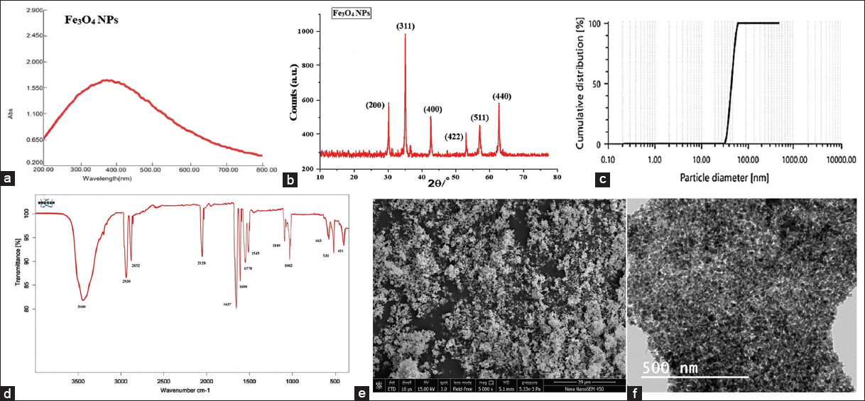 | Figure 2: Characterization of Fe3O4 nanoparticles (NPs) (a) UV-Vis absorption spectra of the synthesized Fe3O4 NPs, (b) X-ray diffraction pattern of synthesized Fe3O4 NPs, (c) particle size distribution image of the synthesized Fe3O4 NPs, (d) Fourier transform infrared spectrum of the synthesized Fe3O4 NPs, (e) scanning electron microscope image of the synthesized Fe3O4 NPs, and (f) transmission electron microscopy image of the synthesized Fe3O4 NPs. [Click here to view] |
4.2. Estimation of Germination Percentage, SL, and RL of Tomato Seedlings
The impacts of Fe3O4 NPs and FeSO4 salt on different biophysical and biochemical parameters of tomato seedlings were studied. The determination of optimal concentration of NPs is critical for stimulating plant growth, while negative or positive effects can be observed at different concentrations. Taking control as a reference, maximum germination % was recorded in tomato seedlings treated with 10 mg/L of Fe3O4 NPs. The germination % was found to rise by 150% at 10 mg/L concentration of Fe3O4 NPs while an 8.69% decrease in germination was reported in seedlings treated with 10 mg/L of FeSO4 salt. Further increase of salt concentration declined the germination % of tomato seedlings. At 100 mg/L of Fe3O4 NPs and FeSO4 salt, the germination % was inhibited by 60% and 84.61%. The overall impact of Fe3O4 NPs on germination % was significantly higher than FeSO4.7H2O treatments [Figure 3 and Table 1]. The maximum SL with a 104.83% increase as compared to control was observed at 10 mg/L while a 7.34% decrease in SL was reported at 50 mg/L of Fe3O4 NPs treatments. The FeSO4 treatments at 10 mg/L showed a 5.99% decrease in SL while maximum inhibition of 28.73% was observed at 100 mg/L as compared to control [Figure 3 and Table 1]. The RL was found to increase by 22.56% at 10 mg/L concentration of Fe3O4 NPs treatments and 8.24% increase in FeSO4.7H2O treated seedlings. At 100 mg/L of Fe3O4 NPs and FeSO4.7H2O RL was found to inhibit by 5.24% and 13.75% with respect to control [Figure 3 and Table 1]. The result was in agreement with other findings of Fe3O4 NPs utilization as nanonutrient for different crop species. The physicomorphological response of finger millets was significantly enhanced with Fe3O4 NPs treatments as compared to FeSO4 salt treatments [49]. Fe3O4 NPs application at 100 mg/L showed prominent improvement in the seed germination %, plant growth, and biomass of barley (Hordeum vulgare L.) crops [32]. At 0.3 mg concentration of biogenic synthesized Fe3O4 NPs showed potent rise in germination %, growth, and yield of maize crops [57]. Nano-Fe3O4 NPs at 20 mg/L were observed to mitigate the effect of cadmium with enhanced SL, RL, and leaf surface area of tomato seedlings [34]. The coated Fe3O4 NPs also reported to increase the bioavailability of Fe content in tomato seeds [21]. Thus, the concentration dose and size of the same synthesized NPs have a different impact on physiological and morphological growth parameters of different crop species.
Table 1: Effect of Fe3O4 NPs on germination, shoot length, and root length of tomato seedlings under Fe salt and Fe3O4 NPs treatments.
| Treatments | Germination (%) | Root length (cm) | Shoot length (cm) |
|---|---|---|---|
| C | 7.66±0.33a | 9.30±0.06e | 10.56±0.03c |
| FeS1 | 6.83±0.56a | 10.06±0.07bc | 9.93±0.13d |
| FeS2 | 4.33±0.33b | 9.93±0.09c | 8.70±0.06f |
| FeS3 | 0.66±0.31c | 8.56±0.07f | 6.20±0.02g |
| FeNP1 | 8.33±0.35a | 10.5±0.06a | 12.70±0.02a |
| FeNP2 | 5.12±0.53b | 10.16±0.07b | 11.76±0.3b |
| FeNP3 | 2.65±0.23c | 9.63±0.03d | 9.0±0.01e |
Data are means±standard error of three independent experiments with three replicates in each experiment. Bars followed by different letters show significant differences at P<0.05 significance level between treatments according to the Duncan’s multiple range test. C=control; FeNP1=10; FeNP2=50; FeNP3=100; FeS1=10; FeS2=50; and FeS3=100 mg/L.
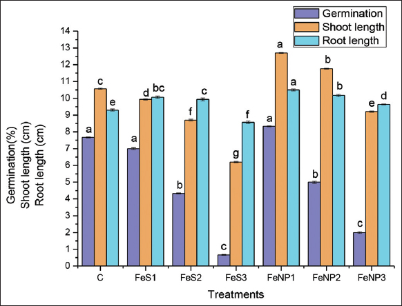 | Figure 3: Effect of Fe3O4 NPs on germination, shoot length and root length of tomato seedlings. Data are means ± standard error of three independent experiments with three replicates in each experiment. Bars followed by different letters show significant differences at p<0.05 significance level between treatments according to the Duncan’s multiple range test. C= control; FeNP1=10; FeNP2= 50; FeNP3= 100; FeS1= 10; FeS2= 50; FeS3= 100 mg/l. [Click here to view] |
4.3. Estimation of Total Chlorophyll Content
Iron is an essential microelement for the formation of chlorophyll and thereby high biomass production. The chlorophyll content showed a significant increase of 42% at 10 mg/L concentration of Fe3O4 NPs treatments while seedlings treated with 10 mg/L FeSO4 salt observed to have a 2% decrease in total chlorophyll content. A contradictory impact of treatment with FeSO4 salt was observed which tends to enhance the total chlorophyll content at 50 mg/L by 7%, while 5% decrease in total chlorophyll content at 100 mg/L of its concentration, as compared to the control [Figure 4]. Similarly, the total chlorophyll content (Chl a and Chl b) was reported to rise in finger millets exposed to Fe3O4 NPs as compared to the bulk form of their salt [49]. Increase in chlorophyll content was also observed in some other crops treated with Fe3O4 NPs such as green-gram [58], wheat [59], and watermelon [14].
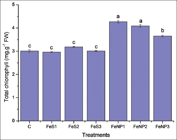 | Figure 4: Effect of Fe3O4 nanoparticles (NPs) on total chlorophyll content of tomato seedlings. Data are means ± standard error of three independent experiments with three replicates in each experiment. Bars followed by different letters show significant differences at P < 0.05 significance level between treatments according to the Duncan’s multiple range test. C= control; FeNP1=10; FeNP2 = 50; FeNP3= 100; FeS1 = 10; FeS2= 50; and FeS3 = 100 mg/L. [Click here to view] |
4.4. Estimation of NR Activity
Iron is one of the subunits present in NR enzyme, a key enzyme for catalyzing nitrate reduction to nitrite (NO3- into NO2-) which is the measure source of plant nitrogen. NR activity regulates plant growth and helps in developing resistance against different biotic and abiotic stresses. The maximum rise of 23.21% in NR activity was observed in seedlings treated with 10 mg/L concentration of Fe3O4 NPs whereas FeSO4 salt at a similar concentration showed a 4.47% increase in NR activity. At 50 mg/L and 100 mg/L of FeSO4 salt treatments, the NR activity declined by 7.02% and 8.52% with respect to control [Figure 5]. At 40 ppm, foliar treatment of Fe3O4 NPs caused significant increases in NR of M. oleifera L. [33]. Nitrogen assimilation and growth of green beans were significantly enhanced with the application of Fe2O3 NPs [60].
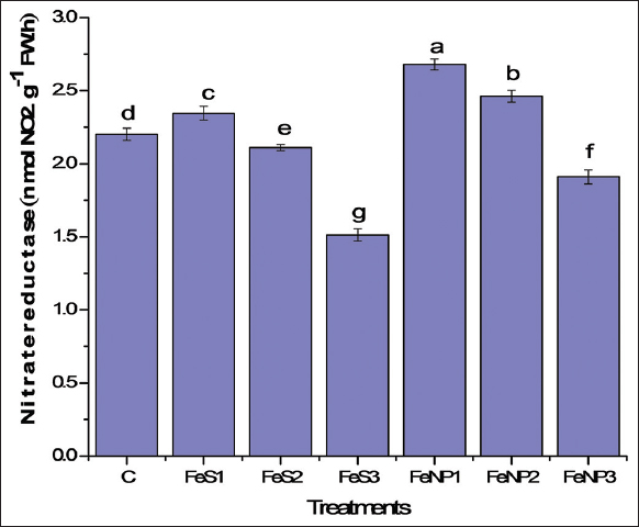 | Figure 5: Effect of Fe3O4 nanoparticles (NPs) on nitrate reductase activity of tomato seedlings. Data are means ± standard error of three independent experiments with three replicates in each experiment. Bars followed by different letters show significant differences at P < 0.05 significance level between treatments according to the Duncan’s multiple range test. C= control; FeNP1=10; FeNP2 = 50; FeNP3= 100; FeS1 = 10; FeS2= 50; and FeS3 = 100 mg/L. [Click here to view] |
4.5. Estimation of LP, EL, Proline Content, and SOD Activity
The indicator of oxidative stress biomarkers in plant growth is rate of LP (MDA content), EL, and proline content. The seedlings exposed to high concentrations of NPs generate enormous amounts of reactive oxygen species (ROS) as a result of high oxidative stress caused to them. The decrease in membrane integrity of plant cells indicates a high EL percentage. The EL at 10 mg/L of Fe3O4 NPs was found to decrease by 52.08% as compared to control. The maximum EL % was reported in seedlings treated with 100 mg/L of FeSO4 salt which was raised by 22.29% while at the same concentration of Fe3O4 NPs EL % was found to rise by 12.97% with respect to the untreated seedlings [Figure 6a]. The MDA content determines the rate of LP and oxidative damage caused in plant cells. The MDA content was observed to decrease by 59.49% on exposure of 10 mg/L of Fe3O4 NPs to tomato seedlings whereas 5.12% decrease of MDA content was reported with 10 mg/L treatment of FeSO4 salt. The maximum rate of LP in tomato seedlings was found at 100 mg/L of FeSO4 salt treatments in which the MDA content was found to rise by 36.20% as compared to control [Figure 6b]. Further, the proline accumulation was found to decrease by 57.23% exposure to 10 mg/L of Fe3O4 NPs and in the case of bulk form of iron salt at 10 mg/L proline content was found to decrease by 5.40% with reference to control. At 100 mg/L, the proline accumulation was found to increase by 42.25% and 25.61% with Fe3O4 NPs and FeSO4 salt treatments, respectively [Figure 6c]. SOD is a first line of defense mechanism in plants, which converts O2•−(superoxide radical) to O2 (oxygen). The 10 mg/L concentration of Fe3O4 NPs tends to reduce the oxidative stress with decline in SOD activity by 10% as compared to the control. The maximum % increase in SOD activity was 6.45% and 2.74% at 100 mg/L of Fe3O4 NPs and FeSO4 salt treatments, respectively [Figure 7]. Fe3O4 NPs under cadmium stress were found effective in regulating oxidative stress biomarkers such as MDA, aldehydes, H2O2, and proline in both shoot and root of tomato seedlings [34]. MDA, H2O2, and proline content tend to decline with the Fe3O4 NPs treatment in M. oleifera L. [33]. The membrane stability was found to improve under salt stress with the application of salicylic acid and Fe3O4 NPs in Trachyspermum ammi L. [61]. Tomato seedling’s response to salinity stress, high EL %, proline content, and antioxidant enzyme activity can be regulated with the iron and zinc oxide NPs treatments, also play a crucial role in callus induction and plant regeneration [62]. Iron (Fe) is an important cofactor of different antioxidant enzyme that plays a critical role in maintaining biological redox reaction system of plant cells [63]. Gupta et al. stated that increased antioxidant enzyme activity leads to homeostatic control of ROS levels in cells which in turn determines the state of the seeds during germination, dormancy, and senescence. They observed that foliar application of ZnO NPs and Fe3O4 NPs in the range of 100–300 mg/L in an open field tends to improve physiobiochemical parameters and antioxidant enzyme activity (SOD, CAT, and POD) of Cucumis sativus L; however, above the optimum concentration of 300 mg/L, it showed a deleterious effect [64]. Treatment of Triticum aestivum plants with Fe3O4 NPs (40–500 mg/L) showed prominent increase of Fe, P, and K content in its leaves with enhanced photosynthetic, respiration, and antioxidant enzyme activity (SOD, APX) thereby promoting the growth of plant [59]. The increase in SOD activity is attributed to activation of SOD isoforms that rely on Fe content and therefore elevated level of iron leads to high SOD activity that minimizes the harmful effect of ROS, increases the level of chlorophyll content that leads to the high photosynthetic activity of seedlings [59]. Fe3O4 nanozymes synthesized from extract of pomegranate fruit peel were observed to be good mimics of natural enzymes as that of peroxidase, catalase, and SOD [65]. Thus, low/optimal concentration of Fe3O4 NPs significantly promoted the overall growth of tomato seedlings which leads to high chlorophyll content, increase in NR and SOD activity, and helps in maintaining the level of EL %, MDA, and proline content as compared to the bulk form of FeSO4 salt.
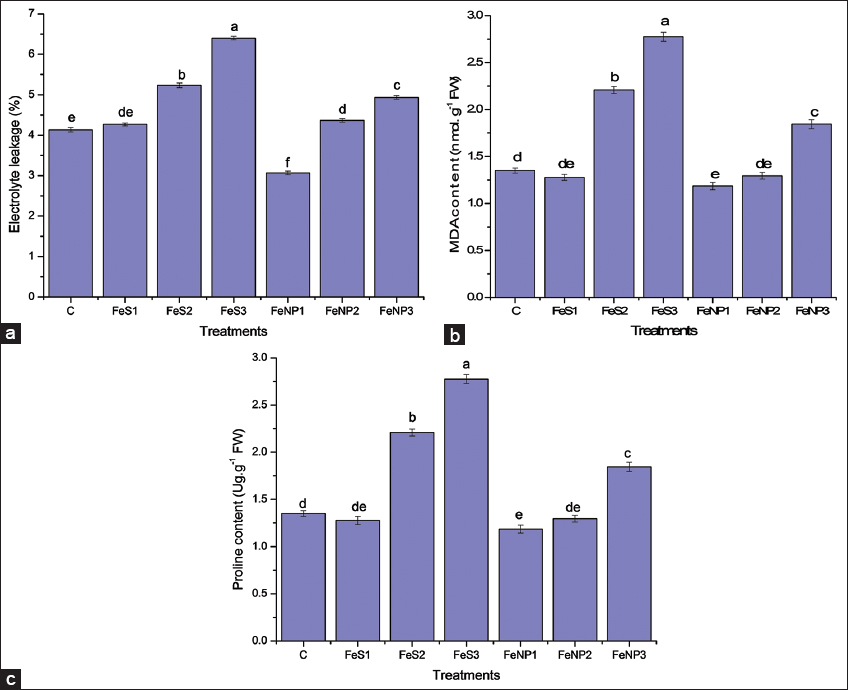 | Figure 6: Effect of Fe3O4 nanoparticles (NPs) on (a) Electrolyte leakage (b) Lipid peroxidation (c) Proline content of tomato seedlings. Data are means ± standard error of three independent experiments with three replicates in each experiment. Bars followed by different letters show significant differences at P < 0.05 significance level between treatments according to the Duncan’s multiple range test. C= control; FeNP1=10; FeNP2 = 50; FeNP3= 100; FeS1 = 10; FeS2= 50; and FeS3 = 100 mg/L. [Click here to view] |
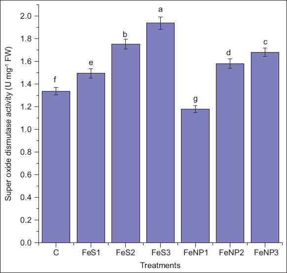 | Figure 7: Effect of Fe3O4 NPs on superoxide dismutase activity of tomato seedlings. Data are means ± standard error of three independent experiments with three replicates in each experiment. Bars followed by different letters show significant differences at P < 0.05 significance level between treatments according to the Duncan’s multiple range test. C= control; FeNP1=10; FeNP2 = 50; FeNP3= 100; FeS1 = 10; FeS2= 50; and FeS3 = 100 mg/l. [Click here to view] |
5. CONCLUSION
In recent decades, the impact of NPs on physicomorphological parameters of different crop species has been observed, which has been signified in our study also. To the best of our knowledge, this is the first kind of study that emphasizes to analyze the influence of Phyllanthus emblica L. mediated biologically synthesized Fe3O4 NPs on biophysical and biochemical parameters of S. lycopersicum L. Fe3O4 NPs formation were confirmed by spectral analysis, that is, UV-visible spectra, FT-IR, XRD, DLS, SEM, and TEM. The optimal concentration of 10 mg/L and 50 mg/L of Fe3O4 NPs was found effective in promoting germination, growth, chlorophyll content, and NR activity of tomato seedlings as compared to the bulk form of FeSO4 salt and control. The lower/optimal concentration of Fe3O4 NPs is found efficacious in reducing MDA content, EL, and proline accumulation. At higher concentration, SOD enzyme activity increases as the cellular defense system is stimulated for protection against ROS generated due to high oxidative stress. Altogether, the findings suggest that the iron absorption, uptake, transport, and its bioavailability to plants can be enhanced through the employment of Fe3O4 NPs as an efficient nanofertilizer to counter iron deficiency in crops.
6. ACKNOWLEDGMENT
The authors are thankful to the Department of Botany, K. S. Saket P. G. College, Dr. Ram Manohar Lohia Avadh University, Ayodhya, India, for providing the necessary laboratory and germination facilities, Department of Chemistry, Dr. HariSingh Gour Vishwavidyalaya, Sagar, Madhya Pradesh, India and Scientium Analyze Solutions, Jaipur, India, for providing necessary spectral facilities.
7. AUTHORS’ CONTRIBUTIONS
All authors made substantial contributions to the conception and design, acquisition of data, or analysis and interpretation of data; took part in drafting the article or revising it critically for important intellectual content; agreed to submit to the current journal; gave final approval of the version to be published; and agreed to be accountable for all aspects of the work. All the authors are eligible to be an author as per the International Committee of Medical Journal Editors (ICMJE) requirements/guidelines.
8. FUNDING
There is no funding to report.
9. CONFLICTS OF INTEREST
The authors report no financial or any other conflicts of interest in this work.
10. ETHICAL APPROVAL
This study does not involve experiments on animals or human subjects.
11. DATA AVAILABILITY
The data analyzed during the present study are included in the manuscript. The raw reading generated during the present study available from the corresponding author on reasonable request.
12. PUBLISHER’S NOTE
This journal remains neutral with regard to jurisdictional claims in published institutional affiliation.
REFERENCES
1. ChichiriccòG, Poma A. Penetration and toxicity of nanomaterials in higher plants. Nanomaterials (Basel) 2015;5:851-73. [https://doi.org/10.3390/nano5020851]
2. Le VN, Rui Y, Gui X, Li X, Liu S, Han Y. Uptake, transport, distribution and bio-effects of SiO2 nanoparticles in Bt-transgenic cotton. J Nanobiotechnology 2014;12:50. [https://doi.org/10.1186/s12951-014-0050-8]
3. Hassan NS, Salah El Din TA, Hendawey MH, Borai IH, Mahdi AA. Magnetite and zinc oxide nanoparticles alleviated heat stress in wheat plants. Curr Nanomater 2018;3:32-43. [https://doi.org/10.2174/2405461503666180619160923]
4. Chen HD, Yada R. Nanotechnologies in agriculture:New tools for sustainable development. Trends Food Sci Technol 2011;22:585-94. [https://doi.org/10.1016/j.tifs.2011.09.004]
5. Azim Z, Singh NB, Khare S, Singh A, Amist N, Yadav RK, et al. Green synthesis of zinc oxide nanoparticles using Vernonia cinerea leaf extract and evaluation as nano-nutrient on the growth and development of tomato seedling. Plant Nano Biol 2022;2:100011. [https://doi.org/10.1016/j.plana.2022.100011]
6. Rui M, Ma C, Hao Y, Guo J, Rui Y, Tang X, et al. Iron oxide nanoparticles as a potential iron fertilizer for peanut (Arachis hypogaea). Front Plant Sci 2016;7:815. [https://doi.org/10.3389/fpls.2016.00815]
7. El-Temsah YS, Oughton DH, Joner EJ. Effects of nano-sized zero-valent iron on DDT degradation and residual toxicity in soil:A column experiment. Plant Soil 2014;368:189-200. [https://doi.org/10.1007/s11104-012-1509-8]
8. Azim Z, Singh NB, Singh A, Amist N, Niharika, Khare S, et al. A review summarizing uptake, translocation and accumulation of nanoparticles within the plants:Current status and future prospectus. J Plant Biochem Biotechnol 2023;32:211-24. [https://doi.org/10.1007/s13562-022-00800-6]
9. Prajitha N, Athira SS, Mohanan PV. Bio-interactions and risks of engineered nanoparticles. Environ Res 2019;172:98-108. [https://doi.org/10.1016/j.envres.2019.02.003]
10. Tombuloglu H, Slimani Y, Tombuloglu G, Almessiere M, Baykal A. Uptake and translocation of magnetite (Fe(3)O(4)) nanoparticles and its impact on photosynthetic genes in barley (Hordeum vulgare L.). Chemosphere 2019;226:110-22. [https://doi.org/10.1016/j.chemosphere.2019.03.075]
11. Nel A, Xia T, Mädler L, Li N. Toxic potential of materials at the nanolevel. Science 2006;311:622-7. [https://doi.org/10.1126/science.1114397]
12. Aziz MK, Chauhan S, Azim Z, Bharati GK, Srivastava S. The biosynthesis of Nickel oxide nanoparticles using watermelon rind extract and their biophysical effects on the germination of Vigna radiata seeds at various concentrations. Int J Sci Res Arch 2022;7:245-54. [https://doi.org/10.30574/ijsra.2022.7.2.0271]
13. Siddiqi KS, Husen A. Engineered gold nanoparticles and plant adaptation potential. Nanoscale Res Lett 2016;11:400. [https://doi.org/10.1186/s11671-016-1607-2]
14. Li J, Chang PR, Huang J, Wang Y, Yuan H, Ren H. Physiological effects of magnetic iron oxide nanoparticles towards watermelon. J Nanosci Nanotechnol 2013;13:5561-7. [https://doi.org/10.1166/jnn.2013.7533]
15. Kah M, Beulke S, Tiede K, Hofmann T. Nanopesticides:State of knowledge, environmental fate, and exposure modeling. Crit Rev Environ Sci Technol 2013;43:1823-67. [https://doi.org/10.1080/10643389.2012.671750]
16. Liu R, Lal R. Potentials of engineered nanoparticles as fertilizers for increasing agronomic productions. Sci Total Environ 2015;514:131-9. [https://doi.org/10.1016/j.scitotenv.2015.01.104]
17. Yoon HY, Lee JG, Esposti LD, Iafisco M, Kim PJ, Shin SG, et al. Synergistic release of crop nutrients and stimulants from hydroxyapatite nanoparticles functionalized with humic substances:Toward a multifunctional nanofertilizer. ACS Omega 2020;5:6598-610. [https://doi.org/10.1021/acsomega.9b04354]
18. Askary M, Talebi SM, Amini F, Bangan AD. Effects of iron nanoparticles on Mentha piperita L. under salinity stress. Biologija 2017a;63:65-7. [https://doi.org/10.6001/biologija.v63i1.3476]
19. Hell R, Stephan UW. Iron uptake, trafficking and homeostasis in plants. Planta 2003;216:541-51. [https://doi.org/10.1007/s00425-002-0920-4]
20. Sanchez-Alcala IS, del Campillo MD, Barrón V, Torrent J. Evaluation of preflooding effects on iron extractability and phytoavailability in highly calcareous soil in containers. J Plant Nutr Soil Sci 2014;177:150-8. [https://doi.org/10.1002/jpln.201200302]
21. Raiesi-Ardali TR, Mamani L, Chorom M, Moezzi A. Improved iron use efficiency in tomato using organically coated iron oxide nanoparticles as efficient bioavailable Fe sources. Chem Biol Technol Agric 2022;9:59. [https://doi.org/10.1186/s40538-022-00318-y]
22. Mortvedt JJ. Correcting iron deficiency in annual and perennial plants:Present technologies and future prospects. Plant Soil 1991;130:273-9. [https://doi.org/10.1007/BF00011883]
23. Li X, Gui X, Rui Y, Ji W, Van Nhan L, Yu Z, et al. Bt-transgenic cotton is more sensitive to CeO2 nanoparticles than its parental non-transgenic cotton. J Hazard Mater 2014;274:173-80. [https://doi.org/10.1016/j.jhazmat.2014.04.025]
24. Askary M, Amirjani MR, Saberi T. Comparison of the effects of nano-iron fertilizer with iron-chelate on growth parameters and some biochemical properties of Catharanthus roseus. JPlant Nutr 2017b;40:974-82. [https://doi.org/10.1080/01904167.2016.1262399]
25. Laurie SH, Tancock NP, Mcgrath SP, Sanders JR. Influence of complexation on the uptake by plants of iron, manganese, copper and zinc. II. Effect of DTPA in a multi-metal and computer simulation study. J Exp Bot 1991;42:515-9. [https://doi.org/10.1093/jxb/42.4.515]
26. Pariona N, Martínez AI, Hernandez-Flores H, Clark-Tapia R. Effect of magnetite nanoparticles on the germination and early growth of Quercus macdougallii. Sci Total Environ 2017;575:869-75. [https://doi.org/10.1016/j.scitotenv.2016.09.128]
27. Elanchezhian R, Kumar D, Ramesh K, Biswas AK, Guhey A, Patra AK. Morpho-physiological and biochemical response of maize (Zea mays L.) plants fertilized with nano-iron (Fe3O4) micronutrient. J Plant Nutr 2017;40:1969-77. [https://doi.org/10.1080/01904167.2016.1270320]
28. Zhu H, Han J, Xiao JQ, Jin Y. Uptake, translocation, and accumulation of manufactured iron oxide nanoparticles by pumpkin plants. J Environ Monit 2008;10:713-7. [https://doi.org/10.1039/b805998e]
29. Barrena R, Casals E, Colón J, Font X, Sánchez A, Puntes V. Evaluation of the ecotoxicity of model nanoparticles. Chemosphere 2009;75:850-7. [https://doi.org/10.1016/j.chemosphere.2009.01.078]
30. Zahra Z, Arshad M, Rafique R, Mahmood A, Habib A, Qazi IA, et al. Metallic nanoparticle (TiO2 and Fe3O4) application modifies rhizosphere phosphorus availability and uptake by Lactuca sativa. J Agric Food Chem 2015;63:6876-82. [https://doi.org/10.1021/acs.jafc.5b01611]
31. López-Luna J, Silva-Silva MJ, Martinez-Vargas S, Mijangos-Ricardez OF, González-Chávez MC, Solís-Domínguez FA, et al. Magnetite nanoparticle (NP) uptake by wheat plants and its effect on cadmium and chromium toxicological behavior. Sci Total Environ 2016;565:941-50. [https://doi.org/10.1016/j.scitotenv.2016.01.029]
32. Tombuloglu H, Albenayyan N, Slimani Y, Akhtar S, Tombuloglu G, Almessiere M, et al. Fate and impact of maghemite (γ-Fe(2)O(3)) and magnetite (Fe(3)O(4)) nanoparticles in barley (Hordeum vulgare L.). Environ Sci Pollut Res Int 2021;29:4710-21. [https://doi.org/10.1007/s11356-021-15965-1]
33. Tawfik MM, Mohamed MH, Sadak MS, Thaloot AT. Iron oxide nanoparticles effect on growth, physiological traits and nutritional contents of Moringa oleifera grown in saline environment. Bull Natl Res Cent 2021;45:177. [https://doi.org/10.1186/s42269-021-00624-9]
34. Zadeh RR, Arvin SM, Jamei R, Mozaffari H, Nejhad FR. Response of tomato plants to interaction effects of magnetic (Fe3O4) nanoparticles and cadmium stress. J Plant Interact 2019;14:474-81. [https://doi.org/10.1080/17429145.2019.1626922]
35. Manzoor N, Ahmed T, Noman M, Shahid M, Nazir MM, Ali L, et al. Iron oxide nanoparticles ameliorated the cadmium and salinity stresses in wheat plants, facilitating photosynthetic pigments and restricting cadmium uptake. Sci Total Environ 2021;769:145221. [https://doi.org/10.1016/j.scitotenv.2021.145221]
36. Konate A, He X, Zhang Z, Ma Y, Zhang P, Alugongo GM, et al. Magnetic (Fe3O4) nanoparticles reduce heavy metals uptake and mitigate their toxicity in wheat seedling. Sustainability 2017;9:790. [https://doi.org/10.3390/su9050790]
37. Bombin S, LeFebvre M, Sherwood J, Xu Y, Bao Y, Ramonell KM. Developmental and reproductive effects of iron oxide nanoparticles in Arabidopsis thaliana. Int J Mol Sci 2015;16:24174-93. [https://doi.org/10.3390/ijms161024174]
38. Li J, Hu J, Xiao L, Wang Y, Wang X. Interaction mechanisms between a-Fe2O3, γ-Fe2O3 and Fe3O4 nanoparticles and Citrus maxima seedlings. Sci Total Environ 2018;625:677-85. [https://doi.org/10.1016/j.scitotenv.2017.12.276]
39. Wang H, Kou X, Pei Z, Xiao JQ, Shan X, Xing B. Physiological effects of magnetite (Fe3O4) nanoparticles on perennial ryegrass (Lolium perenne L.) and pumpkin (Cucurbita mixta) plants. Nanotoxicology 2011;5:30-42. [https://doi.org/10.3109/17435390.2010.489206]
40. Saini R, Sharma N, Oladeji OS, Sourirajan A, Dev K, Zengin G, et al. Traditional uses, bioactive composition, pharmacology, and toxicology of Phyllanthus emblica fruits:A comprehensive review. J Ethnopharmacol 2022;282:114570. [https://doi.org/10.1016/j.jep.2021.114570]
41. Hoagland DR, Arnon DJ. The Water Culture Method for Growing Plants without Soil. Circular 347. California Agriculture Experiment Station. Berkeley, CA:University of California;1950.
42. Arnon DI. Copper enzymes in isolated chloroplasts. Polyphenoloxidase in Beta vulgaris. Plant Physiol 1949;24:1-15. [https://doi.org/10.1104/pp.24.1.1]
43. Lichtenthaler HK. Chlorophylls and carotenoids:Pigments of photosynthetic biomembranes. Methods Enzymol 1987;148:350-82. [https://doi.org/10.1016/0076-6879(87)48036-1]
44. Jaworski EG. Nitrate reductase assay in intact plant tissues. Biochem Biophys Res Commun 1971;43:1274-9. [https://doi.org/10.1016/S0006-291X(71)80010-4]
45. Heath RL, Packer L. Photoperoxidation in isolated chloroplasts. I. Kinetics and stoichiometry of fatty acid peroxidation. Arch Biochem Biophys 1968;125:189-98. [https://doi.org/10.1016/0003-9861(68)90654-1]
46. Lutts S, Kinect JM, Bouharmont J. NaCl-induced senescence in leaves of rice (Oryza sativaL.) cultivars differing in salinity resistance. Ann Bot 1996;78:389-98. [https://doi.org/10.1006/anbo.1996.0134]
47. Bates LS, Waldren RD, Teare ID. Rapid determination of free proline for water-stress studies. Plant Soil 1973;39:205-7. [https://doi.org/10.1007/BF00018060]
48. Beauchamp C, Fridovich I. Superoxide dismutase:Improved assays and an assay applicable to acrylamide gels. Anal Biochem 1971;44:276-87. [https://doi.org/10.1016/0003-2697(71)90370-8]
49. Chandra AK, Pandey D, Tiwari A, Gururani K, Agarwal A, Dhasmana A, et al. Metal based nanoparticles trigger the differential expression of key regulatory genes which regulate iron and zinc homeostasis mechanism in finger millet. J Cereal Sci 2021;100:103235. [https://doi.org/10.1016/j.jcs.2021.103235]
50. Thoidingjam S, Tiku AB. Therapeutic efficacy of Phyllanthus emblica-coated iron oxide nanoparticles in A549 lung cancer cell line. Nanomedicine (Lond) 2019;14:2355-71. [https://doi.org/10.2217/nnm-2019-0111]
51. Dana E, Taha A, Afkar E. Green synthesis of iron nanoparticles by Acacia niloticapods extract and its catalytic, adsorption, and antibacterial activities. Appl Sci 2018;8:1922. [https://doi.org/10.3390/app8101922]
52. Azizi A. Green synthesis of Fe3O4nanoparticles and its application in preparation of Fe3O4/cellulose magnetic nanocomposite:A suitable proposal for drug delivery system. J Inorg Organomet Polym Mater 2020;30:3552-61. [https://doi.org/10.1007/s10904-020-01500-1]
53. Sandhya J, Kalaiselvam S. Biogenic synthesis of magnetic iron oxide nanoparticles using inedible Borassus flabellifer seed coat:Characterization, antimicrobial, antioxidant activity and in vitro cytotoxicity analysis. Mater Res Express 2020;7:015045. [https://doi.org/10.1088/2053-1591/ab6642]
54. Renuka R, Devi KR, Sivakami M, Thilagavathi T, Uthrakumar R, Kaviyarasu K. Biosynthesis of silver nanoparticles using Phyllanthus emblica fruit extract for antimicrobial application. Biocatal Agric Biotechnol 2020;24:101567. [https://doi.org/10.1016/j.bcab.2020.101567]
55. Kiwumulo HF, Muwonge H, Ibingira C, Lubwama M, Kirabira JB, Ssekitoleko RT. Green synthesis and characterization of iron-oxide nanoparticles using Moringa oleifera:A potential protocol for use in low and middle income countries. BMC Res Notes 2022;15:149. [https://doi.org/10.1186/s13104-022-06039-7]
56. Afrouz M, Ahmadi-Nouraldinvand F, Elias SG, Alebrahim MT, Tseng TM, Zahedian H. Green synthesis of spermine coated iron nanoparticles and its effect on biochemical properties of Rosmarinus officinalis. Sci Rep 2023;13:775. [https://doi.org/10.1038/s41598-023-27844-5]
57. Jayarambabu N, Rao KV, Park SH, Rajendar V. Biogenic synthesized Fe3O4nanoparticles affect on growth parameter of maize (Zea Mays L.). Dig J Nanomater Biostruct 2018;13:903-13.
58. Saleem S, Khan MS. Phyto-interactive impact of green synthesized iron oxide nanoparticles and Rhizobium pusense on morpho-physiological and yield components of greengram. Plant Physiol Biochem 2023;194:146-60. [https://doi.org/10.1016/j.plaphy.2022.11.013]
59. Feng Y, Kreslavski VD, Shmarev AN, Ivanov AA, Zharmukhamedov SK, Kosobryukhov A, et al. Effects of iron oxide nanoparticles (Fe3O4) on growth, photosynthesis, antioxidant activity and distribution of mineral elements in wheat (Triticum aestivum) plants. Plants 2022;11:1894. [https://doi.org/10.3390/plants11141894]
60. Gutiérrez-Ruelas NJ, Palacio-Marquez A, Sanchez E, Munoz-Marquez E, Chavez-Mendoza CC, Ojeda-Barrios DL, et al. Impact of the foliar application of nanoparticles, sulfate and iron chelate on the growth, yield and nitrogen assimilation in green beans. Not Bot Horti Agrobot Cluj Napoca 2021;49:12437. [https://doi.org/10.15835/nbha49312437]
61. Abdoli S, Ghassemi-Golezani K, Alizadeh-Salteh S. Responses of ajowan (Trachyspermum ammi L.) to exogenous salicylic acid and iron oxide nanoparticles under salt stress. Environ Sci Pollut Res Int 2020;27:36939-53. [https://doi.org/10.1007/s11356-020-09453-1]
62. Aazami MA, Rasouli F, Ebrahimzadeh A. Oxidative damage, antioxidant mechanism and gene expression in tomato responding to salinity stress under in vitro conditions and application of iron and zinc oxide nanoparticles on callus induction and plant regeneration. BMC Plant Biol 2021;21:597. [https://doi.org/10.1186/s12870-021-03379-7]
63. Torabian S, Zahedi M, Khoshgoftar AH. Effects of foliar spray of nano-particles of FeSO4 on the growth and ion content of sunflower under saline condition. J Plant Nutr 2017;40:615-23. [https://doi.org/10.1080/01904167.2016.1240187]
64. Gupta N, Jain SK, Tomar BS, Anand A, Singh J, Sagar V, et al. Impact of foliar application of ZnO and Fe(3)O(4) nanoparticles on seed yield and physio-biochemical parameters of cucumber (Cucumis sativus L.) seed under open field and protected environment vis a vis during seed germination. Plants (Basel) 2022;11:3211. [https://doi.org/10.3390/plants11233211]
65. Mundekkad D, Alex AV. Analysis of structural and biomimetic characteristics of the green-synthesized Fe3O4nanozyme from the fruit peel extract of Punica granatum. Chem Pap 2022;76:3863-78. [https://doi.org/10.1007/s11696-022-02130-2]