
ARTICLE HIGHLIGHTS
• Successfully purified an anticancer compound from a marine derived P. lentimorbus SAGM 3
• Anticancer compound was chemically confirmed as Chojalactone C based on spectral studies
• First report of Chojalactone C from a bacterium
• Chojalactone C exhibit promising anticancer activities against two breast cancer cell lines
1. INTRODUCTION
Around 70% of the Earth is covered by sea, and researchers have discovered up to 36 phyla residing within it [1]. The marine ecosystem has been speculated to contain a great number of different species [2], and their living resources are well known for their extensive variety of metabolites with unique features [3]. However, they are underexploited for the study of biomedical significance [4,5]. Among the available literature, sea anemones have recently gained attention as a promising new area for research and introducing chemical entities for use in therapeutical domains [6], e.g., actinoporin [7], equinatoxin [8], sea anemone peptide [9], etc. Patients in critical care, especially those suffering from cancer, acquired immunodeficiency syndrome, inflammatory conditions, arthritis, malaria, and many other infections from viruses, bacteria, and fungi, may find new hope in their therapies because of these entities [10].
According to Al-Zereini [11], the renewed interest in marine natural products is the result of rising demand for therapeutic compounds. Cancer is one of the most life-threatening diseases in the world, which needs immediate attention for drug discovery based on new approaches. Hence, metabolites originated in marine basins have become the focus of the current concept in pharmaceutical research [12,13]. At present, one-third of the top twenty therapeutic drugs on the market are derived primarily from plant sources. Molecules expressed by cancer cells, such as those involved in oncogenic signal transduction pathways, are the prime targets of lethal effects caused by natural bioactive compounds [14,15]. It has also been recorded that many marine-derived metabolites can effectively inhibit the growth of human cancer cells using animal and human clinical trials, which makes them significant in the biomedical sciences [16], e.g., Eribulin, Dolastatin 10, Bryostatin-1, etc. [17-19], but very few studies are available, hence this present study.
Paenibacillus is a group of Gram-positive and facultative anaerobic bacteria available in vast natural habitats [20]. Many species have been reported for agricultural applications, which include nitrogen fixation, phosphate solubilization, indole-3-acetic acid production, and siderophore secretion, and biomedical applications with the production of promising antimicrobial compounds such as polymyxins, fusaricidins, etc., further, for the production of exopolysaccharides and many industrially important enzymes [21,22]. In light of the above, Paenibacillus lentimorbus SAGM 3 was studied for anticancer compound synthesis, purification, and structural characterization. To our knowledge, the isolation and extensive structural characterization of an anticancer compound from P. lentimorbus have not been previously reported, which justifies the present research.
2. MATERIALS AND METHODS
2.1. Marine Bacterium and Cultural Conditions
Marine P. lentimorbus SAGM 3 used in this study for the synthesis of an anticancer compound was published earlier by our research team, which has reported the screening, molecular identification, and media formulation [23]. Briefly, P. lentimorbus SAGM 3 was isolated from a sea anemone, Heteractis species, which was identified using the 16S rRNA partial sequence method and submitted to the NCBI GenBank with the accession number MW737456.1. The production of an anticancer compound was performed using seawater-prepared media with the following conditions: 2% glucose, 1% ammonium nitrate, pH = 7.5, 37°C, 150 rpm agitation, and a 60 h incubation period, about 400 mL working volume was used in 1 L conical flasks for fermentation. After centrifugation at 3000 rpm for 15 min, the cell-free filtrate of the cultured broth evidenced appreciable anticancer effects using the MTT assay [24] against MCF7 and MDA-MB-231 breast cancer cell lines. Hence, further research aiming for the structural characterization of the extracted anticancer compound.
2.2. Purification of Anticancer Compound
The production of an anticancer compound was performed in a 1L conical flask with a 400 ml volume of production media, and extraction was done with cell-free supernatant using an equal volume of diethyl ether. After 8 h, the solvent phase was separated and dried under rotary vacuum evaporation. The collected solvent extract was dialyzed against phosphate buffer solution (PBS) at pH 7 for 24 h with intermediate changes of PBS to remove the salt, and the final solution was subjected to lyophilization. The lyophilized compound was confirmed to have anticancer activities against the MCF-7 and MDA-MB-231 cancer cell lines. Further, the lyophilized extract was purified for an anticancer compound at room temperature with reverse phase-column chromatography using C18 silica gel at 230–400 mesh size as the stationary substrate. The lyophilized extract was dispensed in 5ml of acetonitrile and water in a ratio of 3:2. Acetonitrile (A) and water (B) were used as the solvent systems, and 0.5 ml/min elution rate was accomplished using the stepwise gradient, starting with a ratio of 60:40 vol/vol (A: B) and ending with a ratio of 100:0 vol/vol (A: B). The fraction that evidenced appreciable anticancer activities in its purified state was biochemically and structurally identified using various spectral analyses as given below.
2.3. Structural Characterization of Purified Anticancer Compound
2.3.1. Fourier transforms infrared spectroscopy (FTIR)
The purified anticancer compound was characterized for its functional groups using the FTIR spectrum. About 5 mg of purified compound was added to 500 mg of KBr, mixed vigorously in a mortar, and pressurized for 2 min at 6 bars to produce a thin disc, which was then set in a diffuse reflectance accessory. The spectrum was filed using an IR affinity FTIR system from Shimadzu under 4 cm-1 spectral resolution within 400–4000 cm-1 wavelength [25]. All the estimations were based on 500 scans, and a KBr pellet served as a background reference.
2.3.2. Nuclear Magnetic Resonance Spectroscopy (NMR)
The chemistry of the purified anticancer compound was confirmed using the spectral evaluations of the 1H and 13C-NMR spectra. The spectra were analyzed using a spectrophotometer, a Bruker AV 600 NMR in Germany, in which deuterated CDCl3 was used as the solubilizing agent at 25°C and operating at 300.13 MHz. Chemical shifts. Chemical shifts were represented here as ppm (parts/million), and tetramethylsilane was used as an internal standard [26].
2.3.3. Gas chromatography and mass spectroscopy (GC-MS)
For GC analysis, the purified component was dissolved in diethyl ether and methylated [27]. GC and MS/MS spectra were recorded using a Thermo Trace GC Ultra coupled with a Polaris Q MS and TriPlus auto-sampler with helium as the carrier gas in the DB-5 column (0.25 mm × 30 m × 0.22 μm). The initial temperature of 50°C was set for 2 min, the final temperature of 250°C was maintained for 10 min, and the flow rate was held at 1ml/min, which has a total experimental period of 32 min. Mass spectra were recorded in scan mode within 0–300 m/z with an ion trap EI+. The temperature of the ion source was fixed at 200°C. The mass spectrum of the resulted methyl ester was compared with the one available in the NIST database.
2.4. Anticancer Activities
The anticancer activities of the purified compound were evaluated using two breast cancer cell lines, viz., MCF7 and MDA-MB-231 (adenocarcinoma), purchased from the National Centre for Cell Sciences, Pune, India. The cells were cultured in Dulbecco’s modified Eagle’s medium along with 10% fetal bovine serum, 2 mM L-glutamine, and 1% penicillin G/streptomycin under a 5% CO2 incubator at 37°C. During the experiment, cells were treated for 24 h under the same temperature and humid conditions with a purified compound ranging from 100–800 μg/mL dissolved in DMEM and added with 5% fetal calf serum, 2 mM L-glutamine, and 1% antibiotic mixture. Sodium lauryl sulfate was used as a positive control. The assay was carried out in 96-well microtiter plates by plating cancer cell lines at a concentration of 1.5 × 10⁴ cells/well. The activities of the purified compound against the cancer cell lines were evaluated using a 550 nm wavelength on a multimode reader (Biotek Elx 808, WI, USA) with an MTT assay [24]. All the values were represented as percentages from triplicate data. Further, microscopic observations were performed on treated cells using Calcein AM and Ethidium homodimer III, in which Calcein AM is a membrane-permeable fluorescent dye that stains all viable cells and Ethidium homodimer III is a membrane-impermeable dye that stains all dead cells on well plates under a fluorescent microscope (ZEISS LSM 880, Germany). Fluorescence was noted by a 490 nm excitation filter and a 520 emission filter for Calcein AM, a 545 nm excitation filter and a 620 emission filter for Ethidium homodimer III, and a 495 nm excitation filter, which was above the range of the 520 emission filter for the combined stains (Calcein AM and Ethidium homodimer III).
3. RESULTS AND DISCUSSION
3.1. Purification of an Anticancer Compound
Cancer is one of the greatest challenges to modern society and health-care systems [28]. Every year, more than 18 million new cases are recorded, with a more than 50% mortality rate [29,30]. This data reveal the emergency for more research in the discovery of promising anticancer therapeutic drugs, which warrants the present study. In this study, the cell-free supernatant of a marine symbiotic bacterium, P. lentimorbus SAGM 3, isolated from a sea anemone, Heteractis sp., exhibited promising anticancer properties against two cancer cell lines, MCF-7 and MDA-MB-231, and its biochemical components were solvent extracted and purified for extensive research. Among the collected fractions during the purification procedure, the 70:30 and 71:29 fractions showed maximum anticancer activities of 100% against both the two cancer cells at 103 mg, which was further dried using a rotary vacuum evaporator and used for the detailed spectral characterization studies. Supporting this study, lyophilized cell-free extracts of sponge-associated bacteria, Pseudomonas fluorescens BCPBMS-1, and Penicillium citrinum, evidenced cytotoxic activity against HEp-2 cancer cells [31]. Likewise, an alkaloid compound obtained from Saccharomonospora sp. exhibited cytotoxicity against human colon cancer cells (HCT-116) [32].
3.2. FTIR Analysis
FTIR analysis was performed on the purified anticancer compound for the identification of function groups present in it [Figure 1]. Spectral peaks recorded at 2954, 2924, and 2853 cm-1 indicated the presence of aliphatic alkane groups (CH), and peaks at 1647 and 1636 cm-1 stated the aliphatic alkene (CH=CH). Similarly, the functional group of aromatic alkanes was observed from the peaks at 888 and 720 cm-1, and aromatic alkenes were predicted from the 1541, 1458, and 1402 cm-1 peaks. At 1735 and 1717 cm-1, a significant ester group was detected; similarly, 1740 and 1653 cm-1 evidenced a ketone group. All the functional groups predicted in the FTIR spectrum revealed the possible structure of (3R)-3-hydroxy-4-methylidene-3-[(2E, 4E, 6E)-octa-2,4,6-trienoyl]oxolan-2-one. Thus, this study further characterized the structure of this biochemical compound using NMR and GC-MS/MS analysis. Similar to this study, FT-IR spectral analysis was used to figure out the new structures of the bioactive compounds Aneurinifactin and Cybersan, which were extracted from a marine bacterium Aneurinibacillus aneurinilyticus SBP-11 and a marine yeast Cyberlindnera saturnus SBPN-27 [33,34].
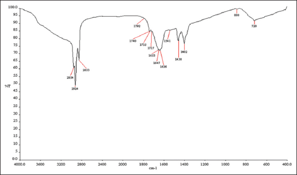 | Figure 1: Fourier transform infrared spectroscopy spectrum of the purified Chojalactone C from a marine Paenibacillus lentimorbus SAGM 3. [Click here to view] |
3.3. 1H and 13C NMR Analysis
1H and 13C NMR spectra of the purified anticancer compound evidenced the possible structure of (3R)-3-hydroxy-4-methylidene-3-([2E,4E,6E]-octa-2,4,6-trienoyl) oxolan-2-one, and the respective spectra are shown in Figure 2a and 2b. 1H NMR spectra indicated the presence of hydrogen atoms in the terminal aliphatic alkane (CHn) at 1.3334–1.8640 ppm and in the aliphatic alkene (CH=CH) between 1.9013 and 2.5983 ppm. Similarly, the chemical shifts between 2.6007 and 2.6431 ppm and 8.0772 and 8.1927 ppm represented the protons of aromatic alkane (CH3) and aromatic alkene (CH=CH). Further, the significant functional groups of this purified anticancer compound were represented by the hydrogen atoms present in the ester group (R-CO-O-CH2), ketone (R-CO-CH), and aldehyde (C-OH) groups, which were represented within the chemical shifts of 3.7228–3.8338 ppm, 3.1990–3.2917 ppm, and 9.8542–9.9731 ppm, respectively.
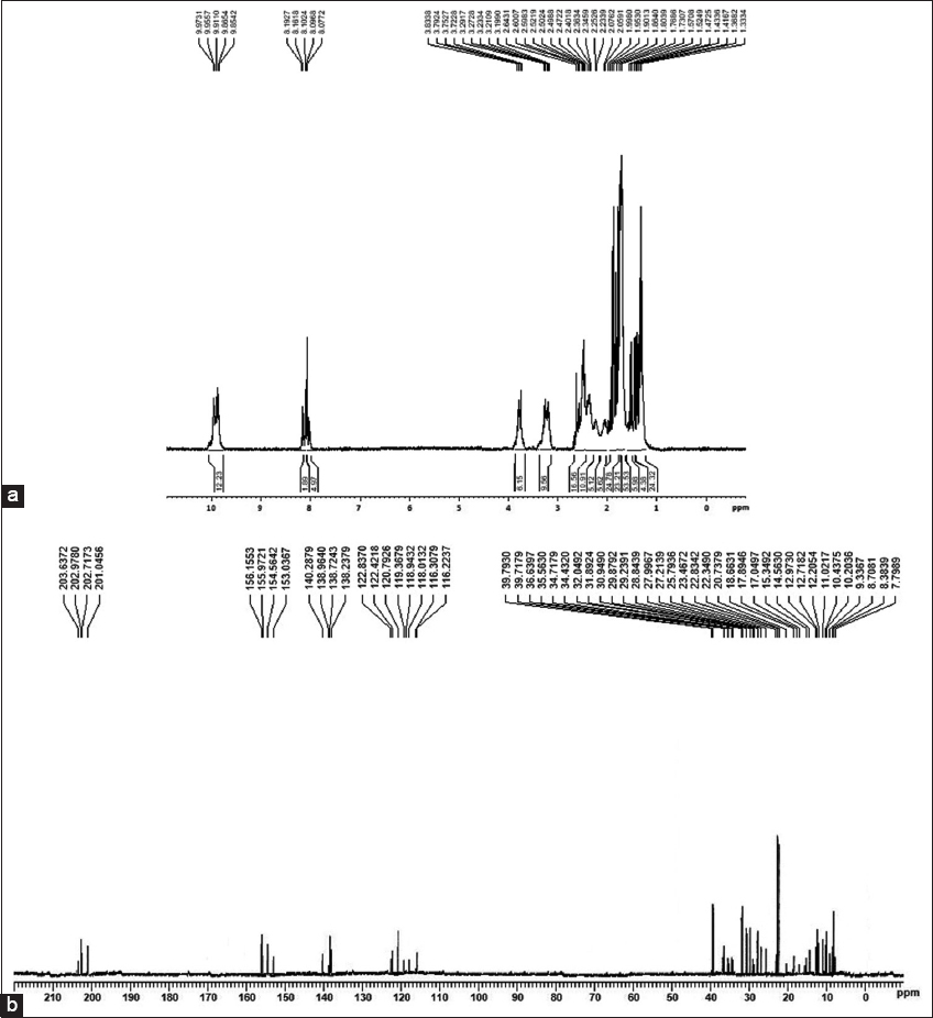 | Figure 2: (a) H1 and (b) C13 NMR spectrum of the purified Chojalactone C from a marine Paenibacillus lentimorbus SAGM 3. [Click here to view] |
Likewise, the 13C NMR spectrum also revealed the possible structure of (3R)-3-hydroxy-4-methylidene-3-([2E,4E,6E]-octa-2,4,6-trienoyl) oxolan-2-one from the analyzed spectra, as shown in Figure 3a and b. The carbon atoms present in the terminal aliphatic alkane (CH3) and aliphatic alkene (CH=CH) were represented between the chemical shifts of 7.7989-18.6631 ppm and 116.2237-122.8370 ppm. Furthermore, the presence of carbon atoms in aromatic alkane (CH2) and aromatic alkene (C=CH2) is evidenced by the chemical shifts between 153.0367–156.1553 ppm and 138.2379–140.2879 ppm. The carbon atom present in the significant functional group of ester (R-CO-O-R) was observed between 20.7379–29.8792 ppm; ketone (R-CO-CH) was observed between 30.9490–39.7930 ppm; and aldehyde (C-OH) within the chemical shifts of 201.0456–203.6372 ppm, respectively.
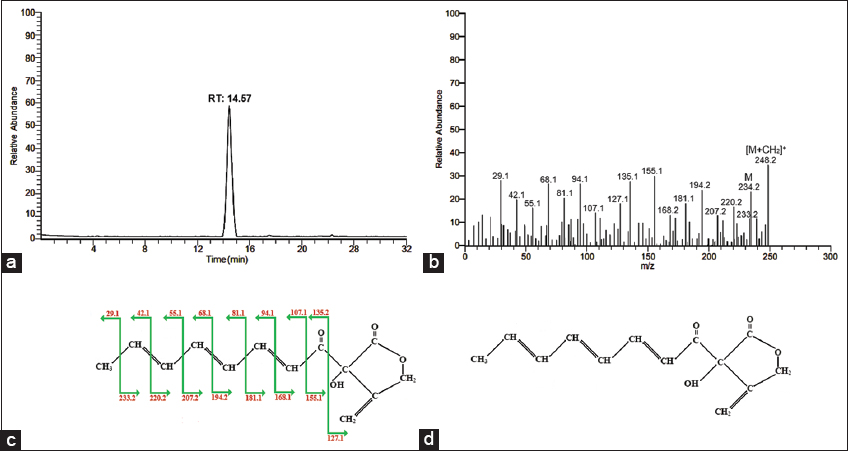 | Figure 3: (a) Gas chromatography, (b) mass spectrometry (MS)/MS spectrum, (c) fragmentation pattern and (d) predicted structure of the purified compound, Chojalactone C ([3R]-3-hydroxy-4-methylidene-3-[(2E,4E,6E)-octa-2,4,6-trienoyl]oxolan-2-one) from a marine Paenibacillus lentimorbus SAGM 3. [Click here to view] |
From the results of the 1H and 13C NMR spectra, the purified anticancer compound revealed all the functional groups of (3R) -3-hydroxy-4-methylidene-3-[(2E,4E,6E)-octa-2,4,6-trienoyl] oxolan-2. Supporting this study, Menéndez et al. [35] purified an anticancer compound, Chromomycin SA, derived from Streptomyces griseus based on the spectral details of NMR. Likewise, NMR spectra were used to elucidate the novel structures of a lipopeptide and a glycolipid bioactive compound, namely Pontifactin and Staphylosan, derived from marine bacterial strains of Pontibacter korlensis SBK-47 and Staphylococcus saprophyticus SBPS [36,37].
3.4. GC and MS/MS Analysis
The biochemical structure of the purified anticancer compound was studied using GC and MS/MS analysis. The sample revealed a single major peak at the retention time of 14.57 min, which revealed the purification of the compound [Figure 3a]. Using MS/MS analysis, the molecular mass (m/z) of the purified compound was identified as 234.2 m/z, and its methylated compound was determined at 248.2 m/z [Figure 3b]. Further, the fragmentation pattern of the purified compound exactly matched the pattern of (3R)-3-hydroxy-4-methylidene-3-([2E,4E,6E]-octa-2,4,6-trienoyl]oxolan-2-one as per the available database in NIST [Figure 3c]. In addition to the GC analysis, the biochemical structure of the anticancer compound was identified using FTIR and NMR analysis as (3R)-3-hydroxy-4-methylidene-3-([2E,4E,6E]-octa-2,4,6-trienoyl)oxolan-2-one, as illustrated in Figure 3d, as evidenced by the overall spectral database performed in this study.
Moreover, this structure was earlier reported from an actinobacterium, Streptomyces sp. CJ-5, which was named as Chojalactone C along with the similar forms of Chojalactone A and B. In the same study, this mixed form exhibits moderate anticancer activity against leukemia P388 cells, and the mechanism of action is unknown till date [38]. In support of this study, an antimicrobial compound, (E)-10-hydroxy-4,6,8,10-tetramethyldodec-4-en-3-one, was identified based on the same systematic spectral profiling of FTIR, NMR, GC, and MS/MS analysis that was purified from a marine Paenibacillus macerans SAM 9 [39]. Likewise, a novel bioactive compound, trigalactomargarate, was identified from a marine Cyberlindnera saturnus SBPN-27 using the same schematic spectral scheme as evidenced in this study and was named “Cybersan” [34].
3.5. Anticancer Activities
The anticancer activities of Chojalactone C were evaluated with two breast cancer cell lines at different concentrations. Chojalactone C showed concentration-dependent anticancer activities in both cancer cell lines tested. Among the tested concentrations, Chojalactone C evidenced complete inhibition of MCF7 at 800 μg/ml, and MDA-MB-231 cells at 700 μg/ml, respectively, and the graphical data of the concentration-dependent activities were illustrated in Figure 4a and b. As per the observation, Chojalactone C was more toxic to MDA-MB-231 cells than MCF7 cells. Anticancer examinations of Chojalactone C against MCF7 and MDA-MB-231 cells were also studied under fluorescence microscopy using two fluorescent dyes: Calcein AM, a fluorescent green dye, denotes viable cells, and Ethidium homodimer III, a fluorescent red dye, denotes dead cells. The appearance of viable cancer cells observed at the positive control without any dead cells and no viable cancer cells were observed at the maximum tested concentrations revealed the cytotoxicity of Chojalactone C against the two breast cancer cell lines.
 | Figure 4: (a and b)Percentage cell growth inhibition of MCF7 and MDA-MB-231 breast cancer cell cell lines against different concentrations of purified Chojalactone C. [Click here to view] |
The representative microscopic examinations are given in Figure 5 and 6. In an earlier investigation, Chojalactone C was reported for its moderate anticancer activity against leukemia P388 cells [38]. Further, no more studies have been performed to date detailing its extensive anticancer potential. Hence, this present study was performed and identified promising anticancer activities of Chojalactone C against MCF7 and MDA-MB-231 breast cell lines. Similar to the present research, a novel flavonoid compound, 6-hydroxykaempferol-3, 6-dimethly ether, isolated from a marine actinobacterium, Streptomyces carpaticus RMS518F, was reported for moderate anticancer activity against human colon carcinoma (HCT-116) and hepatocellular carcinoma (Hep G2) cell lines [40]. Two compounds, cyclo-(L-Pro-D-Pro-L-Tyr-L-Tyr) and 2-hydroxyethyl-3-methyl-1,4-naphthoquinone, isolated from a marine actinobacterium, Actinoalloteichus cyanogriseus 12A22, from the South China Sea, revealed moderate anticancer activity against the human breast cancer (MDA-MB-435) cell line [41]. From the overall observations, this study provides valuable data to enhance more elaborate anticancer studies using Chojalactone C.
 | Figure 5: Anticancer activities of the Chojalactone C against MCF7 cell line under fluorescence microscopy using Calcein AM (fluorescent green colour denotes viable cells) and Ethidium homodimer III (fluorescent red colour denotes dead cells). [Click here to view] |
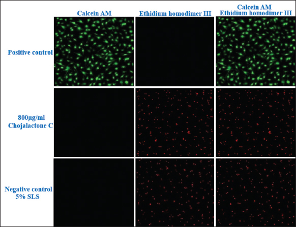 | Figure 6: Anticancer activities of the Chojalactone C against MDA-MB-231 cell line under fluorescence microscopy using Calcein AM (fluorescent green colour denotes viable cells) and Ethidium homodimer III (fluorescent red colour denotes dead cells). [Click here to view] |
4. CONCLUSION
This study purified an appreciable anticancer compound from a marine-derived P. lentimorbus SAGM 3, and it was identified as Chojalactone C based on various spectral examinations. Interestingly, this purified compound exhibits complete inhibition of two breast cancer cell lines, viz., MCF7 and MDA-MB-231 cells. These results hold promising data for a detailed study in the near future regarding its cytotoxicity and in vivo examinations intended for the possible development of a therapeutic drug.
5. ACKNOWLEDGMENT
The authors gratefully acknowledge the Department of Biotechnology, Faculty of Science, Annamalai University, Annamalai Nagar, Chidambaram, 608 002, Tamil Nadu, India, for providing lab facilities and supporting our research.
6. AUTHORS’ CONTRIBUTIONS
All authors made substantial contributions to conception and design, acquisition of data, or analysis and interpretation of data; took part in drafting the article or revising it critically for important intellectual content; agreed to submit to the current journal; gave final approval of the version to be published; and agreed to be accountable for all aspects of the work. All the authors are eligible to be an author as per the International Committee of Medical Journal Editors (ICMJE) requirements/guidelines.
7. FUNDING
There is no funding to report.
8. CONFLICTS OF INTEREST
The authors declare that they have no conflict of interest on publication of this article.
9. ETHICAL APPROVALS
This study does not involve experiments on animals or human subjects.
10. DATA AVAILABILITY
All data generated and analyzed are included within this research article.
11. PUBLISHER’S NOTE
This journal remains neutral with regard to jurisdictional claims in published institutional affiliation.
REFERENCES
1. Pomponi SA. The bioprocess-technological potential of the sea. In:Progress in Industrial Microbiology. Vol. 35. Amsterdam:Elsevier;1999. 5-13. [https://doi.org/10.1016/S0079-6352(99)80092-7]
2. Balan SS, Raffi SM, Jayalakshmi S. Probing of potential luminous bacteria in Bay of Bengal and its enzyme characterization. J Microbiol Biotechnol 2013;23:811-7. [https://doi.org/10.4014/jmb.1206.06020]
3. Balan SS. Production, Characterization, Evaluation of a Glycolipid Biosurfactant from a Marine Strain Bacillus cereus and Development of Non-toxic Skin and Hair Care Cosmetic Formulations, Doctoral Dissertation. India:Annamalai University;2014. 63-98.
4. Jimeno J, Faircloth G, Sousa-Faro JM, Scheuer P, Rinehart K. New marine derived anticancer therapeutics-a journey from the sea to clinical trials. Mar Drugs 2004;2:14-29. [https://doi.org/10.3390/md201014]
5. Senthilkumar K, Kim SK. Marine invertebrate natural products for anti-inflammatory and chronic diseases. Evid Based Complement Alternat Med 2013;2013:572859. [https://doi.org/10.1155/2013/572859]
6. Thangaraj S, Bragadeeswaran S. Assessment of biomedical and pharmacological activities of sea anemones Stichodactyla mertensii and Stichodactyla gigantea from Gulf of Mannar biosphere reserve, Southeast coast of India. J Venom Anim Toxins Incl Trop Dis 2012;18:53-61. [https://doi.org/10.1590/S1678-91992012000100007]
7. Fedorov S, Dyshlovoy S, Monastyrnaya M, Shubina L, Leychenko E, Kozlovskaya E, et al. The anticancer effects of actinoporin RTX-A from the sea anemone Heteractis crispa (=Radianthus macrodactylus). Toxicon 2010;55:811-7. [https://doi.org/10.1016/j.toxicon.2009.11.016]
8. Soletti RC, de Faria GP, Vernal J, Terenzi H, Anderluh G, Borges HL, et al. Potentiation of anticancer-drug cytotoxicity by sea anemone pore-forming proteins in human glioblastoma cells. Anticancer Drugs 2008;19:517-25. [https://doi.org/10.1097/CAD.0b013e3282faa704]
9. Moreels L, Peigneur S, Galan DT, De Pauw E, Béress L, Waelkens E, et al. APETx4, a novel sea anemone toxin and a modulator of the cancer-relevant potassium channel KV10.1. Mar Drugs 2017;15:287. [https://doi.org/10.3390/md15090287]
10. Rocha J, Peixe L, Gomes NC, Calado R. Cnidarians as a source of new marine bioactive compounds--an overview of the last decade and future steps for bioprospecting. Mar Drugs 2011;9:1860-86. [https://doi.org/10.3390/md9101860]
11. Al-Zereini W. Natural Products from Marine Bacteria, Doctoral Dissertation. Germany: Technical University of Kaiserslautern;2006.
12. Feinberg AP, Ohlsson R, Henikoff S. The epigenetic progenitor origin of human cancer. Nat Rev Genet 2006;7:21-33. [https://doi.org/10.1038/nrg1748]
13. Balan SS, Jayalakshmi S. Glycolipid biosurfactant production using low cost medium from marine bacterium Pseudomonas aeruginosa of Mudasalodai coast. Int J Green Chem Bioprocess 2013;3:33-7.
14. Howitz KT, Sinclair DA. Xenohormesis: Sensing the chemical cues of other species. Cell 2008;133:387-91. [https://doi.org/10.1016/j.cell.2008.04.019]
15. Newman DJ, Cragg GM. Natural products as sources of new drugs over the last 25 years. J Nat Prod 2007;70:461-77. [https://doi.org/10.1021/np068054v]
16. Häßler T, Schieder D, Pfaller R, Faulstich M, Sieber V. Enhanced fed-batch fermentation of 2,3-butanediol by Paenibacillus polymyxa DSM 365. Bioresour Technol 2012;124:237-44. [https://doi.org/10.1016/j.biortech.2012.08.047]
17. Newman DJ, Cragg GM. Advanced preclinical and clinical trials of natural products and related compounds from marine sources. Curr Med Chem 2004;11:1693-713. [https://doi.org/10.2174/0929867043364982]
18. Da Rocha AB, Lopes RM, Schwartsmann G. Natural products in anticancer therapy. Curr Opin Pharmacol 2001;1:364-9. [https://doi.org/10.1016/S1471-4892(01)00063-7]
19. Khalifa SA, Elias N, Farag MA, Chen L, Saeed A, Hegazy MF, et al. Marine natural products:A source of novel anticancer drugs. Mar Drugs 2019;17:491. [https://doi.org/10.3390/md17090491]
20. Zhang L, Chen S, Xie H, Tian Y, Hu K. Efficient acetoin production by optimization of medium components and oxygen supply control using a newly isolated Paenibacillus polymyxa CS107. J Chem Technol Biotechnol 2012;87:1551-7. [https://doi.org/10.1002/jctb.3791]
21. Daud NS, Din AR, Rosli MA, Azam ZM, Othman NZ, Sarmidi MR. Paenibacillus polymyxa bioactive compounds for agricultural and biotechnological applications. Biocatal Agric Biotechnol 2019;18:101092. [https://doi.org/10.1016/j.bcab.2019.101092]
22. Grady EN, MacDonald J, Liu L, Richman A, Yuan ZC. Current knowledge and perspectives of Paenibacillus:A review. Microb Cell Fact 2016;15:203. [https://doi.org/10.1186/s12934-016-0603-7]
23. Vinothkumar N, Pugalendhi P. Isolation and characterization of anticancer compound producing marine Paenibacillus lentimorbus SAGM 3 collected from a sea anemone, Heteractis species. Biosci Biotechnol Res Asia 2023;20:69-78. [https://doi.org/10.13005/bbra/3069]
24. Wilson AP. Cytotoxicity and viability assays. In:Animal Cell Culture - A Practical Approach, Vol. 1, New York:Oxford University Press;2000, 175-219. [https://doi.org/10.1093/oso/9780199637973.003.0007]
25. Dent G. Preparation of samples for IR spectroscopy as KBr disks. Internet J Vib Spectrosc 1996;1:1-2.
26. Mani P, Dineshkumar G, Jayaseelan T, Deepalakshmi K, Ganesh Kumar C, Senthil Balan S. Antimicrobial activities of a promising glycolipid biosurfactant from a novel marine Staphylococcus saprophyticus SBPS 15. 3 Biotech 2016;6:163. [https://doi.org/10.1007/s13205-016-0478-7]
27. Jenkins TC, Thies EJ, Mosley EE. Direct methylation procedure for converting fatty amides to fatty acid methyl esters in feed and digesta samples. J Agric Food Chem 2001;49:2142-5. [https://doi.org/10.1021/jf001356x]
28. Pacheco C, Baião A, Ding T, Cui W, Sarmento B. Recent advances in long-acting drug delivery systems for anticancer drug. Adv Drug Deliv Rev 2023;194:114724. [https://doi.org/10.1016/j.addr.2023.114724]
29. Huang J, Liu W, Kang W, He Y, Yang R, Mou X, et al. Effects of microbiota on anticancer drugs:Current knowledge and potential applications. EBioMedicine 2022;83:104197. [https://doi.org/10.1016/j.ebiom.2022.104197]
30. Yang S, Li D, Liu W, Chen X. Polysaccharides from marine biological resources and their anticancer activity on breast cancer. RSC Med Chem 2023;14:1049-59. [https://doi.org/10.1039/D3MD00035D]
31. Vasanthabharathi V, Jayalakshmi S. Bioactive potential from marine sponge Callyspongia diffusa associated Psedumonas fluorescens BCPBMS-1 and Penicillium citrinum. Microb Bioact 2018;1:8-13. [https://doi.org/10.25163/microbbioacts.11002A2221300318]
32. Maloney KN, Macmillan JB, Kauffman CA, Jensen PR, Dipasquale AG, Rheingold AL, et al. Lodopyridone, a structurally unprecedented alkaloid from a marine actinomycete. Org Lett 2009;11:5422-4. [https://doi.org/10.1021/ol901997k]
33. Balan SS, Kumar CG, Jayalakshmi S. Aneurinifactin, a new lipopeptide biosurfactant produced by a marine Aneurinibacillus aneurinilyticus SBP-11 isolated from Gulf of Mannar:Purification, characterization and its biological evaluation. Microbiol Res 2017;194:1-9. [https://doi.org/10.1016/j.micres.2016.10.005]
34. Balan SS, Kumar CG, Jayalakshmi S. Physicochemical, structural and biological evaluation of Cybersan (trigalactomargarate), a new glycolipid biosurfactant produced by a marine yeast, Cyberlindnera saturnus strain SBPN-27. Process Biochem 2019;80:171-80. [https://doi.org/10.1016/j.procbio.2019.02.005]
35. Menéndez N, Nur-e-Alam M, Braña AF, Rohr J, Salas JA, Méndez C. Biosynthesis of the antitumor chromomycin A3 in Streptomyces griseus:Analysis of the gene cluster and rational design of novel chromomycin analogs. Chem Biol 2004;11:21-32. [https://doi.org/10.1016/S1074-5521(03)00283-7 https://doi.org/10.1016/j.chembiol.2003.12.011]
36. Balan SS, Kumar CG, Jayalakshmi S. Pontifactin, a new lipopeptide biosurfactant produced by a marine Pontibacter korlensis strain SBK-47:Purification, characterization and its biological evaluation. Process Biochem 2016;51:2198-207. [https://doi.org/10.1016/j.procbio.2016.09.009]
37. Balan SS, Mani P, Kumar CG, Jayalakshmi S. Structural characterization and biological evaluation of Staphylosan (dimannooleate), a new glycolipid surfactant produced by a marine Staphylococcus saprophyticus SBPS-15. Enzyme Microb Technol 2019;120:1-7. [https://doi.org/10.1016/j.enzmictec.2018.09.008]
38. Hoshino S, Wakimoto T, Onaka H, Abe I. Chojalactones A-C, cytotoxic butanolides isolated from Streptomyces sp. cultivated with mycolic acid containing bacterium. Org Lett 2015;17:1501-4. [https://doi.org/10.1021/acs.orglett.5b00385]
39. Bharathi T, Sambandan K, Sivasubramani K. Structural characterization of (E)-10-hydroxy-4,6,8,10-tetramethyldodec-4-en-3-one bioactive molecule from marine Paenibacillus macerans isolate. Biosci Biotechnol Res Commun 2021;14:1228-33. [https://doi.org/10.21786/bbrc/14.3.34]
40. Selim RM, Shaaban M, Hamdy AA, Abou Zeid AA, Ata A. Viscosine:A new microbial flavonoid from marine-derived, Streptomyces sp. RMS518F. Vietnam J Chem 2019;57:288-95. [https://doi.org/10.1002/vjch.201900034]
41. Zhang X, Song C, Bai Y, Hu J, Pan H. Cytotoxic and antimicrobial activities of secondary metabolites isolated from the deep-sea-derived Actinoalloteichus cyanogriseus 12A22. 3 Biotech 2021;11:283. [https://doi.org/10.1007/s13205-021-02846-0]